

Loading, please wait...
Loading, please wait...

Clinicians recognize that Duchenne muscular dystrophy progressively compromises respiratory musculature. Consequently, affected individuals experience gradual diaphragmatic weakness and reduced chest wall compliance long before overt respiratory failure occurs. Early detection of ventilation inhomogeneity in DMD provides crucial insight into subtle physiological changes that precede severe hypoventilation. Specifically, microatelectasis develops in peripheral lung units as inspiratory forces decline. Furthermore, patients frequently adopt a rapid, shallow breathing pattern to compensate for diaphragmatic fatigue. Because peripheral airway clearance diminishes, retained secretions exacerbate uneven gas distribution across dependent alveoli. In addition, standard pulmonary function tests often fail to capture these focal mechanics because spirometry only reflects global lung performance. Therefore, understanding regional gas maldistribution allows clinicians to intervene before acute respiratory crises develop. Overall, recognizing these subtle changes enhances proactive clinical management. Moreover, progressive respiratory muscle weakness reduces functional residual capacity. As a result, small airway collapse occurs during resting tidal breathing. Clinicians must identify these regional abnormalities early because subclinical atelectasis predisposes vulnerable patients to recurrent lower respiratory tract infections. Ultimately, characterizing ventilation inhomogeneity in DMD bridges the gap between early structural muscle degradation and overt ventilatory insufficiency.
Assessing regional lung mechanics traditionally required computed tomography or radioisotope scintigraphy. However, both modalities expose vulnerable patients to ionizing radiation and require transport away from the bedside. Fortunately, electrical impedance tomography offers an exceptional alternative by providing dynamic, non-invasive imaging of regional ventilation. The technology applies minute, alternating electrical currents through an array of electrodes mounted on an elastic chest belt. Consequently, the device tracks alterations in thoracic bioimpedance as regional gas volumes change during the respiratory cycle. In addition, electrical impedance tomography operates entirely radiation-free, allowing repeated assessments at the bedside without safety concerns. Furthermore, the procedure does not require active patient effort or forced respiratory maneuvers, making it ideal for young patients with neuromuscular limitations. Therefore, clinicians can evaluate tidal breathing continuously under resting conditions. Moreover, advanced analytical algorithms generate cross-sectional functional images that accurately illustrate dynamic gas distribution. Thus, this diagnostic technology transforms neuromuscular respiratory assessment by capturing regional heterogeneity in real time. Similarly, clinicians obtain reproducible data without causing patient discomfort or fatigue. As a result, bedside electrical impedance monitoring provides objective physiologic indices that standard spirometric testing cannot deliver.
To investigate regional mechanics before ventilatory failure, investigators conducted an observational trial evaluating eighteen young men with confirmed Duchenne muscular dystrophy. Crucially, none of these participants had initiated long-term mechanical ventilatory assistance prior to study enrolment. Furthermore, the research team recruited eleven healthy, age-matched male medical residents to serve as a comparative control cohort. The investigators collected dynamic thoracic electrical impedance tomography measurements during resting spontaneous breathing across the entire calendar year. Specifically, the authors designated the Global Inhomogeneity Index as their primary functional parameter. This quantitative index calculates the relative variation of tidal impedance values across all responsive lung pixels. Consequently, lower values denote uniform gas distribution, whereas elevated scores indicate significant ventilation mismatch. In addition, the researchers performed comprehensive statistical comparisons to evaluate regional discrepancies between the two cohorts. Therefore, this rigorous methodology allowed precise quantification of subtle mechanical impairments during unassisted resting respiration. Moreover, the investigators standardized resting positions to eliminate gravitational confounders. In addition, standardizing these testing parameters ensured high data integrity across all longitudinal measurements. As a result, the protocol isolated intrinsic respiratory mechanics from external postural influences, thereby delivering robust comparative data between healthy individuals and neuromuscular patients.
The analytical results demonstrated striking functional differences between the two study populations. Notably, patients with Duchenne muscular dystrophy exhibited a significantly higher Global Inhomogeneity Index compared with healthy control subjects. The dystrophic cohort displayed a mean Global Inhomogeneity Index of 0.44, whereas the control group averaged 0.37. Furthermore, the wide distribution range among patients highlighted substantial individual variability in subclinical lung compromise. Consequently, these findings confirm that significant ventilation mismatch develops long before patients manifest daytime hypercapnia or obvious clinical failure. In addition, peripheral airway closure and localized microatelectasis clearly contribute to this measured heterogeneity. Because shallow breathing prevents complete lung expansion, dependent alveoli experience repetitive collapse and uneven refilling during tidal breaths. Therefore, the study proves that standard clinical assessment tools underestimate early respiratory decline. Moreover, these objective findings demonstrate that neuromuscular weakness impairs regional pulmonary ventilation during the initial stages of disease progression. Importantly, healthy control subjects maintained consistent, homogeneous air distribution throughout all lung zones. In contrast, dystrophic patients showed pronounced regional disparities despite maintaining seemingly adequate resting oxygenation. Furthermore, these insights emphasize why clinical teams must remain vigilant even when physical examinations appear entirely reassuring. Thus, functional imaging uncovers latent physiological abnormalities that conventional bedside monitoring routinely overlooks.
These observational insights carry profound clinical implications for neuromuscular pulmonary care. Firstly, identifying regional ventilation disparities enables clinicians to reconsider the optimal timing for proactive respiratory interventions. Currently, medical guidelines often rely on vital capacity declines or daytime hypercapnia to trigger ventilatory support. However, waiting for severe restriction delays treatment until extensive atelectasis and chest wall stiffness have already occurred. Consequently, incorporating electrical impedance tomography could identify candidates who benefit from early lung volume recruitment therapy. Furthermore, targeted airway clearance techniques, such as mechanical insufflation-exsufflation, can restore alveolar recruitment in poorly ventilated zones. In addition, starting nighttime non-invasive ventilation before overt respiratory exhaustion might prevent secondary chest wall deformities and recurrent pulmonary infections. Therefore, objective physiological indices allow pulmonologists to personalize therapeutic regimens according to actual regional mechanics. Ultimately, adopting early functional monitoring improves long-term respiratory trajectories and enhances patient quality of life. Similarly, regular monitoring of ventilation distribution helps clinicians assess the therapeutic efficacy of cough-assist protocols over time. As a result, proactive teams can adjust respiratory therapies dynamically, ensuring optimal lung inflation throughout every stage of neuromuscular disease progression.
Ventilation inhomogeneity develops primarily due to progressive respiratory muscle weakness, particularly affecting the diaphragm and intercostal musculature. Consequently, patients adopt rapid, shallow breathing patterns that fail to expand peripheral alveoli fully. In addition, weakened cough mechanics reduce effective secretion clearance, producing regional airway occlusion. Together, these mechanical factors promote patchy microatelectasis and altered local compliance. Therefore, resting tidal air distributes unevenly across dependent lung zones before hypoxemia or hypercapnia becomes clinically apparent.
Electrical impedance tomography measures dynamic changes in electrical conductivity across cross-sectional thoracic planes during tidal breathing. An elastic belt containing evenly spaced electrodes injects painless, imperceptible alternating currents into the chest. Because air-filled lung tissue exhibits higher electrical resistance than surrounding fluid or muscle, impedance fluctuations directly mirror regional volume changes. Consequently, specialized mathematical algorithms reconstruct real-time functional tomograms. These images allow clinicians to quantify ventilation distribution and calculate indices like the Global Inhomogeneity Index without radiation.
Early detection of regional inhomogeneity is vital because it reveals asymptomatic pulmonary deterioration long before conventional spirometry or blood gas tests detect abnormalities. Consequently, clinicians can initiate timely preventive therapies, including lung volume recruitment and assisted airway clearance, before extensive atelectasis becomes irreversible. Furthermore, identifying focal ventilation defects helps physicians optimize the exact timing of nocturnal non-invasive ventilation. Ultimately, early proactive intervention preserves chest wall mobility, mitigates recurrent chest infections, and substantially enhances long-term survival in vulnerable patients.
Disclaimer: This content is for informational and educational purposes only... Refer to the latest local and national guidelines for clinical practice.
References

Read summarized clinical updates, watch expert medical content, and earn CME certifications right from your smartphone.
Open your smartphone camera to scan and install.





A new study demonstrates that electrical impedance tomography can detect significant ventilation inhomogeneity in Duchenne muscular dystrophy patients before overt respiratory failure occurs, offering a non-invasive bedside tool to guide earlier pulmonary care and ventilatory support planning.
Today
A novel two-sample Mendelian randomization study demonstrates a causal genetic relationship between gout and an increased risk of epilepsy, highlighting the systemic neuro-inflammatory pathways connecting metabolic dysfunction and seizure disorders.
Today

A KGMU study reveals over half of women with breast abnormalities fail to undergo diagnostic confirmation, leading to advanced-stage detection. Discover the socio-cultural barriers, clinical risks of incomplete resection, and community-level solutions needed to bridge the critical screening-to-diagnosis gap in India.
Today

A comparative non-contact study reveals that high-power radiofrequency delivery with the QDOT MICRO catheter induces an artifactual contact force rise. Electrophysiologists must understand this power-dependent thermal and electrical phenomenon to avoid inadvertent catheter withdrawal and ineffective ablation.
Today
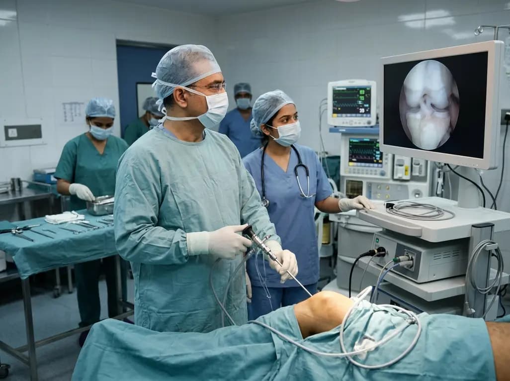
Healthium Medtech has transitioned its arthroscopy vertical into SIRONIX Orthopedics, aiming to capture key segments of the global joint care market. The strategic shift introduces dedicated portfolios in sports medicine, joint regeneration, surgical reconstruction, and digital operating technologies.
Today

Caffeoylquinic acids, such as chlorogenic acid, offer broad antioxidant, antidiabetic, and anti-inflammatory properties. Advanced plant cell and organ culture methods now enable scalable, sustainable, and pure production of these high-value therapeutic metabolites for pharmaceutical and clinical applications.
Today
