.webp&w=384&q=75)

Loading, please wait...
Loading, please wait...
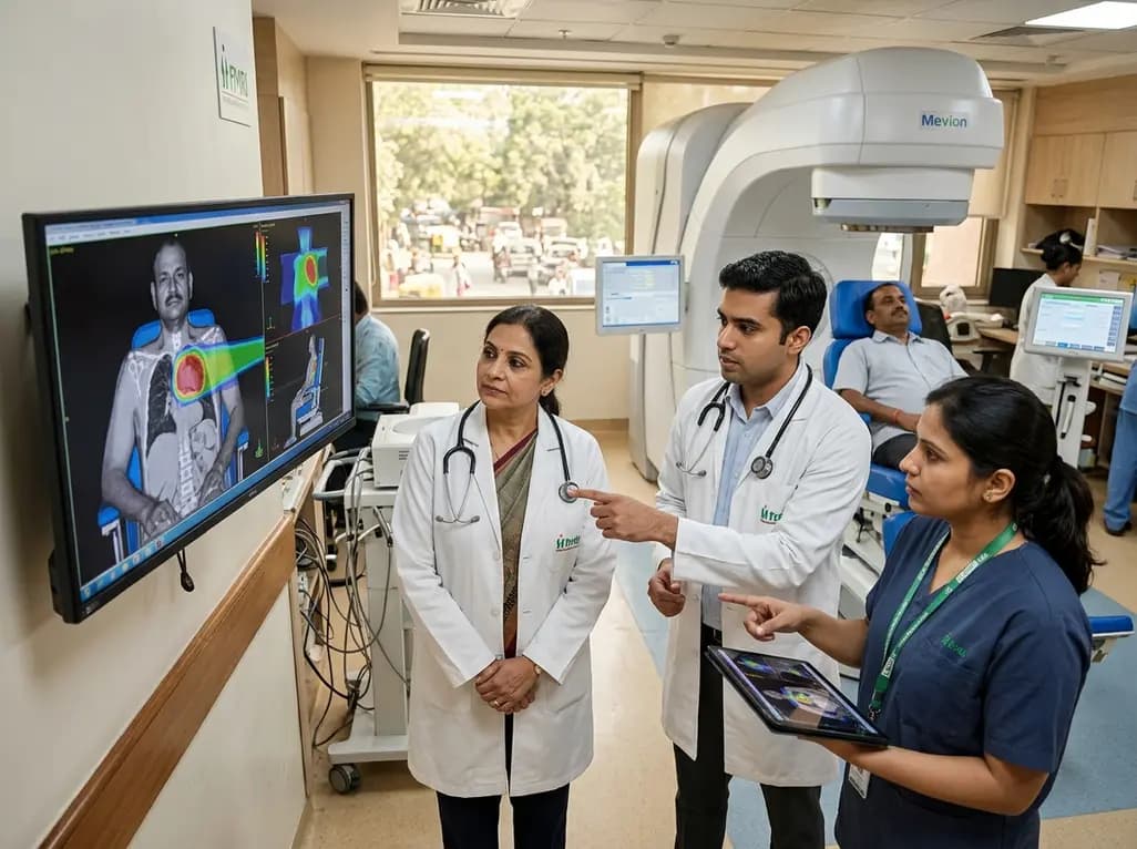
The landscape of precision radiation oncology is witnessing a major technological leap. Fortis Memorial Research Institute in Gurugram announced plans to introduce upright proton therapy, marking an unprecedented milestone for Indian oncology. Radiation oncologists constantly strive to maximize tumoricidal dose while protecting adjacent healthy tissues. Conventional photon radiotherapy delivers radiation along its entire trajectory, depositing significant exit doses into normal organs. In contrast, protons exploit the physical phenomenon of the Bragg peak. Consequently, proton beams deposit the maximum radiation dose precisely within the tumor and stop abruptly. By introducing a seated delivery paradigm, clinicians can elevate precision cancer care across diverse clinical scenarios. As Fortis Memorial Research Institute prepares for installation, this modality represents a monumental shift for Indian healthcare.
For decades, proton therapy facilities relied on massive, multi-story rotating gantries to direct proton beams around recumbent patients. These conventional installations require specialized infrastructure, immense concrete shielding, and significant financial capital. In contrast, upright proton therapy reimagines this delivery model by rotating the patient rather than the beam line. The Mevion S250-FIT Proton Therapy System integrates an ultra-compact synchrocyclotron accelerator with a fixed-beam delivery configuration. Furthermore, this compact system pairs seamlessly with the Marie upright positioning and imaging platform from Leo Cancer Care. Rather than rotating a gantry weighing over one hundred tons, the system rotates the seated patient with sub-millimeter mechanical accuracy. Additionally, RayStation treatment planning software provides real-time computational power to optimize complex beam trajectories. Therefore, hospitals can install advanced particle therapy inside vaults previously built for standard linear accelerators. As a result, this architectural innovation substantially lowers construction costs and site preparation barriers. Most importantly, clinical centers can dramatically expand patient access without necessitating gargantuan facility footprints. Furthermore, this streamlined physical setup allows seamless integration into busy tertiary hospital campuses. Medical physicists also benefit from simplified quality assurance protocols and reduced mechanical wear.
The clinical superiority of particle therapy stems fundamentally from proton physics. Standard high-energy photon beams exhibit an entrance dose, reach a shallow peak, and gradually attenuate as they traverse healthy tissue. Consequently, exit radiation inevitably damages normal anatomical structures beyond the target volume. In contrast, protons exhibit a characteristic Bragg peak, where particles travel through tissue with low energy deposition until reaching a defined depth. At that designated range, the protons release nearly their entire therapeutic dose and stop completely. Thus, zero exit dose reaches healthy tissues distal to the tumor margin. Moreover, advanced pencil beam scanning paints the prescribed dose layer by layer across complex target geometries. When combined with upright positioning, gravitational shifts optimize internal organ separation. For instance, gravity pulls pelvic bowel loops superiorly away from seated prostate targets, which reduces rectal and intestinal radiation exposure. Similarly, an upright posture naturally expands lung capacity and stabilizes diaphragmatic excursion during thoracic irradiation. Therefore, medical physicists can sculpt steep dose gradients that protect critical organs at risk. Consequently, radiation oncologists can safely treat deep-seated tumors with unprecedented margins of safety. Ultimately, these dosimetric advantages reduce acute toxicity and minimize long-term secondary malignancy risks.
Proton beam therapy delivers exceptional clinical utility across a wide spectrum of challenging malignancies. In pediatric oncology, tissues undergo active growth and exhibit profound radiosensitivity. Consequently, collateral radiation exposure during childhood cancer treatment can cause severe developmental delays, endocrine abnormalities, neurocognitive deficits, and secondary cancers. By eliminating exit dose, proton radiation spares critical pediatric structures including the developing brain, spinal cord, and heart. Furthermore, upright proton therapy offers vital advantages for complex adult tumors located near sensitive neural structures. Specifically, clinicians utilize proton beams for skull base chordomas, chondrosarcomas, central nervous system tumors, and recurrent head and neck cancers. For these patients, preserving adjacent optic nerves, brainstem tissue, and cochlear apparatus remains paramount to maintain functional quality of life. Additionally, thoracic malignancies such as locally advanced non-small cell lung cancer and mediastinal lymphomas benefit substantially. When treating left-sided breast cancer, proton therapy limits incidental cardiac radiation exposure, which prevents late ischemic heart disease. Therefore, oncologists can escalate radiation dose to aggressive tumors while maintaining acceptable therapeutic safety margins across diverse patient populations. Moreover, ongoing clinical protocols continue to demonstrate improved survival outcomes and fewer treatment interruptions when using particle beams.
Traditional radiation therapy forces patients to lie completely supine on a rigid carbon-fiber couch for prolonged periods. However, many cancer patients suffer from severe dyspnea, orthopnea, musculoskeletal pain, or superior vena cava syndrome. For these vulnerable individuals, lying flat induces immense physical discomfort and severe respiratory distress. In contrast, upright seating positions naturally facilitate diaphragm excursion and ease breathing mechanics. Patients suffering from excessive oral secretions or head and neck tumors experience far less aspiration risk when seated upright. Furthermore, the upright configuration significantly mitigates claustrophobia, which frequently complicates treatments inside narrow gantry bores. Patients maintain natural eye-level visual contact with radiation therapy technologists before beam delivery begins. Consequently, this dignified positioning fosters greater psychological comfort and substantially reduces treatment-associated anxiety. In addition, robust immobilization systems maintain rigorous sub-millimeter positional reproducibility throughout each treatment fraction. The integrated diagnostic-quality fan-beam CT scanner captures three-dimensional verification images directly at the treatment isocenter. Thus, clinicians confirm accurate tumor targeting without requiring uncomfortable patient repositioning, which markedly improves treatment compliance and overall clinical throughput. Therefore, patients remain calm and relaxed throughout treatment delivery.
Seamless clinical implementation of upright particle therapy requires sophisticated computational and imaging infrastructure. The integrated setup pairs Leo Cancer Care's Marie system with RayStation, an industry-leading radiation therapy planning system developed by RaySearch Laboratories. First, the treatment team performs volumetric simulation using an upright diagnostic-quality computed tomography scanner. RayStation's advanced algorithms then evaluate tissue stopping power ratios and account for density variations within the upright anatomy. Moreover, the software optimizes pencil beam scanning spot placement with robust Monte Carlo dose calculation algorithms. Because physiological motion differs between supine and seated orientations, adaptive planning capabilities play a pivotal role. RayStation enables clinicians to track anatomical deformations and execute rapid online replanning whenever tumors shrink or internal organs shift. Additionally, the system correlates surface tracking data with daily volumetric imaging to detect minute setup errors. Therefore, radiation oncologists can safely reduce planning target volume margins. As a result, treatment teams deliver highly conformal dose distributions directly to tumor targets every single day. Ultimately, this seamless technological synergy ensures uncompromising precision throughout the entire therapeutic regimen.
Q1: How does upright proton therapy differ from traditional proton therapy systems?
Traditional proton therapy uses massive multi-story rotating gantries that weigh over one hundred tons to direct radiation beams around a supine patient. In contrast, upright proton therapy utilizes a stationary fixed beam while rotating the seated patient on a precision platform. This engineering shift dramatically reduces vault size, lowers construction costs, and allows seamless installation in existing hospital suites while maintaining exceptional sub-millimeter delivery accuracy.
Q2: Why is upright proton therapy especially beneficial for pediatric cancer patients?
Pediatric cancer patients possess developing organs that are exceptionally vulnerable to radiation-induced damage and secondary malignancies. Because proton therapy eliminates exit doses through the Bragg peak, it prevents unnecessary radiation to surrounding normal organs. Furthermore, the upright positioning platform provides a less intimidating, more ergonomic environment for children. This design reduces anxiety, minimizes the duration of immobilization, and frequently decreases sedation requirements during daily fractions.
Q3: What clinical advantages does seated patient positioning offer during radiation delivery?
Seated positioning provides substantial anatomical and psychological advantages for oncology patients. Patients with compromised respiratory function, such as lung cancer sufferers, breathe significantly easier upright than lying flat. Furthermore, gravity naturally pulls abdominal organs inferiorly, separating critical bowel structures from pelvic tumor targets. Moreover, maintaining an upright posture alleviates claustrophobia, fosters reassuring eye contact with clinicians, and significantly improves overall comfort during lengthy therapeutic sessions.
Disclaimer: This content is for informational and educational purposes only. It does not constitute medical advice or replace professional judgment. Refer to the latest local and national guidelines for clinical practice.
References

Read summarized clinical updates, watch expert medical content, and earn CME certifications right from your smartphone.
Open your smartphone camera to scan and install.





Fortis Memorial Research Institute is set to install India's first upright proton therapy system. Combining the Mevion S250-FIT accelerator with Leo Cancer Care's Marie positioning solution, this clinical breakthrough enhances dose precision, reduces normal tissue toxicity, and improves seated patient comfort.
last month

Failing to account for competing events like mortality in longitudinal geriatric studies produces biased risk estimates and misleading inferences. This review highlights key methodological principles, insights from the Precipitating Events Project, and practical approaches for clinical research appraisal.
Today

A study in JAMIA Open shows lightweight open-source reasoning LLMs achieve up to 95% sensitivity and accelerate evidence synthesis screening by nearly twentyfold, presenting a cost-effective, scalable AI solution for biomedical research and systematic reviews.
Today
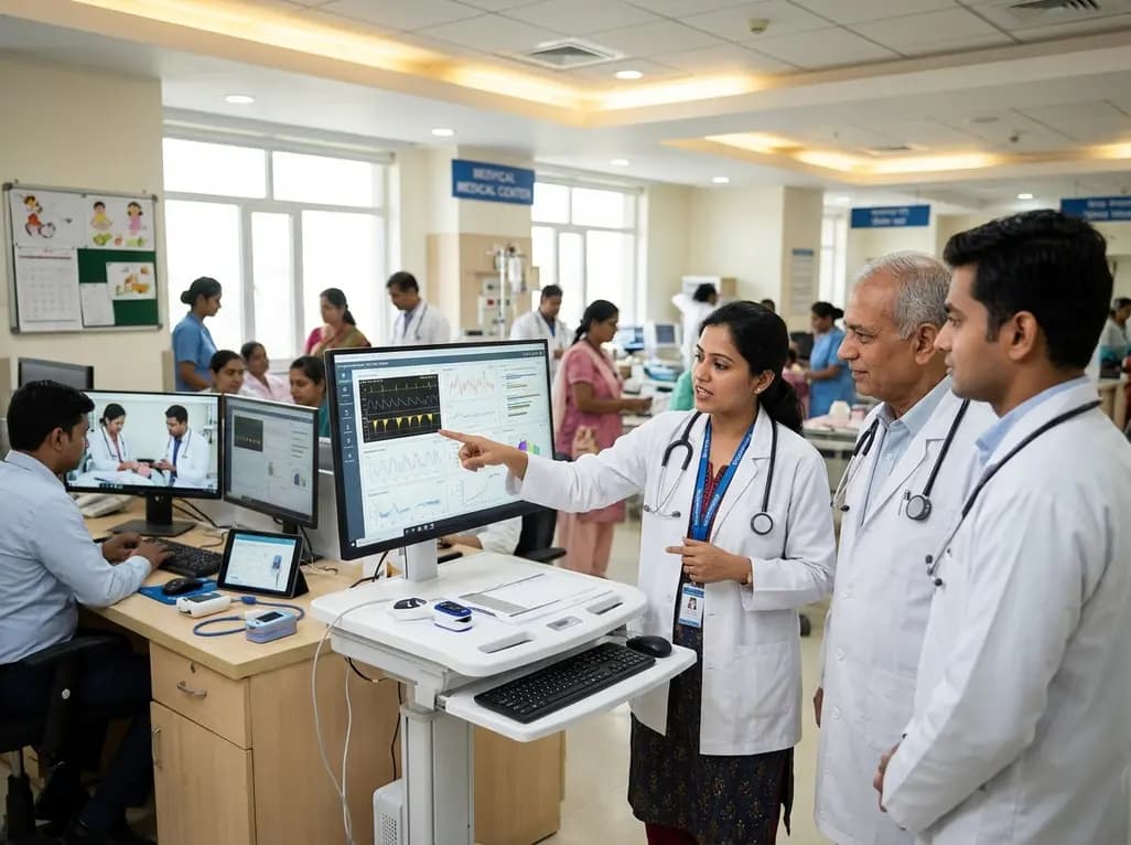
A landmark study details how an enterprise pediatric remote patient management model expanded from a single cardiology pilot to 16 diverse specialty programs by combining centralized digital operations with specialty clinical leadership.
Today
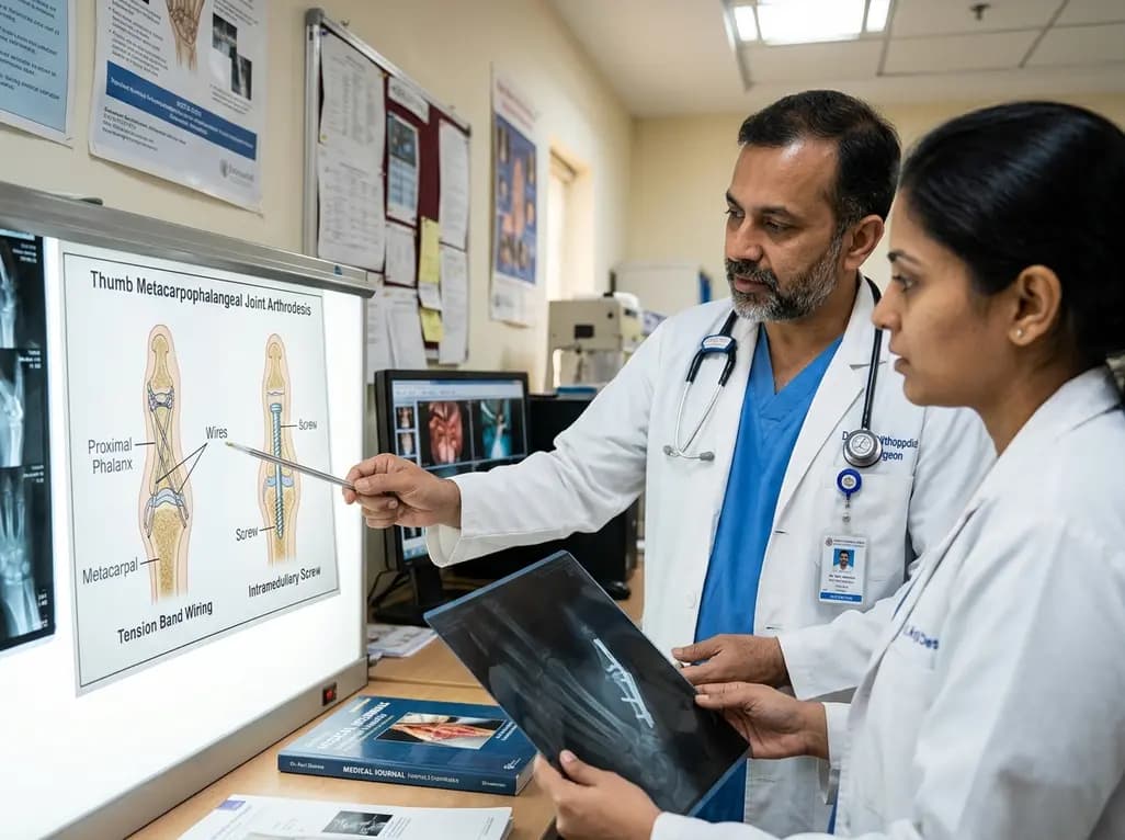
A comparative review of tension band wiring and intramedullary screw fixation in thumb metacarpophalangeal arthrodesis, evaluating union rates, hardware complications, and clinical outcomes.
Yesterday
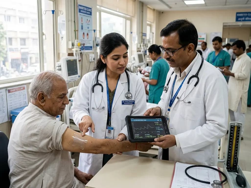
A breakthrough ultrathin multimodal sensor decouples physical hemodynamic monitoring from disposable electrochemical calibration, achieving long-term wearable glucose prediction with 81.36% accuracy and a 9.12% MARD. This offers a robust, drift-free solution for outpatient metabolic surveillance.
Today
