

Loading, please wait...
Loading, please wait...

Surgical management of degenerative, inflammatory, or post-traumatic thumb pathologies frequently necessitates definitive stabilization. Specifically, thumb metacarpophalangeal arthrodesis remains an essential reconstructive procedure that restores key pinch strength while eliminating disabling focal discomfort. Surgeons traditionally choose between tension band constructs and modern compression devices to achieve durable osseous healing. Therefore, evaluating clinical differences between these methodologies assists hand surgeons in optimizing reconstructive outcomes.
Painful arthrosis and severe capsuloligamentous laxity represent the primary clinical indications for joint fusion. For instance, chronic gamekeeper thumb injuries frequently produce recalcitrant joint instability. Similarly, patients with advanced rheumatoid arthritis or primary osteoarthritis often experience debilitating subluxation. When conservative splinting and therapeutic injections fail, joint fusion offers predictable stability.
Surgeons historically prioritized joint mobility; however, stability at the first metacarpophalangeal junction remains considerably more critical for functional power. Sacrificing this small arc of motion rarely causes noticeable disability. Instead, it significantly bolsters key pinch and power grasp. Consequently, accurate joint positioning during surgical fixation determines long-term success. Surgeons typically aim for ten to fifteen degrees of flexion and slight pronation. This functional alignment maximizes lateral pinch efficiency against the adjacent fingers. Proper preoperative planning ensures that patient expectations match functional post-surgical gains.
Tension band wiring represents a time-tested technique in orthopedic hand surgery. Biomechanically, this construct converts distracting tensile forces on the dorsal joint surface into dynamic compressive forces across the fusion site during active pinch. Surgeons typically employ two parallel, smooth Kirschner wires driven across the prepared joint surfaces. Subsequently, they loop stainless steel wire in a figure-of-eight configuration through a transverse osseous drill hole.
This arrangement provides exceptional rotational control and rigid structural stability. Furthermore, tension band wiring accommodates poor bone stock effectively. Surgeons can manually adjust wire tension to compress irregular articular margins. In addition, the equipment requirements remain modest and cost-effective, which benefits resource-conscious surgical settings. Nevertheless, prominent subcutaneous hardware frequently causes dorsal skin irritation. Patients often report localized tenderness when the wire ends impinge against the overlying extensor hood. Consequently, secondary hardware removal represents a recognized limitation of this classic technique.
Headless intramedullary screw fixation has gained widespread adoption as an attractive alternative. This low-profile technique utilizes a cannulated, variable-pitch compression screw buried entirely beneath the subchondral bone. Surgeons insert the implant retrograde or antegrade across the denuded articular surfaces under fluoroscopic guidance. The differing thread pitch automatically draws the opposing cortices together, generating substantial interfragmentary compression.
Because the screw sits completely countersunk within the medullary canal, soft tissue irritation drops substantially. Thus, patients rarely experience extensor tendon adhesions or painful subcutaneous protrusions. Moreover, the rigid internal fixation allows immediate soft tissue mobilization of unaffected adjacent joints. Physical therapists can initiate targeted rehabilitation protocols earlier, thereby reducing overall post-operative hand stiffness. However, achieving proper trajectory demands meticulous technical precision. Errant drilling can compromise cortical integrity or misalign the intended angle of fusion. Additionally, revising a failed intramedullary screw can present substantial reconstructive challenges.
Comparative clinical investigations demonstrate high overall union rates exceeding ninety percent for both fixation techniques. Despite these favorable healing metrics, distinct differences emerge regarding post-operative complications and secondary reoperations. Tension band wiring consistently achieves dependable bony consolidation, yet it demonstrates higher rates of superficial surgical site irritation. Hardware prominence often necessitates secondary surgical excision once solid osseous bridging occurs.
Conversely, intramedullary screws largely eliminate hardware prominence issues. Patients rarely require implant extraction unless an overt nonunion or chronic infection develops. However, some clinical series note marginally higher incidences of delayed union or nonunion with screw fixation if rotational torque overcomes the single-point fixation construct. In addition, the presence of severe osteopenia may reduce screw purchase, diminishing compressive integrity. Infection rates remain comparably low between both cohorts when surgeons employ meticulous soft tissue handling. Therefore, balancing implant cost, soft tissue tolerance, and osseous quality dictates the final choice.
Selecting the optimal fixation method requires careful assessment of individual patient factors. Surgeons must evaluate bone mineral density, baseline functional demands, and systemic inflammatory status. For example, manual laborers who subject the thumb to intense torsional loading may benefit from the auxiliary rotational rigidity provided by crossed wires. Conversely, patients with thin soft tissue envelopes and high cosmetic expectations favor headless intramedullary implants.
Furthermore, intraoperative technique profoundly influences long-term durability. Surgeons must meticulously prepare the articular surfaces down to bleeding subchondral bone, regardless of the chosen implant. Planar joint resections or cup-and-cone contouring provide broad, congruous contact surfaces that accelerate primary osteogenesis. Postoperative immobilization should continue until preliminary radiographic consolidation appears. Once bridging trabeculae span the fusion interface, progressive strengthening can commence safely. Ultimately, surgeon familiarity and individualized patient anatomy should guide operative decision-making.
The primary goal is to eliminate pain and establish a stable, rigid pillar for thumb opposition and pinch activities. While the procedure sacrifices joint motion, it significantly enhances hand grip power and lateral pinch strength. Consequently, patients regain predictable function without the debilitating instability caused by advanced arthritis or severe ligamentous rupture.
Tension band wiring carries a higher incidence of hardware prominence and extensor tendon irritation, frequently requiring secondary implant removal. In contrast, headless intramedullary screws remain buried within the bone, minimizing soft tissue complications. However, intramedullary screws may carry a slightly higher risk of nonunion if bone purchase is suboptimal or rotational forces exceed fixation strength.
Patients typically wear a protective thumb spica splint for four to six weeks while bone healing occurs. Surgeons obtain follow-up radiographs to confirm progressive osseous union before permitting unsupported hand use. Unrestricted pinching, gripping, and active loading generally resume between eight and twelve weeks postoperatively, depending on patient healing and construct stability.
Disclaimer: This content is for informational and educational purposes only. It is not intended to be a substitute for professional medical advice, diagnosis, or treatment. Always seek the advice of your physician or other qualified health provider with any questions you may have regarding a medical condition. Never disregard professional medical advice or delay in seeking it because of something you have read here. The authors and publishers are not responsible for any adverse effects or consequences resulting from the use of any suggestions, preparations, or procedures discussed in this article. Refer to the latest local and national guidelines for clinical practice.
References
Arendale CR et al. Arthrodesis of the Thumb Metacarpophalangeal Joint: Tension Band Wiring Versus Intramedullary Screw Fixation. J Hand Surg Am. 2026 Oct 23. doi: undefined. PMID: 42909447.
Bennice J, Gallucci G, Zaidenberg EE, De Carli P, Boretto JG. Comparative study between tension band and compression screw fixation in metacarpophalangeal and proximal interphalangeal joint arthrodesis. Rev Asoc Argent Ortop Traumatol. 2020;85(4):329-336.
Proubasta I, Lamas C, Trullols L, Itarte J. Arthrodesis of the metacarpophalangeal joint of the thumb using a Herbert screw. Tech Hand Up Extrem Surg. 2006;10(2):93-98.

Read summarized clinical updates, watch expert medical content, and earn CME certifications right from your smartphone.
Open your smartphone camera to scan and install.





A comparative review of tension band wiring and intramedullary screw fixation in thumb metacarpophalangeal arthrodesis, evaluating union rates, hardware complications, and clinical outcomes.
Today
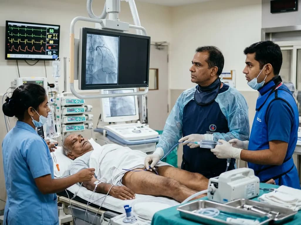
An elderly man with partially repaired Tetralogy of Fallot presented with STEMI caused by coronary embolism rather than atherosclerosis. Successful treatment with sustained mechanical aspiration thrombectomy underscores the vital need for high clinical vigilance in adult congenital heart disease.
Today

Demanding exhaustive hormonal profiling can inadvertently discourage clinical studies involving females. Methodological standards must align with the specific scientific question to expand, not restrict, equitable evidence for women.
Today
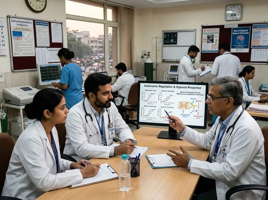
A recent study demonstrates that spinal oxygen sensors do not require mitochondrial electron transport to trigger sympathetic excitation during acute hypoxia, revealing new insights into how mitochondrial oxidative stress modulates autonomic tone in cardiovascular and neurological diseases.
Today
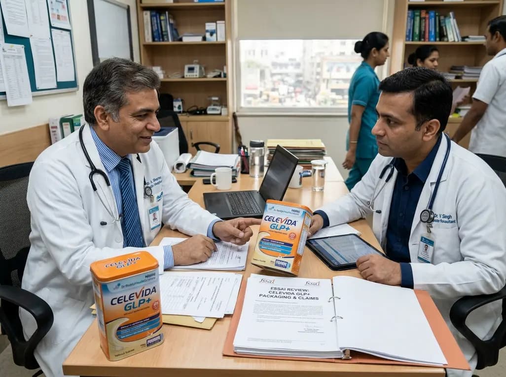
Following regulatory scrutiny from FSSAI regarding promotional representations and lean muscle preservation claims, Dr. Reddy's Laboratories and Nestlé Health Science are currently reviewing the branding and marketing strategy for their joint nutritional supplement Celevida GLP+.
Today
