.webp&w=384&q=75)

Loading, please wait...
Loading, please wait...
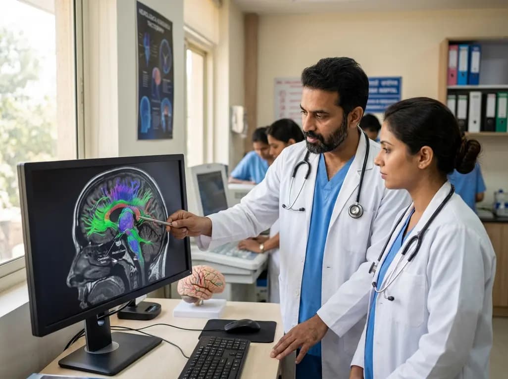
Modern skull base neurosurgery demands extreme precision to minimize morbidity while maximizing tumor resection. Tumors involving the cavernous sinus and Meckel's cave frequently distort regional neurovascular anatomy. Consequently, operating surgeons must anticipate the altered trajectory of cranial nerves before making an incision. Preoperative diffusion tensor imaging, particularly trigeminal nerve tractography, provides a noninvasive solution to this diagnostic challenge. Historically, standard anatomical magnetic resonance imaging struggled to distinguish thin, compressed nerves from adjacent neoplastic margins. In contrast, advanced tractography leverages the anisotropic diffusion of water molecules along neural axons to map three-dimensional fiber tracts. As a result, neurosurgeons gain critical spatial insights that predict intraoperative anatomy with remarkable clarity. Recent clinical validation studies confirm that tractography reliably visualizes cranial nerve pathways even during severe parasellar distortion. Furthermore, this imaging modality allows surgical teams to formulate individualized operative strategies tailored to unique patient anatomy. By avoiding unexpected nerve manipulation and excessive mechanical traction, tractography plays a vital role in safeguarding neurological function during skull base operations.
The parasellar compartment represents one of the most intricate corridors in human skull base anatomy. Specifically, the cavernous sinus and Meckel's cave host critical structures, including the internal carotid artery and multiple cranial nerves. Pathologies arising in this confined region commonly include trigeminal schwannomas, meningiomas, and chordomas. Because these lesions originate within or adjacent to the trigeminal cistern, they exert substantial compressive forces on the nerve trunk and its divisions. Moreover, chronic neoplastic expansion displaces, stretches, and thins the trigeminal nerve against rigid dural boundaries and skull base bone. In many instances, the tumor envelope completely encases the sensory rootlets and Gasserian ganglion. Therefore, surgeons encounter significant technical hurdles when establishing a safe dissection plane. Accidental traction or direct thermal injury can provoke severe facial numbness, painful dysesthesias, or neurotrophic keratitis. Consequently, obtaining dependable preoperative knowledge regarding the precise displacement vector of the nerve is essential. Such information allows surgeons to avoid direct entry into displaced nerve fibers and ensures safe exposure of the tumor core.
Systematic classification of nerve displacement patterns establishes an objective framework for operative planning. High-resolution tractography categorizes trigeminal nerve shifts into three distinct spatial patterns: medial displacement, lateral displacement, and circumferential encasement. In recent clinical evaluations involving twenty-eight patients, tractography successfully predicted the true surgical position in twenty-six individuals, achieving an impressive 92.9% concordance rate. Among these surgical cases, lateral displacement emerged as the most frequent configuration, appearing in thirteen patients. Conversely, medial displacement occurred in seven individuals, while circumferential encasement characterized the remaining eight patients. When tumors push the nerve medially, the lesion typically expands from the lateral cavernous wall or petrous apex. In contrast, tumors driving the nerve laterally often arise from the sphenoid body, petroclival synchondrosis, or medial cavernous compartment. Circumferential encasement represents the most technically challenging presentation, wherein neoplastic tissue completely surrounds fragile nerve bundles. Thus, identifying these distinct spatial relationships before surgical exploration provides surgical teams with unmatched visual clarity, transforming abstract preoperative imaging into actionable surgical guidance.
The specific pattern of nerve displacement directly guides the selection of the optimal surgical approach. Neurosurgeons fundamentally choose between lateral trajectories, such as transcranial or transorbital corridors, and medial trajectories, such as endoscopic endonasal routes. When tractography reveals medial nerve displacement, surgeons almost universally prefer a lateral approach. In clinical series, lateral approaches deployed for medially displaced nerves achieved an 85.7% favorable outcome rate by accessing the tumor without crossing the nerve. Conversely, when the nerve experiences lateral displacement, medial corridors offer an exquisite anatomical pathway. Surgeons utilizing endoscopic endonasal corridors in lateral displacement cohorts recorded a remarkable 100% favorable outcome rate. In contrast, performing a lateral approach in the presence of lateral nerve displacement achieved only a 66.7% favorable outcome rate. Under these circumstances, lateral approaches force the surgeon to dissect past the vulnerable, displaced nerve to reach deeper tumor components. Therefore, aligning the surgical trajectory with the vector of nerve displacement substantially reduces mechanical trauma and optimizes neurological preservation.
Although tractography-derived displacement patterns offer foundational guidance, tumor consistency and histological characteristics frequently alter surgical execution. Soft, aspirable tumors, such as cystic schwannomas or soft pituitary adenomas, readily lend themselves to medial endoscopic endonasal debulking. However, fibrous, calcified, or highly vascular tumors, including many petroclival meningiomas, present substantial technical resistance within narrow endoscopic corridors. Consequently, neurosurgeons often select transcranial lateral approaches even when dealing with laterally displaced nerves. Lateral microsurgical windows provide expansive visualization, allow robust two-handed instrumentation, and permit dynamic manipulation of firm neoplastic tissue. Furthermore, cases characterized by circumferential nerve encasement pose formidable technical hurdles regardless of trajectory. In clinical investigations, encased tumors managed exclusively through lateral approaches yielded only a 25% favorable outcome rate. This sobering outcome reflects the dense fibrous adhesion between the tumor matrix and the compromised epineurium. Ultimately, DTI tractography functions not as an autonomous decision-maker, but as an indispensable decision-support tool that must harmonize with intraoperative judgment, tumor consistency, and microsurgical expertise.
Preoperative DTI tractography demonstrates outstanding diagnostic reliability for mapping displaced cranial nerves. In surgical cohorts examining cavernous sinus and Meckel's cave lesions, tractography achieved an impressive 92.9% concordance rate with direct intraoperative findings. Consequently, skull base neurosurgeons can reliably visualize anatomical trajectories before skin incision. This predictive fidelity empowers surgical teams to tailor trajectories, minimize unexpected nerve manipulation, and preserve neurological function during complex microsurgical or endoscopic cranial base resections.
Surgical teams preferentially select a medial trajectory, such as the endoscopic endonasal approach, when tumors displace the trigeminal nerve laterally. Under these precise anatomical circumstances, a medial corridor provides direct access to the pathology without requiring nerve retraction. Indeed, clinical studies demonstrate a 100% favorable outcome rate when surgeons use medial access for lateral displacement. However, surgeons must evaluate tumor firmness, because fibrous lesions often necessitate lateral access for safer mechanical manipulation.
Circumferential tumor encasement severely compromises nerve preservation because neoplastic tissue infiltrates the epineurium and regional vascular structures. When tumors completely surround the nerve, surgeons invariably encounter dense adhesions that preclude an anatomically safe dissection plane. Therefore, extensive tumor debulking frequently demands delicate dissection directly on thinned nerve bundles. Consequently, encased lesions demonstrate markedly reduced favorable outcomes, often achieving acceptable neurological preservation in only 25% of treated complex skull base cases.
Disclaimer: This content is for informational and educational purposes only and should not be considered medical advice. Always consult a qualified healthcare professional regarding any medical conditions or treatment plans. Refer to the latest local and national guidelines for clinical practice.
References
Lee JS et al. Tractography-Based Prediction of Trigeminal Nerve Displacement Patterns in Cavernous Sinus and Meckel's Cave Tumors: Correlation With Intraoperative Findings. Oper Neurosurg. 2026 Sep 02. doi: 10.1227/ons.0000000000002186. PMID: 42684067.
Jacquesson T et al. Probabilistic Tractography to Predict the Position of Cranial Nerves Displaced by Skull Base Tumors: Value for Surgical Strategy Through a Case Series of 62 Patients. Neurosurgery. 2019 Jul;85(4):E716-E726. doi: 10.1093/neuros/nyy538. PMID: 30476219.
Bao A et al. Quantitative Anatomical Comparison of Surgical Approaches to Meckel's Cave. J Clin Med. 2023 Oct;12(21):6850. doi: 10.3390/jcm12216850.

Read summarized clinical updates, watch expert medical content, and earn CME certifications right from your smartphone.
Open your smartphone camera to scan and install.





Preoperative DTI tractography achieved a 92.9% concordance rate in predicting trigeminal nerve displacement in cavernous sinus and Meckel's cave tumors. Displacement patterns guide medial versus lateral surgical approaches, though tumor consistency remains decisive for achieving safe resection.
4 weeks back

A landmark biomonitoring study reveals that chronic pesticide exposure significantly alters buccal microRNA expression and increases micronucleus frequency in agricultural workers. These findings uncover actionable molecular signatures of oxidative stress, inflammation, and genomic instability.
Today

India's national digital health service achieved a historic milestone of fifty crore virtual consultations. Notably, female patients account for over fifty-seven percent of utilisation, demonstrating improved healthcare accessibility for remote communities, chronic disease management, and primary triage.
Today

A large UK Biobank study identified four routine biomarker subtypes that predict cardiometabolic disease progression to multimorbidity. Integrating multi-state modelling, metabolomics, and proteomics revealed distinct biological pathways, offering new avenues for tailored clinical risk stratification.
Today

Dual-nanofiber interpenetrating network scaffolds combining silica and PLLA/gelatin nanofibers with sodium alginate achieve complementary mechanical stability, immunomodulatory M2 macrophage polarization, and enhanced osteogenesis for advanced bone repair.
Today

Researchers at IISER Berhampur have engineered a covalent organic framework immunosensor that detects urinary NGAL. This novel nanotechnology platform distinguishes diabetic nephropathy from other renal conditions, enabling rapid, minimally invasive stratification and earlier clinical intervention for patients.
Today
