.webp&w=384&q=75)

Loading, please wait...
Loading, please wait...

Diabetic kidney disease detection remains one of the greatest clinical hurdles in modern diabetology and nephrology across India. Chronic hyperglycemia inflicts insidious, progressive microvascular damage across renal nephrons. Consequently, millions of diabetic individuals face an elevated lifetime risk of kidney failure. Traditional functional tests, such as serum creatinine and estimated glomerular filtration rate, detect damage only after significant parenchymal injury occurs. Furthermore, microalbuminuria exhibits high biological variability and cannot distinguish diabetic nephropathy from non-diabetic etiologies. To address this challenge, researchers at IISER Berhampur have developed an innovative diagnostic platform. Specifically, the team engineered a nanotechnology-based fluorescent immunosensor that analyzes urine samples. This breakthrough technology provides a rapid, minimally invasive approach for disease stratification. Consequently, it promises to transform bedside diagnostic assessments.
Clinicians regularly encounter significant difficulties when evaluating renal impairment in individuals with chronic diabetes. Traditionally, persistent proteinuria has served as the classic hallmark of diabetic glomerulosclerosis. However, clinicians now recognize non-proteinuric phenotypes with increasing frequency across clinical practice. Therefore, patients may lose glomerular filtration capacity without presenting notable albumin excretion. Additionally, secondary glomerulonephritis, hypertensive nephrosclerosis, and tubulointerstitial nephritis frequently mimic classic diabetic nephropathy. Overlapping clinical manifestations often obscure the primary underlying pathology. While renal biopsy remains the definitive gold standard, it entails invasive risks including severe bleeding. Consequently, many vulnerable patients cannot safely undergo histopathological evaluation. Furthermore, secondary care centers often lack rapid access to nephropathology infrastructure. Therefore, clinical teams frequently initiate empirical therapy without precise pathological confirmation. These clinical dilemmas highlight the urgent need for molecular tools that identify tubular stress early. Non-invasive diagnostic surrogates can reveal distinct tubular epithelial alterations before irreversible scarring occurs. Recent clinical guidelines prioritize early intervention to arrest renal parenchymal decline. Nevertheless, delayed diagnosis consistently undermines therapeutic protocols. A targeted biosensing method could eliminate diagnostic uncertainty and stratify disease risk with exceptional speed.
To overcome standard diagnostic limitations, the IISER Berhampur research team turned to advanced porous nanomaterials. Specifically, the scientists designed a covalent organic framework, commonly known as a COF, to construct a highly sensitive immunosensor. Covalent organic frameworks represent crystalline porous polymers featuring well-defined structures and high surface areas. Because of these distinct physicochemical features, COFs provide exceptional scaffold support for anchoring biological recognition elements. The research group functionalized the synthetic framework to create an intelligent optical reporter system. When the target biomarker binds to the sensor, it modulates the intrinsic fluorescence profile. Consequently, this chemical interaction generates a distinct optical readout that laboratory devices can quantify rapidly. Assistant Professor Parikshit Moitra from the Department of Chemical Sciences led this groundbreaking investigation. Additionally, research scholar Satyabrata Senapati contributed extensively to developing the sensing architecture. Published in the Royal Society of Chemistry journal Chemical Communications, their proof-of-concept study demonstrates remarkable analytical precision. The sensor achieved over ninety percent diagnostic sensitivity, specificity, and accuracy in experimental trials. Therefore, this nanotechnology framework represents a robust platform for modern biochemical assay development. Furthermore, the material demonstrates structural stability and optical reproducibility across multiple sample batches. This chemical stability makes the platform highly suitable for translational development.
The biosensing platform focuses specifically on detecting neutrophil gelatinase-associated lipocalin, abbreviated as NGAL. Physiologically, tubular epithelial cells express this twenty-five kilodalton protein at very low baseline concentrations. However, acute ischemic or toxic insults induce rapid NGAL synthesis within the loop of Henle and collecting tubules. In diabetic kidney disease, persistent metabolic stress, hyperglycemia, and local inflammation drive sustained tubular damage. Consequently, urinary NGAL concentrations rise substantially well before serum creatinine levels elevate. Traditional markers only flag functional filtration failure after extensive parenchymal nephron loss. In contrast, NGAL functions as an active structural damage marker that reflects cellular distress. The IISER Berhampur platform selectively captures urinary NGAL molecules without cross-reacting with unrelated proteins. By quantifying this tubular biomarker, the sensor effectively distinguishes diabetic kidney disease samples from other renal pathologies. Moreover, urine collection offers a completely non-invasive sample source that minimizes patient discomfort. Thus, patients can provide samples readily during routine clinic visits without venous puncture. This unique biological target provides nephrologists with direct visibility into tubular health and disease activity. Additionally, monitoring biomarker fluctuations could help clinicians track acute tubular decompensation. Therefore, measuring urinary NGAL empowers physicians to evaluate disease progression with greater clinical precision.
The clinical implications of this diagnostic innovation are particularly vital for healthcare delivery in India. Currently, India carries an enormous national burden of type 2 diabetes mellitus. Consequently, millions of individuals remain vulnerable to progressive diabetic kidney disease and end-stage renal failure. Many patients reside in rural and semi-urban settings where advanced hospital infrastructure remains scarce. In these resource-constrained environments, standard biochemical testing often experiences delayed processing times. In contrast, a portable fluorescence sensor could deliver rapid point-of-care testing in primary health centers. Early detection enables general practitioners to initiate lifestyle modifications and glucose control earlier. Furthermore, clinicians can prescribe kidney-protective medications, including SGLT2 inhibitors and modern renin-angiotensin-aldosterone blockers, before irreversible fibrosis develops. Timely medical intervention can substantially delay or prevent the eventual necessity for chronic hemodialysis and kidney transplantation. Therefore, point-of-care diagnostic tools can dramatically reduce long-term healthcare expenditures for both families and public health systems. Additionally, stratifying high-risk cohorts allows nephrologists to optimize referral networks across tertiary medical institutions. This molecular technology offers substantial potential to strengthen diabetic renal surveillance nationwide. Ultimately, accessible biosensors could democratize specialized screening across rural communities. Such decentralized diagnostics ensure that high-risk individuals receive timely therapeutic guidance regardless of their geographic location.
Although these laboratory findings offer tremendous promise, the sensor requires further development before routine clinical deployment. As Dr. Parikshit Moitra emphasized, the technology currently represents a robust proof-of-concept rather than an immediate diagnostic replacement. First, researchers must conduct large-scale prospective clinical validation trials involving diverse patient cohorts. These clinical studies must include patients across various stages of chronic kidney disease and diabetic duration. Second, biomedical engineers must optimize device fabrication to ensure affordable mass production. Developing portable, battery-operated optical readers will prove critical for real-world primary care use. Third, investigators must establish standardized urinary threshold values that correlate with histological damage and clinical outcomes. Concurrently, regulatory authorities will demand rigorous analytical verification to guarantee testing precision under variable environmental conditions. Ambient temperature and humidity fluctuations across Indian clinics must not degrade sensor stability. Nevertheless, interdisciplinary collaboration between chemical scientists, biological researchers, and hospital nephrologists is accelerating this translational journey. If future clinical trials corroborate these preliminary results, point-of-care fluorescence sensing will establish a new diagnostic benchmark in nephrology. Furthermore, automated microfluidic cartridges could streamline sample handling for community health workers. Consequently, ongoing clinical refinement will bridge the vital gap between laboratory innovation and community healthcare delivery.
Q1: What makes the IISER Berhampur biosensor different from standard kidney function tests?
Standard renal tests like serum creatinine and urine albumin reflect functional filtration changes, which typically manifest after substantial nephron damage occurs. In contrast, this nanotechnology biosensor utilizes a fluorescent covalent organic framework to quantify urinary NGAL. Because NGAL indicates active tubular injury, the sensor detects cellular damage much earlier. Furthermore, it achieves over ninety percent accuracy in differentiating diabetic nephropathy from other kidney disorders non-invasively.
Q2: Why is urinary NGAL an effective biomarker for diabetic kidney disease?
Neutrophil gelatinase-associated lipocalin is an intracellular protein produced by renal tubular epithelial cells during ischemic, metabolic, or inflammatory stress. In diabetic nephropathy, tubular injury occurs early alongside microvascular changes. Consequently, urinary NGAL concentrations rise well before serum creatinine levels elevate. Measuring NGAL enables clinicians to detect ongoing tubular epithelial stress promptly. This sensitivity allows timely pharmacological intervention before irreversible renal scarring becomes established.
Q3: Is this nanotechnology sensor ready for immediate use in medical clinics?
The sensor currently functions as a validated proof-of-concept platform developed in an academic laboratory setting. Although experimental results demonstrate exceptional sensitivity and specificity, the technology is not yet approved for daily clinical practice. Researchers must first conduct extensive multicenter clinical validation trials across diverse patient populations. Additionally, manufacturing engineers must standardize reader devices and secure regulatory approvals before commercial healthcare adoption occurs.
Disclaimer: This content is for informational and educational purposes only. It does not constitute medical advice or replace professional judgment. Refer to the latest local and national guidelines for clinical practice.
References

Read summarized clinical updates, watch expert medical content, and earn CME certifications right from your smartphone.
Open your smartphone camera to scan and install.





Researchers at IISER Berhampur have engineered a covalent organic framework immunosensor that detects urinary NGAL. This novel nanotechnology platform distinguishes diabetic nephropathy from other renal conditions, enabling rapid, minimally invasive stratification and earlier clinical intervention for patients.
Today

A large UK Biobank study identified four routine biomarker subtypes that predict cardiometabolic disease progression to multimorbidity. Integrating multi-state modelling, metabolomics, and proteomics revealed distinct biological pathways, offering new avenues for tailored clinical risk stratification.
Today
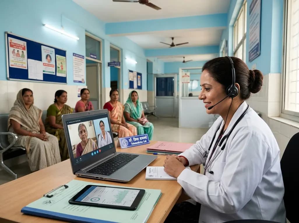
India's national digital health service achieved a historic milestone of fifty crore virtual consultations. Notably, female patients account for over fifty-seven percent of utilisation, demonstrating improved healthcare accessibility for remote communities, chronic disease management, and primary triage.
Today

Advances in pediatric cardiology have allowed most patients with congenital heart defects to reach adulthood. However, acquired cardiovascular risk factors like hypertension and diabetes now significantly drive late mortality in adult congenital heart disease, requiring proactive cardiometabolic intervention.
Today
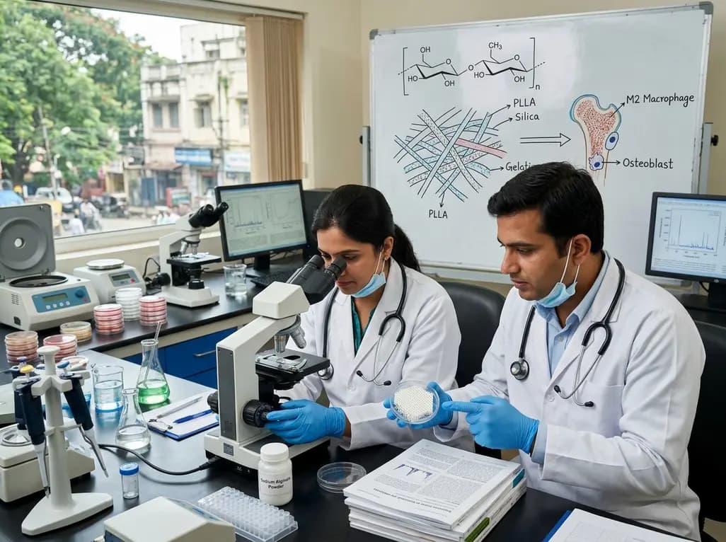
Dual-nanofiber interpenetrating network scaffolds combining silica and PLLA/gelatin nanofibers with sodium alginate achieve complementary mechanical stability, immunomodulatory M2 macrophage polarization, and enhanced osteogenesis for advanced bone repair.
Today
