.webp&w=384&q=75)

Loading, please wait...
Loading, please wait...

Orthopedic and reconstructive surgeons frequently encounter critical-sized bone defects resulting from high-energy vehicular trauma, non-unions, and oncological resections. Traditional autografts remain the therapeutic benchmark. However, donor-site morbidity and restricted harvest volumes complicate patient management. Consequently, regenerative medicine focuses on biomimetic bone tissue engineering scaffolds that replicate native extracellular architecture. A recent investigation in Advanced Healthcare Materials highlights an innovative dual-nanofiber interpenetrating network design. This engineering strategy successfully resolves historical trade-offs between load-bearing structural integrity and proactive osteoinductive biology. By synergizing organic and inorganic polymers, the construct optimizes cellular behavior and promotes rapid skeletal healing.
Skeletal reconstruction requires biomaterial implants that withstand physiological loads while simultaneously orchestrating cell differentiation. In routine clinical practice, orthopedic specialists encounter major hurdles with conventional synthetic bone substitutes. For example, ceramic blocks exhibit substantial brittleness, which increases fracture risks under shear loading. Conversely, biodegradable hydrogels often lack adequate compressive strength, collapsing prematurely before vascularized osteogenesis matures. Furthermore, single-component inorganic nanofibrous scaffolds frequently exhibit structural instability when exposed to continuous dynamic stresses in vivo. Because of these mechanical shortcomings, standard implants struggle to maintain appropriate structural space for vascular invasion. In addition, surgeons must consider the local biological microenvironment during bone repair. Chronic local inflammation often impairs osteoblast differentiation and delays osseous union. When biomaterials fail to guide host immune responses, fibroblastic encapsulation occurs instead of robust trabecular bone formation. Therefore, clinicians urgently require advanced composite constructs that combine elastic energy dissipation with dynamic cellular signaling. Fabricating scaffolds that replicate the natural organic-inorganic balance of human bone represents a promising path forward. Ultimately, solving these interdependent biological and biomechanical requirements remains vital for optimizing surgical outcomes in complex skeletal defects.
To overcome historical material constraints, investigators developed 3D-printed composite constructs featuring a dual-nanofiber interpenetrating network. Specifically, the engineering team integrated inorganic silica nanofibers alongside organic poly(L-lactic acid) and gelatin nanofibers into a cohesive hydrogel. Sodium alginate provided the surrounding three-dimensional printing matrix, ensuring precise anatomical geometry through additive manufacturing. Furthermore, researchers chemically crosslinked the poly(L-lactic acid) and gelatin fibers directly with the sodium alginate polymer network. This covalent stabilization firmly anchored the organic fibrous components within the hydrogel framework. As a result, the construct prevented structural collapse and eliminated the structural instability common in single-component fiber systems. The architecture mimics the hierarchical organization of native human bone, which blends inorganic mineralized crystals with organic collagenous fibrils. Moreover, the 3D-printed layout preserves interconnected micropores, ensuring efficient nutrient perfusion and metabolic waste evacuation. Surgeons require predictable scaffold degradation that aligns precisely with de novo bone regeneration rates. By tuning dual-nanofiber bone tissue engineering scaffolds, clinicians can offer structural shielding while facilitating rapid osteoprogenitor recruitment across the defect interface. Consequently, this manufacturing platform represents a notable technological leap in custom biofabrication.
Systematic comparative analyses in this investigation revealed distinct, complementary functions for each nanofiber component within the matrix. Specifically, the poly(L-lactic acid) and gelatin organic nanofibers primarily reinforced mechanical stability and structural resilience. These polymeric fibers resisted tensile stresses and prevented macroscopic crack propagation during compressive deformation. Interestingly, experimental data demonstrated that organic nanofibers did not exhibit osteogenic bioactivity superior to standard poly(L-lactic acid) microparticles. In contrast, inorganic silica nanofibers served as the primary biological drivers for osteogenesis. These silica nanofibers steadily release bioactive silicon ions into the surrounding extracellular space. Bioactive silicon directly upregulates osteogenic transcription factors in resident stem cells, stimulating rapid mineralization. Furthermore, silica nanofibers showed substantially greater osteoinductive capacity than conventional spherical silica nanoparticles. This superior outcome stems from their high aspect ratio, which presents biomimetic nanotopographical cues to migrating osteoblasts. Consequently, the combination of organic structural scaffolding and inorganic ionic signaling establishes an optimal niche for robust bone formation. Thus, each component provides a specialized biomechanical or biochemical contribution that the other component lacks. Because of this synergy, the hybrid scaffold achieves superior performance compared to homogeneous constructs.
Successful osseous regeneration depends heavily on early immunoregulation at the host-biomaterial interface. When clinicians implant synthetic bone grafts, resident macrophages initiate an immediate inflammatory response. Pro-inflammatory M1 macrophages initially clear cellular debris, but prolonged M1 persistence triggers pathological fibrous encapsulation. Fortunately, the silica nanofibers in this interpenetrating network actively promote pro-reparative M2 macrophage polarization. These M2 phenotypes release essential anti-inflammatory cytokines, including interleukin-10 and transforming growth factor-beta. Consequently, the local microenvironment transitions smoothly from destructive inflammation toward regenerative osteogenesis. In addition, M2 macrophages secrete robust angiogenic growth factors that stimulate capillary sprouting into the porous construct. Without rapid microvascular ingrowth, implanted bone substitutes suffer central necrosis and graft failure. The dual-nanofiber architecture coordinates this immunomodulatory cascade without requiring expensive exogenous recombinant growth factors. Therefore, modulating host immune cells through material composition provides a cost-effective, reproducible strategy for clinical reconstructive applications. Furthermore, dampening destructive chronic inflammation protects surrounding soft tissues during the initial healing phases. As a result, patients experience accelerated graft integration and reduced postoperative complications.
These biomaterial findings present substantial clinical implications for orthopedic surgeons, traumatologists, and maxillofacial specialists. In complex trauma reconstruction, managing massive bone loss frequently exhausts autologous donor sites. Synthetic composite scaffolds offering both immediate mechanical support and active osteoinduction could replace autografts in high-risk patients. For instance, elderly patients with osteoporosis exhibit compromised healing capacity and reduced stem cell potency. By continuously delivering silicon ions, these scaffolds stimulate sluggish osteoblast populations even in impaired systemic environments. Moreover, 3D printing enables custom patient-specific geometries, matching intricate mandibular or pelvic resections precisely. However, successful clinical translation will require rigorous evaluation in large animal load-bearing models before human trials. Researchers must evaluate long-term biodegradation kinetics and mechanical fatigue under cyclic loading conditions. Additionally, establishing standardized production protocols aligned with regulatory quality controls remains essential for commercial manufacturing. Clinicians must also assess sterilization techniques to ensure they do not compromise nanofiber surface nanotopography. As biomaterial engineering continues to evolve, dual-nanofiber scaffolds offer an exciting blueprint for next-generation orthobiologics.
Dual-nanofiber scaffolds integrate complementary organic and inorganic components into an interpenetrating polymer network. Poly(L-lactic acid) and gelatin nanofibers chemically crosslink with sodium alginate hydrogel, providing elasticity and preventing crack propagation under load. Concurrently, silica nanofibers contribute compressive stiffness while reinforcing the hydrogel architecture. This synergistic mechanical behavior resolves the severe structural fragility and premature collapse commonly seen when using single-component inorganic fibers or unreinforced hydrogels in load-bearing surgical defects.
Silica nanofibers outperform nanoparticles primarily because of their high aspect ratio, which mimics the natural extracellular matrix architecture. This fibrous topography provides superior contact guidance for cell adhesion, spreading, and directional migration. In addition, silica nanofibers release therapeutic silicon ions continuously rather than through a sudden burst. These sustained ionic signals upregulate osteogenic genes and promote pro-reparative M2 macrophage polarization, significantly accelerating physiological bone healing in vivo.
Macrophages dictate early tissue responses following biomaterial implantation. Pro-inflammatory M1 macrophages clear debris initially, but sustained M1 activation leads to chronic inflammation and fibrous encapsulation. Conversely, pro-reparative M2 macrophages resolve local inflammation, release osteogenic growth factors, and stimulate angiogenesis. Scaffolds that proactively drive M2 polarization establish a pro-healing microenvironment. Consequently, evaluating macrophage polarization provides essential prognostic data regarding whether an engineered implant will integrate successfully or fail prematurely.
Disclaimer: This content is for informational and educational purposes only and does not constitute medical advice. It is not intended to substitute for professional clinical judgment, diagnosis, or treatment. Healthcare providers should verify all scientific data, regulatory notices, drug dosages, and surgical procedures against authoritative local protocols and official medical resources. Refer to the latest local and national guidelines for clinical practice.
References

Read summarized clinical updates, watch expert medical content, and earn CME certifications right from your smartphone.
Open your smartphone camera to scan and install.





Dual-nanofiber interpenetrating network scaffolds combining silica and PLLA/gelatin nanofibers with sodium alginate achieve complementary mechanical stability, immunomodulatory M2 macrophage polarization, and enhanced osteogenesis for advanced bone repair.
Today
New research reveals that amylin receptor signalling in the laterodorsal tegmental nucleus controls food intake through inhibitory projections to the ventral tegmental area. This study demonstrates how midbrain reward circuits regulate feeding, presenting crucial targets for next-generation obesity pharmacotherapy.
Today

A ten-year cohort study of ABU case logs reveals that URPS fellowship training significantly drives higher annual case volumes, advanced surgical complexity, and increased female representation in pelvic floor reconstruction, underscoring the vital need for structured subspecialty training.
Today
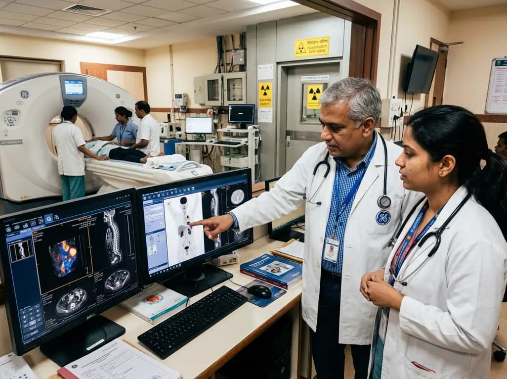
GE HealthCare has agreed to acquire Sofie Biosciences in a 945 million dollar all-cash transaction. The deal secures global rights to the investigational pan-cancer tracer FAPI-74 and expands cyclotron manufacturing infrastructure to accelerate precision theranostics and clinical molecular imaging workflows.
Today

India's food safety authority has cracked down on high-caffeine beverages sold under stimulant branding, prompting legal challenges from major beverage corporations. This review examines FSSAI regulations, cardiac and metabolic complications, pediatric risks, and actionable counseling strategies for physicians.
Today
