

Loading, please wait...
Loading, please wait...

Microsurgical management of vascular lesions in the spinal cord represents one of the most demanding frontiers in neurosurgery. Historically, broad classification schemes provided limited guidance for intradural approaches. A recent investigation in the Journal of Neurosurgery introduces an anatomical classification system designed to guide operative strategy. Specifically, this framework categorizes complex spinal arteriovenous malformations using preoperative digital subtraction angiography and magnetic resonance imaging. By establishing precise structural types, the authors link vascular anatomy directly to specific surgical techniques. Consequently, neurosurgeons can tailor their operative corridors with greater anatomical precision, avoiding excessive cord manipulation and preserving critical neural pathways.
Surgical success in spinal cord vascular disease depends heavily on accurate diagnostic mapping. Historically, classifications separated malformations into simplified intramedullary and perimedullary categories. However, these traditional categories often obscured critical nuances regarding feeder distribution and parenchymal penetration. In this retrospective study spanning 17 years, researchers analyzed 52 consecutive intra- and perimedullary lesions resected by a single experienced neurosurgeon. The team combined digital subtraction angiography, magnetic resonance imaging, and intraoperative photography to evaluate each lesion. Furthermore, they scrutinized three fundamental anatomical variables: arterial supply, depth within the spinal cord, and spatial orientation. Arterial feeders originated from the anterior spinal artery, the posterior spinal artery, or both vessels. Additionally, researchers categorized depth based on purely pial or deeper intramedullary involvement. Spatial positioning revealed anterior, posterior, concentric, or eccentric orientations. Therefore, this multidimensional analysis provides clear insight into nidus architecture. By distinguishing superficial pial surfaces from true parenchymal networks, the framework eliminates ambiguity. Consequently, surgical teams can anticipate operative angles and arterial control points prior to surgery.
The proposed classification system expands previous binary descriptions into five distinct anatomical configurations. First, Type 1 lesions represent posterior pial malformations, accounting for 21 percent of analyzed cases. These lesions sit along the dorsal cord surface and receive blood supply primarily from the posterior spinal artery. Second, Type 2 lesions consist of anterior pial malformations, which also comprised 21 percent of the surgical cohort. These ventral surface malformations rely predominantly on anterior spinal artery branches. Third, Type 3 lesions encompass anterior intramedullary malformations, observed in 13 percent of patients. These anomalies deeply invade the ventral parenchyma, presenting steep technical challenges. Fourth, Type 4 lesions represent concentric intramedullary malformations, which formed 8 percent of the series. Notably, these malformations reside entirely within the cord parenchyma without an obvious surface nidus. Finally, Type 5 lesions comprise eccentric extra- and intramedullary malformations, representing 33 percent of cases. These lesions embed partly within the parenchyma while projecting through the pial surface. Thus, this five-tiered paradigm captures the complete clinical spectrum.
Tailoring the operative approach to each anatomical subtype directly enhances technical efficacy while reducing neural trauma. In this series, extramedullary dissection proved successful for superficial lesions, including 95 percent of Types 1 and 2 pial malformations. Similarly, surgical teams treated all eccentric Type 5 malformations with an initial extramedullary exposure. However, ventral access required specialized positioning maneuvers. Consequently, surgeons performed gentle spinal cord rotation via denticulate ligament traction in 73 percent of Type 2 anterior pial cases and 76 percent of Type 5 eccentric malformations. When addressing true parenchymal disease, myelotomy selection proved essential. Surgeons utilized midline posterior myelotomies for all Type 3 and Type 4 intramedullary malformations, establishing direct visualization through the posterior median sulcus. In contrast, surgeons performed targeted perinidal myelotomies for 82 percent of Type 5 eccentric lesions, capitalizing on the visible pial presentation. Furthermore, holomedullary lesions received selective in situ feeder occlusion rather than complete resection. Overall, surgeons applied recommended algorithmic maneuvers in 86 percent of procedures.
Objective clinical metrics substantiate the practical utility of this structured operative framework. Overall, surgical teams achieved complete nidus resection or total angiographic obliteration in 83 percent of treated patients. Furthermore, long-term functional assessments demonstrated that 65 percent of surviving individuals remained neurologically intact or exhibited only mild deficits, corresponding to McCormick Grade 1 or 2. Conversely, 35 percent of patients experienced moderate to severe deficits or paraplegia at final follow-up. Most importantly, only 6 percent of patients suffered true neurological deterioration relative to their preoperative baseline status. These robust clinical figures demonstrate that meticulous preoperative planning mitigates iatrogenic spinal cord injury. Nevertheless, the authors emphasize a crucial conceptual distinction regarding this model. Specifically, the system serves strictly as an operative planning guide rather than a predictive prognostic scale. It clarifies pathoanatomy and guides surgical steps, but it does not predict spontaneous rupture or natural history. Therefore, clinicians must interpret the staging system exclusively as an actionable surgical roadmap.
This classification offers immediate practical benefits for neurosurgical teams managing complex vascular anomalies. Traditional classifications frequently combined perimedullary and intramedullary lesions, leaving practitioners without standardized dissection protocols. In contrast, this expanded model establishes reproducible operative milestones for every anatomical variant. For example, recognizing a Type 5 eccentric lesion alerts the surgeon that extramedullary surface control should precede parenchymal dissection. Similarly, identifying a Type 4 concentric lesion confirms that deep midline entry is mandatory because no surface landmark exists. Moreover, the integration of multimodal neuroimaging allows surgical teams to coordinate intraoperative vascular imaging, such as indocyanine green videoangiography, with preoperative roadmaps. Additionally, continuous intraoperative neurophysiological monitoring remains paramount during cord rotation and myelotomy. As tertiary centers across India and globally treat these rare vascular pathologies, adopting standardized classifications will facilitate multi-institutional comparisons. Ultimately, linking structural morphology to specific microsurgical maneuvers elevates patient safety and promotes surgical excellence.
The system classifies spinal arteriovenous malformations into five distinct categories based on arterial supply, parenchymal depth, and spatial location. Type 1 represents posterior pial lesions, Type 2 denotes anterior pial lesions, and Type 3 comprises anterior intramedullary lesions. Additionally, Type 4 consists of concentric intramedullary malformations, whereas Type 5 encompasses eccentric extra- and intramedullary malformations. This structural framework directly guides microsurgical approaches for each anatomical variant.
Surgeons apply gentle spinal cord rotation primarily when managing ventral and ventrolateral pathologies. Specifically, denticulate ligament release and gentle traction facilitate rotation in anterior pial lesions (Type 2) and eccentric malformations (Type 5). In the landmark series, surgeons performed cord rotation in 73 percent of Type 2 cases and 76 percent of Type 5 cases. This maneuver provides adequate microsurgical access without requiring excessive traction on vulnerable neural tissue.
No, this classification system functions strictly as a surgical strategy tool rather than a predictive outcome index. It clarifies vascular pathoanatomy and guides specific technical choices, such as myelotomy selection and feeder coagulation. However, it does not assess baseline physiological reserve, natural rupture risk, or long-term clinical prognosis. Neurosurgeons must therefore combine this structural roadmap with established clinical scales to evaluate overall patient prognosis comprehensively.
Disclaimer: This content is for informational and educational purposes only and should not be construed as medical advice. Healthcare professionals should make clinical decisions based on their independent medical judgment and patient-specific factors. Refer to the latest local and national guidelines for clinical practice.
References
Taylor DG et al. A proposed classification system for intramedullary spinal cord arteriovenous malformations that informs surgical strategy. J Neurosurg. 2026 Nov 20. doi: 10.3171/2026.6.JNS26183. PMID: 43063481.
Spetzler RF, Detwiler PW, Riina HA, Porter RW. Modified classification of spinal cord vascular lesions. J Neurosurg Spine. 2002;96(2 Suppl):145-156. doi:10.3171/spi.2002.96.2.0145.
Kim LJ, Spetzler RF. Classification and surgical management of spinal arteriovenous lesions: arteriovenous fistulae and arteriovenous malformations. Neurosurgery. 2006;59(5 Suppl 3):S195-S201. doi:10.1227/01.NEU.0000237335.82234.CE.

Read summarized clinical updates, watch expert medical content, and earn CME certifications right from your smartphone.
Open your smartphone camera to scan and install.





A newly proposed 5-type anatomical classification for spinal arteriovenous malformations correlates preoperative imaging with specific microsurgical approaches, improving resection accuracy while preserving neurological function.
Today

A validated natural language processing pipeline leverages clinical notes and intermediate annotations to identify risky alcohol use across surgical patients, offering automated preoperative risk stratification.
Today

The GESS-AEEM consensus establishes a clear clinical implementation framework for testosterone therapy in postmenopausal women with hypoactive sexual desire disorder, focusing on transdermal dosing, precise laboratory monitoring, and safety communication.
Yesterday
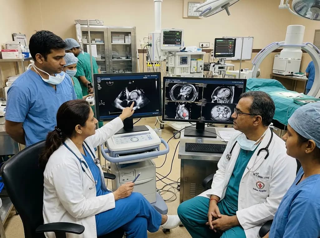
A rare case of an asymptomatic left ventricular hemangioma highlights the vital role of multimodal imaging in differentiating vascular tumors from thrombus. Complete surgical resection under transesophageal echocardiography guidance prevents thromboembolism and delivers optimal outcomes.
Today

A landmark study evaluates long-term quality of life in adolescents and adults with brachial plexus birth injury. Severe functional limitation and limited access to care significantly worsen outcomes, underscoring the urgent need for persistent, multidisciplinary management across adult healthcare transitions.
Today

An analysis of 1,000 posts by macro- and mega-influencers on Instagram and TikTok revealed that 78.5% contained nutrition misinformation, while only 24% of creators held valid credentials. This article examines the clinical risks of viral dietary fads and provides actionable guidance for healthcare professionals.
Today
