

Loading, please wait...
Loading, please wait...

Primary cardiac tumors represent an exceptionally rare clinical finding in routine medical practice. Among benign cardiac neoplasms, hemangiomas account for less than three percent of reported cases. A left ventricular hemangioma occurs even less frequently, as most cardiac hemangiomas develop in the right atrium or ventricles. Consequently, clinicians often fail to identify these lesions during routine cardiac assessments. Patients frequently remain entirely asymptomatic throughout the initial stages of disease progression. For instance, a recent report highlighted an asymptomatic 72-year-old woman diagnosed during an incidental cardiac workup. However, despite an absence of overt clinical symptoms, these benign vascular lesions pose significant health hazards. Tumors inside the ventricular cavity can trigger lethal ventricular arrhythmias, mechanical outflow tract obstruction, or catastrophic systemic embolization. Furthermore, dynamic flow patterns across the left ventricle may provoke platelet aggregation and secondary thrombus formation over the vascular endothelia. Therefore, early identification of an intracardiac mass demands rapid and thorough investigation. Cardiologists must systematically evaluate ventricular intracavitary lesions to exclude malignancy and assess imminent embolic risk. Recognizing atypical presentations ensures that surgical teams intervene proactively before irreversible cardiac complications or systemic cerebrovascular events emerge.
Distinguishing a left ventricular hemangioma from a non-neoplastic intracardiac thrombus presents a persistent diagnostic dilemma. Clinicians frequently encounter ventricular mural thrombi following myocardial infarction or in dilated cardiomyopathies with severe ventricular dyskinesia. In contrast, benign vascular tumors can emerge in structurally normal hearts without regional wall motion abnormalities. Because both lesions appear as intraventricular filling defects on unenhanced echocardiography, misdiagnosis remains common. Moreover, administering empiric anticoagulant therapy for a suspected thrombus provides zero therapeutic benefit for a true vascular neoplasm. In fact, prolonged anticoagulation may elevate the patient's hemorrhage risk without eliminating the underlying neoplastic architecture. Consequently, clinicians must evaluate specific vascular characteristics rather than assuming every ventricular mass represents an organized clot. Left ventricular contrast echocardiography plays a pivotal role in this distinction by demonstrating internal microvascular perfusion. Thrombi remain completely avascular and appear as prominent perfusion voids during contrast administration. Conversely, cavernous and capillary hemangiomas demonstrate brisk internal contrast opacification due to their rich sinusoidal vascular channels. Thus, early microvascular assessment prevents prolonged, ineffective anticoagulation regimens and guides appropriate surgical scheduling.
Relying on a single diagnostic modality rarely yields sufficient morphological detail for intracardiac vascular tumors. Therefore, clinicians must adopt an integrated multimodal imaging strategy to characterize the lesion accurately. Transthoracic echocardiography provides rapid baseline anatomical data, including chamber dimensions, lesion mobility, and valvular competence. However, transthoracic acoustic windows often lack adequate spatial resolution to delineate deep myocardial invasion. To overcome this limitation, physicians employ coronary computed tomography angiography alongside cardiac magnetic resonance imaging. Coronary computed tomography angiography accurately defines the tumor's coronary blood supply and identifies feeding vessels derived from epicardial arteries. Meanwhile, cardiac magnetic resonance serves as the definitive reference standard for soft-tissue characterization. Hemangiomas typically display intermediate signal intensity on T1-weighted sequences and marked hyperintensity on T2-weighted imaging. Furthermore, first-pass perfusion and late gadolinium enhancement sequences reveal sustained, heterogeneous contrast uptake reflecting hypervascular cavernous spaces. These distinctive radiologic findings clearly differentiate hemangiomas from fibrous fibromas, avascular thrombi, or gelatinous myxomas. Consequently, comprehensive multimodal imaging offers cardiac surgeons a reliable preoperative roadmap, minimizing operative surprises and refining surgical strategies.
Surgical resection provides definitive management for accessible left ventricular hemangiomas. However, operating within the dense left ventricular cavity presents substantial technical challenges due to limited visualization and complex subvalvular structures. Intraoperative transesophageal echocardiography, commonly known as TEE, provides real-time guidance during each phase of cardiopulmonary bypass. Before cardioplegic arrest, the echocardiographer confirms the exact spatial attachment, base dimension, and relationship to the papillary muscles. This continuous visual assessment prevents inadvertent chordal injury and protects mitral valve geometry during resection. Additionally, intraoperative TEE assists the surgical team in achieving negative margins while preserving adequate ventricular wall thickness. Following excision, the team immediately utilizes TEE to confirm complete tumor clearance from the ventricular chamber. Real-time Doppler interrogation further verifies that mitral leaflet coaptation and systemic left ventricular outflow remain completely uncompromised. Moreover, intraoperative TEE ensures the complete evacuation of intraventricular debris, thereby mitigating catastrophic post-bypass cerebral emboli. Consequently, real-time echocardiographic monitoring transforms high-risk intraventricular maneuvers into controlled, predictable surgical procedures with minimal postoperative morbidity.
Histopathological evaluation remains the gold standard for confirming tumor etiology after complete surgical extirpation. Gross examination typically reveals a soft, well-circumscribed, dark red or purplish mass with spongy vascular architecture. Microscopically, pathologists identify endothelial-lined vascular channels that categorize the neoplasm as cavernous, capillary, or arteriovenous subtype. Cavernous hemangiomas feature large, dilated, blood-filled sinusoidal spaces separated by delicate fibrous connective tissue septa. Immunohistochemical staining confirms benign endothelial markers, including CD31, CD34, and vascular endothelial growth factor. Following surgical excision, patients generally experience an uneventful hospital recovery and rapid restoration of normal functional capacity. However, long-term postoperative surveillance remains indispensable for detecting potential recurrence or delayed mechanical complications. Clinicians recommend periodic transthoracic echocardiography at six-month and annual intervals to monitor ventricular systolic performance and cavity integrity. Because cardiac hemangiomas carry a benign histology, complete surgical resection essentially confers a curative prognosis without requiring adjuvant oncologic therapy. Ultimately, early multidisciplinary collaboration among cardiologists, imagers, pathologists, and surgeons ensures optimal clinical outcomes and prevents life-threatening thromboembolic events.
Differentiating these lesions is challenging because both conditions appear as intracavitary filling defects on unenhanced echocardiography. Ventricular thrombi typically develop in akinesic segments after infarction, but distinguishing them from vascular tumors requires specialized functional imaging. While thrombi lack internal vascularity, hemangiomas possess extensive capillary networks. Clinicians rely on contrast echocardiography and cardiac magnetic resonance imaging to detect microvascular flow, thereby distinguishing hypervascular neoplasms from completely avascular, organized blood clots.
Complete surgical resection is strongly recommended because intracavitary ventricular tumors carry significant thromboembolic risks regardless of symptom status. High-velocity ventricular blood flow across fragile neoplastic endothelia promotes spontaneous platelet aggregation and lethal systemic emboli. Furthermore, hemangiomas can enlarge unpredictably, provoking malignant ventricular arrhythmias or mechanical intracardiac flow obstruction. Surgical excision completely eliminates these life-threatening hazards, facilitates definitive histopathological confirmation, and provides an excellent long-term prognosis with minimal risk of postoperative recurrence.
Intraoperative transesophageal echocardiography plays a vital role by providing real-time anatomical visualization throughout the operative procedure. It allows cardiac surgeons to verify exact tumor localization, define subvalvular attachments, and inspect relationships with papillary muscles prior to cardiotomy. Following resection, the echocardiographer assesses hemodynamic competence, confirms complete lesion extirpation, and rules out residual intracardiac debris. Consequently, this continuous guidance minimizes structural valve trauma, protects ventricular function, and reduces perioperative systemic complications.
Disclaimer: This content is for informational and educational purposes only and does not constitute medical advice, diagnosis, or treatment. Healthcare professionals must exercise independent clinical judgment. Refer to the latest local and national guidelines for clinical practice.
References

Read summarized clinical updates, watch expert medical content, and earn CME certifications right from your smartphone.
Open your smartphone camera to scan and install.





A rare case of an asymptomatic left ventricular hemangioma highlights the vital role of multimodal imaging in differentiating vascular tumors from thrombus. Complete surgical resection under transesophageal echocardiography guidance prevents thromboembolism and delivers optimal outcomes.
Today

A prospective UK Biobank study of 118,724 adults reveals that high dietary anthocyanidin intake reduces incident autoimmune hypothyroidism risk by 25%. Multi-omic data link these flavonoids to improved lipidomic profiles and reduced inflammation, highlighting promising nutritional strategies for thyroid health.
Today

The GESS-AEEM consensus establishes a clear clinical implementation framework for testosterone therapy in postmenopausal women with hypoactive sexual desire disorder, focusing on transdermal dosing, precise laboratory monitoring, and safety communication.
Today
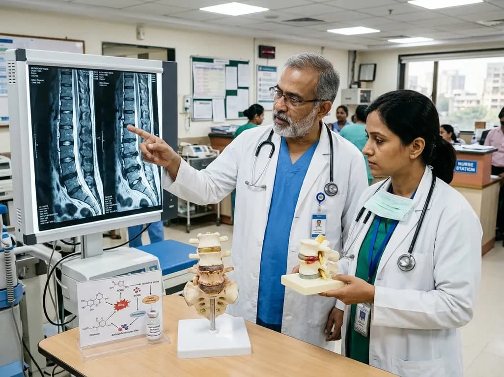
Intervertebral disc degeneration is a primary driver of chronic low back pain. Emerging evidence highlights reactive oxygen species and redox imbalance as central mechanisms. Targeted antioxidant strategies and biomaterial delivery systems offer promising disease-modifying therapies for spine health.
Today

A multicenter cohort study reveals that the 30-minute C-peptide response during OGTT independently predicts cystic fibrosis-related diabetes in patients with normal glucose tolerance, offering an early risk-stratification biomarker.
Today
