.webp&w=384&q=75)

Loading, please wait...
Loading, please wait...
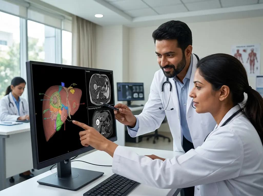
Accurate hepatic tissue evaluation represents a cornerstone of modern abdominal diagnostic imaging. Clinicians frequently encounter diagnostic dilemmas when assessing focal hepatic lesions in patients suffering from chronic liver parenchymal diseases. For decades, multi-parametric protocols have depended on high-resolution tissue contrasts to distinguish benign regenerative nodules from hepatocellular carcinoma. However, respiratory motion often degrades standard sequences and complicates radiological interpretation. Recent technological advancements introducing single-shot T2 liver MRI provide an effective solution to these longstanding diagnostic hurdles. By acquiring complete slice data within a fraction of a second, this approach drastically suppresses physiological motion artifacts. Consequently, radiologists can achieve significantly sharper anatomic margins and confident tissue characterization. Furthermore, pairing ultrafast acquisitions with artificial intelligence establishes a powerful diagnostic pathway for complex oncology patients.
Conventional liver magnetic resonance imaging historically relies on free-breathing turbo spin-echo sequences to deliver high signal contrast. Although these acquisitions yield satisfactory tissue contrast in ideal conditions, they require prolonged scan durations often exceeding two and a half minutes. Consequently, respiratory fluctuations and involuntary diaphragmatic excursions routinely introduce phase-encoding motion artifacts. These persistent artifacts severely degrade edge sharpness and blur subtle morphological characteristics of small nodules.
In addition, patients with end-stage cirrhosis, advanced age, or severe dyspnea frequently struggle to maintain steady breathing patterns. As a result, respiratory-triggered sequences often suffer from extended acquisition windows and unpredictable image quality. Blurring across tissue interfaces obscures mild hyperintensity, which represents a crucial hallmark for characterizing early malignancy. Furthermore, motion artifacts frequently mimic pathological lesions or mask true architectural features. Radiologists facing degraded datasets must frequently contend with indeterminate radiological classifications, potentially delaying targeted therapeutic interventions. Therefore, clinical teams require motion-robust sequences that maintain exquisite diagnostic fidelity without overburdening vulnerable patients.
Single-shot turbo spin-echo sequences fundamentally transform abdominal imaging by collecting all required spatial frequencies during a single radiofrequency excitation train. This ultrafast acquisition captures each anatomical slice in merely fractions of a second, slashing total examination time from several minutes to under fourteen seconds. Therefore, single-shot T2 liver MRI practically eliminates the disruptive influence of breathing motion and bowel peristalsis.
Moreover, rapid volumetric coverage improves patient tolerance, particularly among critically ill or uncooperative individuals. The rapid acquisition ensures uniform tissue depiction across the entire liver volume without slice misalignment. Additionally, clinicians observe superior lesion conspicuity, which aids in discriminating benign biliary cysts and cavernous hemangiomas from malignant masses. Because the sequence rapidly freezes physiologic motion, hepatic margins and intrahepatic vascular bifurcations remain crisp. Consequently, radiologists can evaluate subtle signal intensities without the confounding ghosting artifacts typical of standard free-breathing protocols. This dramatic reduction in acquisition time streamlines busy clinical workflows while delivering dependable diagnostic images across diverse patient cohorts.
Historically, single-shot acquisitions suffered from inherent trade-offs, such as elevated image noise, T2 blurring, and lower baseline spatial resolution. However, the integration of deep learning-based reconstruction algorithms effectively resolves these historical limitations. Deep neural networks, trained on vast datasets of pristine magnetic resonance images, selectively remove background noise while restoring high-frequency spatial boundaries.
Specifically, applying deep learning reconstruction to single-shot data dramatically elevates the signal-to-noise ratio and lesion-to-liver contrast-to-noise ratio. Quantitative assessments confirm marked enhancements in parenchymal signal homogeneity and tissue differentiation. Furthermore, qualitative evaluations reveal substantial improvements in hepatic edge delineation, small vessel clarity, and overall artifact suppression. Deep learning algorithms perform this denoising process in real time, preventing reconstruction delays during acute clinical assessments. Consequently, radiologists obtain images that combine the motion immunity of single-shot techniques with the superior aesthetic and diagnostic sharpness of conventional multi-shot acquisitions. This synergy elevates routine liver evaluation to unprecedented levels of diagnostic certainty.
Accurate nodule characterization holds paramount importance when evaluating cirrhotic livers under standardized Liver Imaging Reporting and Data System frameworks. Standard free-breathing sequences often generate ambiguous findings, leading to misinterpretations where benign entities receive false malignancy scores. In contrast, ultrafast single-shot techniques preserve genuine T2 signal characteristics and prevent deceptive motion-induced intensity changes.
Notably, clinical evidence demonstrates that single-shot protocols, especially when boosted by deep learning, substantially reduce non-identifiable T2 features in malignant lesions. By accurately unmasking mild-to-moderate T2 hyperintensity, these advanced sequences facilitate the appropriate upgrading of indeterminate LR-3 observations to definitive LR-4 or LR-5 categories. Simultaneously, clear visualization of marked hyperintensity enables confident classification of benign hemangiomas and simple cysts. Thus, the sequence prevents unwarranted invasive biopsies or premature oncologic interventions. Ultimately, refined categorization sharpens clinical decision-making, ensuring that patients with confirmed hepatocellular carcinoma receive timely locoregional therapy while preserving benign cases from overtreatment.
The clinical adoption of accelerated T2-weighted imaging protocols offers substantial practical benefits for multidisciplinary liver tumor boards. Because single-shot acquisitions take less than fifteen seconds, imaging departments can markedly shorten total scan appointments. This rapid efficiency reduces scanner backlog, lowers operational costs, and minimizes patient discomfort inside the bore.
Furthermore, dependable image quality simplifies diagnostic interpretation for radiologists, hepatologists, and gastrointestinal surgeons alike. Clinicians can confidently plan surgical resections, radiofrequency ablations, or liver transplantations based on accurate nodule localization and precise vascular mapping. Additionally, the elimination of motion artifacts reduces the need for repeated sequences or supplementary contrast administrations. In busy hospital settings, such efficiency directly translates into faster turnaround times for critical oncologic staging. As artificial intelligence continues to refine abdominal MRI protocols, adopting motion-free reconstruction techniques will establish a new benchmark for patient-centered hepatic care and precision oncology workflows worldwide.
Single-shot T2 sequences acquire all necessary spatial k-space lines in a continuous echo train following a single radiofrequency excitation pulse. This ultrafast mechanism captures each slice in under a second, completing the entire liver survey within fourteen seconds. In contrast, free-breathing protocols require several minutes, exposing data acquisition to respiratory motion and diaphragmatic excursion. Consequently, the single-shot technique effectively freezes physiologic movement, eliminating ghosting artifacts and significantly improving tissue sharpness.
Deep learning reconstruction utilizes convolutional neural networks trained on extensive imaging libraries to suppress background noise and enhance fine anatomical structures. While standard single-shot sequences often suffer from elevated image noise and spatial blurring, deep learning algorithms selectively reconstruct sharp tissue interfaces. Consequently, this computational approach significantly increases the signal-to-noise ratio and lesion-to-liver contrast. This optimization enables radiologists to clearly visualize tiny focal lesions and delineate subtle vascular margins with high diagnostic confidence.
Under LI-RADS criteria, T2 signal intensity serves as a critical ancillary feature for categorizing indeterminate liver nodules. Mild-to-moderate T2 hyperintensity strongly suggests hepatocellular carcinoma, whereas marked hyperintensity typically indicates benign lesions such as cysts or hemangiomas. When motion artifacts obscure these features, nodules are frequently misclassified as indeterminate LR-3. Accurate T2 characterization facilitates appropriate upgrading to LR-4 or LR-5, ensuring timely oncological treatment while preventing unnecessary invasive biopsies in benign cases.
Disclaimer: This content is for informational and educational purposes only. It is not intended to provide medical advice, diagnosis, or treatment guidelines. Healthcare professionals should exercise independent clinical judgment. Refer to the latest local and national guidelines for clinical practice.
References

Read summarized clinical updates, watch expert medical content, and earn CME certifications right from your smartphone.
Open your smartphone camera to scan and install.





Single-shot T2-weighted MRI combined with deep learning reconstruction significantly outperforms conventional free-breathing acquisitions, enhancing liver lesion conspicuity and LI-RADS categorization accuracy in chronic liver disease.
last month

A landmark biomonitoring study reveals that chronic pesticide exposure significantly alters buccal microRNA expression and increases micronucleus frequency in agricultural workers. These findings uncover actionable molecular signatures of oxidative stress, inflammation, and genomic instability.
Today

India's national digital health service achieved a historic milestone of fifty crore virtual consultations. Notably, female patients account for over fifty-seven percent of utilisation, demonstrating improved healthcare accessibility for remote communities, chronic disease management, and primary triage.
Today

A large UK Biobank study identified four routine biomarker subtypes that predict cardiometabolic disease progression to multimorbidity. Integrating multi-state modelling, metabolomics, and proteomics revealed distinct biological pathways, offering new avenues for tailored clinical risk stratification.
Today
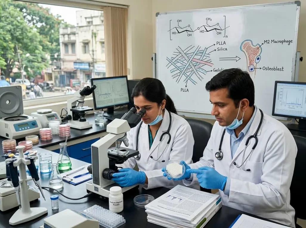
Dual-nanofiber interpenetrating network scaffolds combining silica and PLLA/gelatin nanofibers with sodium alginate achieve complementary mechanical stability, immunomodulatory M2 macrophage polarization, and enhanced osteogenesis for advanced bone repair.
Today

Researchers at IISER Berhampur have engineered a covalent organic framework immunosensor that detects urinary NGAL. This novel nanotechnology platform distinguishes diabetic nephropathy from other renal conditions, enabling rapid, minimally invasive stratification and earlier clinical intervention for patients.
Today
