.webp&w=384&q=75)

Loading, please wait...
Loading, please wait...

Minimally invasive transforaminal lumbar interbody fusion has transformed spine care by decreasing muscle morbidity and blood loss. However, achieving adequate segmental lordosis in MIS-TLIF remains a persistent technical challenge for spinal surgeons. Traditional transforaminal techniques frequently risk iatrogenic hypolordosis or flat-back deformity because static interbody cages often seat without restoring anatomical sagittal tilt. Consequently, contemporary reconstructive spine surgery increasingly emphasizes sagittal balance alongside neurological decompression. A groundbreaking clinical investigation in Spine evaluates an innovative surgical protocol combining intraoperative mechanical table hinging with bilateral facetectomies. This hybrid technique specifically addresses sagittal malalignment while preserving the hallmark benefits of minimal surgical access.
Historically, lumbar arthrodesis procedures prioritized neural decompression and solid fusion over regional alignment. Today, clinicians recognize that maintaining segmental lordosis in MIS-TLIF directly influences patient-reported outcomes and adjacent segment longevity. When surgeons insert an interbody cage via a unilateral transforaminal portal, the posterior column often resists lordotic restoration. Furthermore, standard operating tables maintain static patient positioning, which restricts anterior disc opening during trial insertion. As a result, impaction of standard static spacers can inadvertently produce segmental kyphosis or inadequate lordotic gain. Over time, persistent sagittal malalignment accelerates mechanical wear at adjacent levels and increases chronic axial low back discomfort. Additionally, insufficient segmental correction forces compensatory hyperlordosis in adjacent motion segments, triggering premature facet arthrosis. Addressing these biomechanical limitations therefore requires intentional posterior column release paired with dynamic operating table manipulation. By deliberately uncoupling posterior bony constraints, surgeons create an adaptable mechanical environment for genuine lordotic recreation. This strategic shift moves MIS-TLIF from a purely decompressive intervention into a powerful tool for sagittal alignment correction.
To overcome rigid anatomical barriers, the described surgical technique integrates bilateral facetectomies with dynamic table manipulation. Initially, the surgeon performs an ipsilateral facetectomy through a tubular retractor to decompress neural structures and prepare the disc space. Subsequently, the surgical team performs a contralateral facetectomy or Smith-Petersen osteotomy to mobilize the posterior column completely. This extensive posterior release detaches mechanical tension bands that otherwise resist lordotic expansion. Following bilateral osteotomies, the surgical team activates the mechanical hinging operating table into deliberate flexion. This table flexion opens the posterior interbody space widely, facilitating safe discectomy and effortless cage insertion without neural traction. Once the surgeon places the interbody spacer into the anterior column, the team transitions the table into extension. Consequently, table extension compresses the posterior osteotomy sites and pivots the vertebrae over the anterior spacer fulcrum. Percutaneous pedicle screws and contoured rods then secure the construct in this lordotic posture. Therefore, dynamic hinging converts the table itself into an active surgical instrument during interbody reconstruction. This deliberate coordination substantially elevates lordotic correction compared to conventional stationary patient positioning.
The retrospective study analyzed 202 consecutive adult patients undergoing MIS-TLIF with dynamic mechanical hinging and bilateral facet releases. Radiographic assessments evaluated segmental lordosis preoperatively, immediately postoperatively, and at six-month follow-up visits. For single-level fusions, the operative protocol achieved an impressive mean immediate postsurgical correction of 5.0 degrees. Moreover, patients maintained a robust mean correction of 4.6 degrees at their six-month radiographic evaluation. Two-level fusions demonstrated similarly robust sagittal improvements, achieving a mean immediate correction of 4.6 degrees. Interestingly, two-level procedures showed an increased mean correction of 6.6 degrees at the six-month postoperative milestone. Statistical evaluations revealed highly significant lordosis enhancements between preoperative and postoperative imaging for single-level and two-level cohorts. Furthermore, single-level cases maintained statistically significant improvements when researchers compared preoperative baseline imaging directly to six-month scans. These radiographic gains markedly surpass historical benchmarks reported for standard unilateral MIS-TLIF approaches, which typically yield negligible lordosis changes. Consequently, combining bilateral facet mobilization with mechanical extension reliably restores segmental alignment across single and multi-level lumbar pathologies.
Understanding baseline anatomical predictors helps surgeons customize their reconstructive goals for each individual patient. Notably, the study identified a significant negative correlation between preoperative segmental lordosis and immediate postsurgical correction. Patients presenting with severe baseline hypolordosis or segmental kyphosis achieved the greatest angular correction following surgery. Similarly, researchers noted a negative correlation between baseline lordosis and the sustained correction observed at six months. Thus, collapsed or flat segments harbor the highest potential for meaningful lordotic gain through dynamic osteotomy closure. However, aggressive posterior compression requires meticulous surgical technique to avoid adjacent soft-tissue complications. For instance, postoperative Baastrup disease can theoretically develop if adjacent spinous processes contact during hyperlordotic compression. The authors systematically tracked patient demographics, including age, body mass index, diabetes status, and smoking history. Fortunately, metabolic variables and tobacco use did not compromise the immediate mechanical restoration of sagittal curvature. Nevertheless, optimizing bone mineral density and metabolic health remains vital for maintaining long-term interbody stability and avoiding implant subsidence.
Restoring physiological lumbar lordosis represents a cornerstone of modern spinal deformity surgery and degenerative spine management. For spine surgeons in India and globally, mastering dynamic table maneuvers provides substantial clinical advantages without requiring expensive novel implants. Surgeons can utilize standard interbody cages while achieving the lordotic profile of costlier expandable devices. Furthermore, adequate sagittal restoration relieves compensatory stresses on adjacent segments, potentially reducing adjacent segment breakdown and reoperation rates. Before initiating mechanical table extension, surgeons must verify complete neural decompression across both exit pathways. In addition, surgeons must ensure thorough disc space clearance and proper anterior cage placement to establish an effective mechanical fulcrum. Bilateral facetectomies demand precise hemostasis, particularly when operating within narrow minimally invasive corridors. Operating room teams must also practice coordinated table adjustments to ensure patient stability and prevent airway tube displacement. Ultimately, integrating bilateral posterior column osteotomies with coordinated mechanical hinging equips surgeons to correct focal deformities reliably and safely.
Executing mechanical table hinging requires rigorous safety protocols to prevent positioning injuries and implant malposition. Before applying surgical table flexion, the operating team must position the patient securely on padded bolsters. This meticulous padding prevents abdominal compression, minimizes epidural venous engorgement, and reduces intraoperative blood loss. Furthermore, the surgeon must inspect neural structures under high magnification before and after adjusting table orientation. Because table extension actively closes the posterior column osteotomy, surgeons must confirm that no dorsal neural tissue suffers impingement. Additionally, surgeons must verify that pedicle screws maintain firm purchase during posterior compression maneuvers. Excessive compression across osteopenic bone could cause screw loosening or pedicle fracture rather than controlled lordotic closure. Therefore, preoperative assessment of vertebral bone density using opportunistic Hounsfield units remains highly advisable. Once the surgeon confirms secure screw locking in extended posture, intraoperative fluoroscopy provides immediate verification of the achieved lordosis. Consequently, structured surgical workflows ensure that mechanical hinging enhances lordotic correction while preserving patient safety throughout the entire minimally invasive procedure.
Conventional MIS-TLIF relies on a unilateral transforaminal approach with intact contralateral posterior structures that resist lordotic expansion. Furthermore, surgeons often insert interbody cages on stationary flat tables without mobilising posterior tension bands. Consequently, static cages frequently seat without restoring anterior column height, occasionally producing flat-back deformity or segmental kyphosis.
Mechanical table hinging dynamically alters spinal curvature during surgery. The team flexes the table initially to open the posterior disc space, allowing safe discectomy and cage placement. Subsequently, shifting the table into extension compresses the bilateral facetectomy gaps over the anterior cage fulcrum, effectively locking the segment in lordosis.
Bilateral facetectomies eliminate the rigid dorsal bony constraints that restrict sagittal correction. By resecting both inferior articular processes, surgeons release the posterior column completely. This release enables significant lordotic closure when the surgical team applies table extension, achieving genuine multi-degree correction without requiring aggressive anterior longitudinal ligament resection.
Disclaimer: This content is for informational and educational purposes only... Refer to the latest local and national guidelines for clinical practice.
References

Read summarized clinical updates, watch expert medical content, and earn CME certifications right from your smartphone.
Open your smartphone camera to scan and install.





A retrospective cohort study demonstrates that combining dynamic intraoperative mechanical table hinging with bilateral facetectomies achieves significant, durable segmental lordosis in MIS-TLIF across single- and two-level fusions, overcoming the traditional kyphosing tendency of transforaminal interbody procedures.
Today
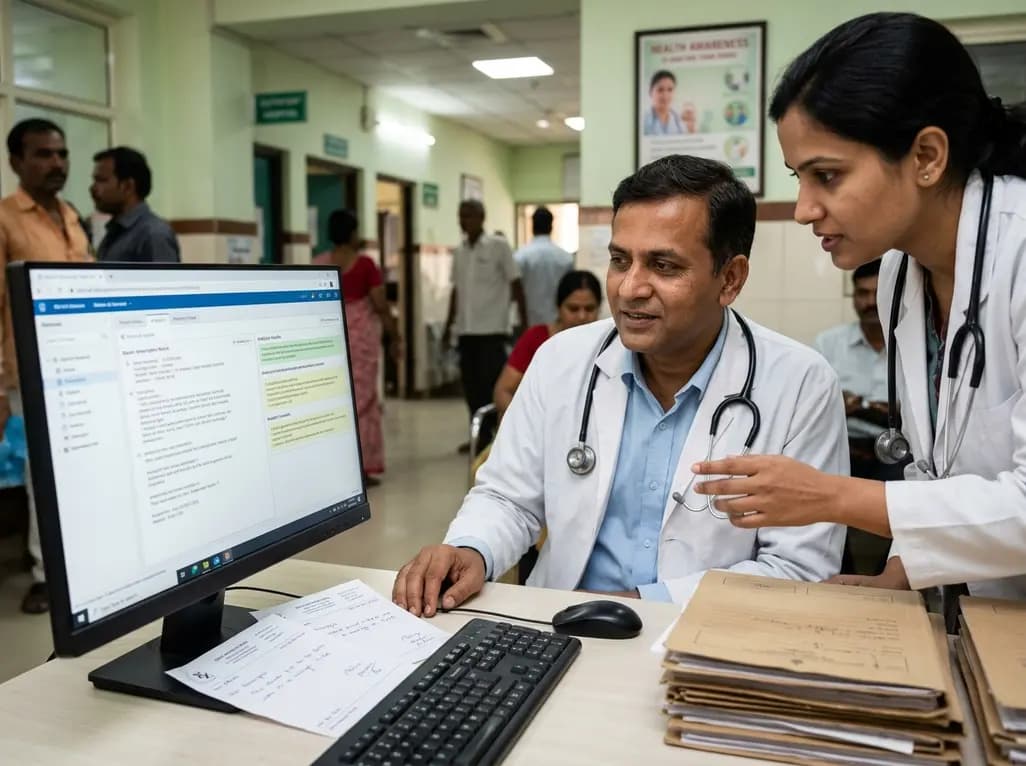
A crossover study demonstrates that locally deployed large language models with exact-match knowledge augmentation significantly elevate outpatient prescription review accuracy to 97.2%, curbing hallucinations and securing patient data without requiring complex cloud infrastructure.
Today
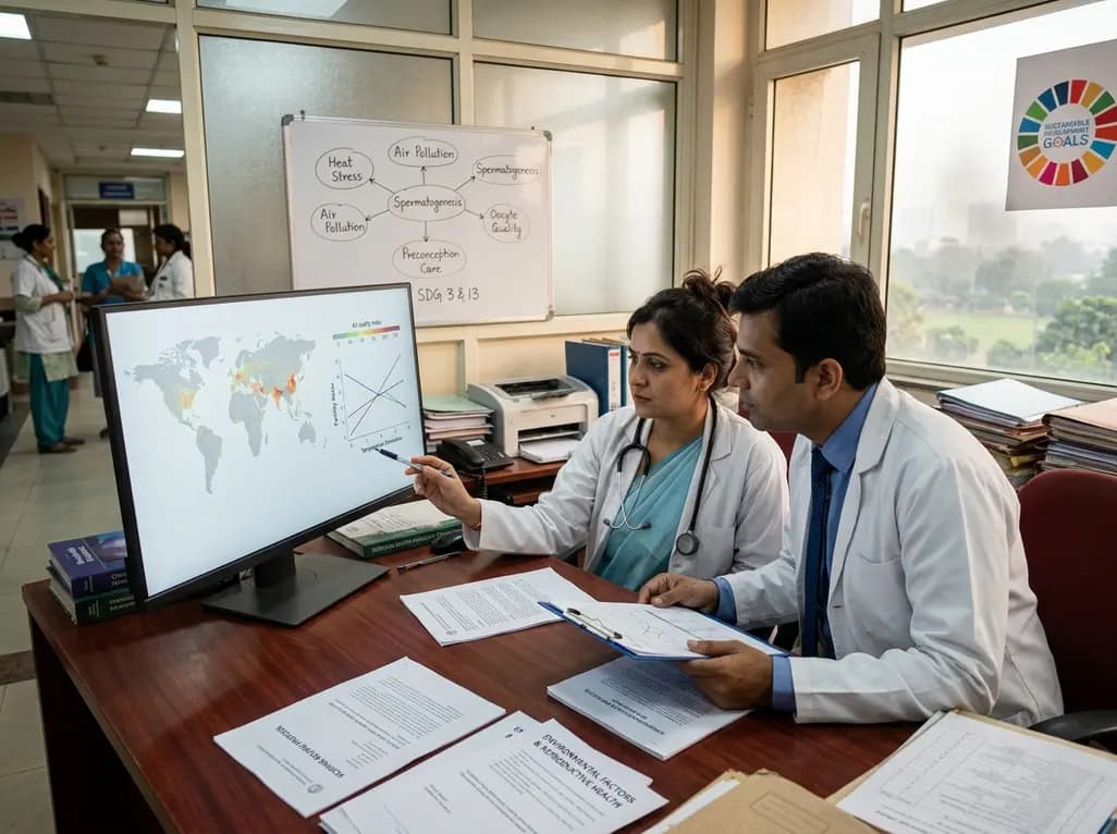
A new scoping review protocol systematically maps global evidence on how climatic shifts, ambient heat, and pollution impact human fertility. Discover key physiological mechanisms, clinical preconception strategies, and critical public health alignments under Sustainable Development Goals 3 and 13.
Today
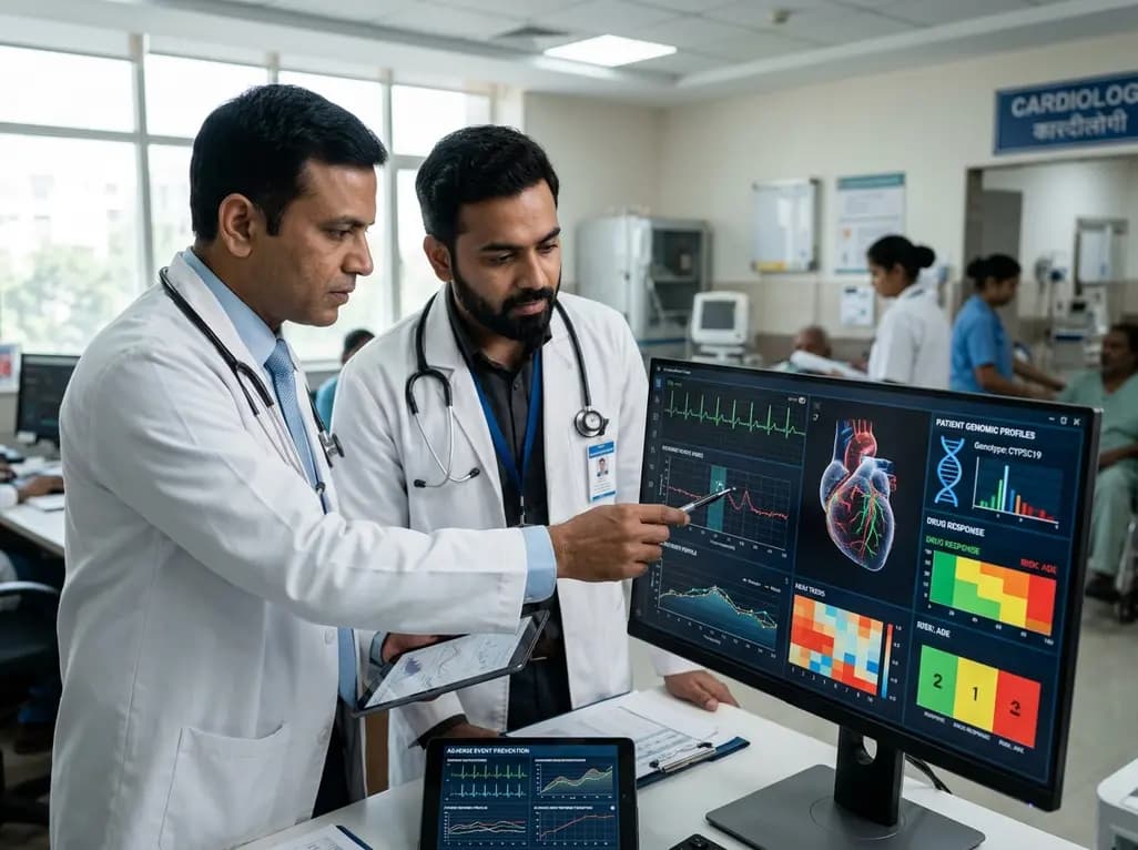
Pharmacogenomics and artificial intelligence are revolutionizing cardiology by tailoring therapies to individual genetic profiles and clinical data, reducing adverse drug events, and improving cardiovascular patient outcomes.
Today
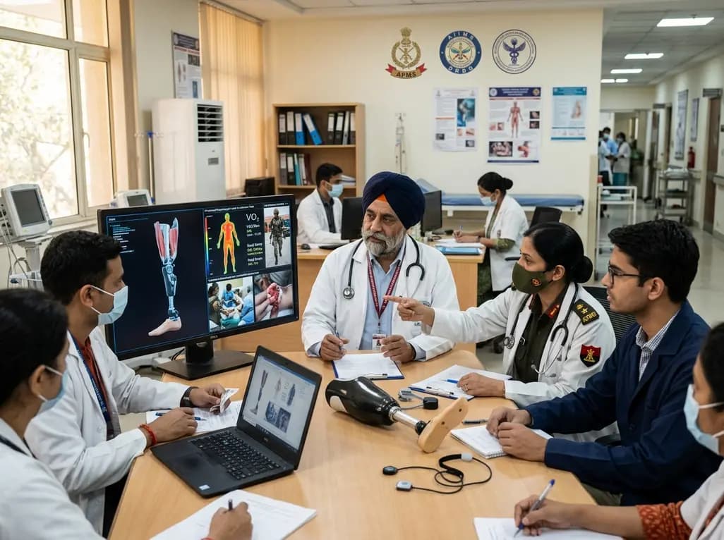
India's premier scientific bodies have joined the Armed Forces Medical Services to solve unique physiological and operational challenges faced by soldiers. The national collaboration covers combat casualty care, artificial intelligence diagnostics, bionic prosthetics, and extreme-environment physiological resilience.
Today
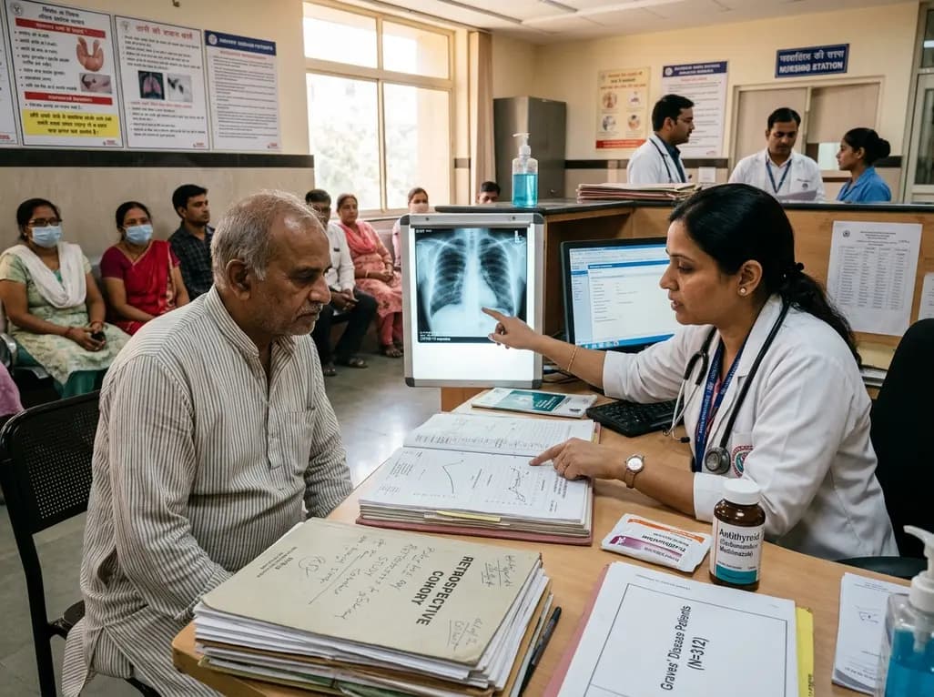
A retrospective cohort study evaluates the short- and long-term impacts of SARS-CoV-2 infection on patients with Graves' disease receiving antithyroid drug therapy, highlighting thyroid status destabilization, clinical symptom exacerbations, and post-viral sequelae.
Today
