

Loading, please wait...
Loading, please wait...

Cranial radiotherapy represents a cornerstone of modern neuro-oncology, yet it carries long-term neurovascular sequelae. Among these delayed complications, radiation-induced cavernous malformations have emerged as significant clinical entities that demand careful vigilance. Physicians historically regarded cerebral cavernous malformations as primarily congenital vascular anomalies. However, growing clinical evidence demonstrates that therapeutic cranial irradiation can induce de novo cavernoma formation. In a groundbreaking comparative cohort investigation, researchers evaluated the distinct clinical behavior of these secondary lesions against sporadic presentations. The investigators analyzed 612 patients with magnetic resonance imaging confirmed lesions treated between 1987 and 2024. Within this cohort, 16 individuals harbored radiation-induced lesions, whereas 596 exhibited non-radiation-associated malformations. The data revealed that secondary vascular lesions behave with elevated clinical volatility. Consequently, clinicians must recognize radiation exposure as a distinct risk modifier rather than a coincidental background factor. Identifying these secondary vascular lesions early allows multidisciplinary neurovascular teams to tailor therapeutic strategies appropriately. Furthermore, understanding the distinct biological trajectory of these malformations helps clinicians avoid catastrophic hemorrhagic events in cancer survivors. Therefore, this comparative analysis provides essential benchmark data for neurosurgeons, oncologists, and neurologists managing long-term survivors of cranial radiation.
The comparative study revealed substantial differences in hemorrhage dynamics between cohorts. Most notably, multivariable logistic regression demonstrated that prior radiation exposure significantly increased the odds of symptomatic hemorrhage. Patients who had received radiation faced more than five times higher odds of intracranial bleeding compared to those with sporadic malformations, yielding an adjusted odds ratio of 5.23. Moreover, the hemorrhage incidence rate reached 7.77 per 100 person-years among irradiated patients. In contrast, patients with sporadic cavernomas experienced an incidence rate of 4.36 per 100 person-years. This notable divergence established an incidence rate ratio of 1.78. Interestingly, Cox proportional hazards regression showed that radiation exposure was not significantly associated with accelerated time to hemorrhage. Hence, while the cumulative probability of bleeding remains markedly elevated, the temporal occurrence exhibits prolonged variability. The timeline of lesion emergence represents another vital clinical consideration. Patients received their cranial irradiation at a median age of 11.9 years. Subsequently, clinicians diagnosed the resulting cavernous malformations a median of 13.5 years later. This extended latency period clearly highlights the persistent life-long risk in pediatric oncology survivors.
The pathogenesis of radiation-associated vascular malformations involves complex microvascular injury and delayed tissue remodeling. Radiation damages delicate endothelial cells, which induces chronic focal inflammation and hyalinization of small vessels. In addition, radiation promotes aberrant neoangiogenesis through the dysregulated release of vascular endothelial growth factor. Consequently, fragile, thin-walled, sinusoidal capillary channels develop without normal intervening brain parenchyma. In the study cohort, patients received a mean radiation dose of 47.8 Gy, with a standard deviation of 15.1 Gy. Furthermore, arteriovenous malformations constituted the most common primary indication for radiation therapy, representing 31 percent of cases. Other patients received cranial radiation for primary pediatric brain tumors, including medulloblastomas and astrocytomas. Radiation fields often encompass developing cerebral vasculature, which renders pediatric brain tissue unusually susceptible to secondary vascular damage. Because pediatric endothelial cells exhibit higher mitotic activity, young patients remain particularly vulnerable to ionizing radiation effects. Therefore, radiation oncologists must carefully optimize conformal delivery techniques to limit unnecessary dose exposure to surrounding normal brain tissue. Minimizing unnecessary field margins preserves vulnerable microvasculature and reduces delayed cerebrovascular pathology.
The clinical presentation patterns between irradiated and sporadic groups displayed meaningful differences and unexpected similarities. Specifically, 68.8 percent of radiation-associated patients presented incidentally during routine follow-up neuroimaging. Meanwhile, non-seizure-associated hemorrhage constituted the initial presentation in 31.3 percent of irradiated individuals. Conversely, the sporadic malformation cohort presented through a broader spectrum of clinical symptoms. In the non-irradiated cohort, 56.4 percent were incidental, 20.6 percent suffered non-seizure-associated hemorrhages, and 23.0 percent developed new-onset seizures. Significantly, multivariable regression indicated that the initial presentation mode did not independently predict subsequent hemorrhagic events. This key observation underscores that incidental detection does not imply benign future behavior. Accordingly, clinicians must implement structured, long-term magnetic resonance imaging protocols for all irradiated individuals. Routine surveillance should utilize gradient-echo or susceptibility-weighted sequences, which offer superior sensitivity for detecting occult microbleeds and tiny hemosiderin deposits. Detecting silent cavernomas early empowers clinicians to provide proactive counseling regarding symptom vigilance and blood pressure regulation. Furthermore, regular serial imaging enables neurosurgical teams to monitor lesion growth and intervene swiftly before catastrophic mass effect or debilitating neurological deficits emerge.
Despite a substantially higher hemorrhage rate among radiation-exposed patients, surgical resection rates remained statistically similar between both cohorts. Overall, 25.0 percent of patients with radiation-induced malformations underwent microsurgical resection, compared with 21.1 percent of patients with sporadic lesions. Furthermore, primary surgical indications showed striking concordance across the study population. Symptomatic intracranial hemorrhage served as the predominant surgical indication in both groups, accounting for 75.0 percent of surgeries in irradiated patients and 43.7 percent in non-irradiated patients. Therefore, neurosurgeons apply consistent operative thresholds regardless of lesion etiology. When a cavernoma causes progressive neurological deficits, intractable epilepsy, or recurrent hemorrhages in accessible brain areas, complete microsurgical excision offers definitive cure. However, surgeons must navigate irradiated tissues with heightened caution due to parenchymal scarring and fragile adjacent parenchyma. Additionally, conservative observation remains appropriate for stable, asymptomatic lesions located in eloquent cortex or deep brainstem regions. Long-term multidisciplinary follow-up between neurosurgeons, neurologists, and oncologists ensures optimized care trajectories for these complex patients over decades. Consequently, personalized clinical decision-making balances operative risks against the elevated lifetime hemorrhage hazard.
Radiation-induced cavernous malformations develop as delayed secondary complications years after cranial radiation therapy, especially in individuals irradiated during childhood. In contrast, sporadic cavernous malformations typically arise congenitally without external triggers. While both types display identical histology with clustered capillary caverns on microscopy, radiation-induced lesions carry a significantly higher subsequent hemorrhage incidence rate. Furthermore, patients with radiation-associated lesions frequently present incidentally decades after initial cancer treatment, requiring prolonged neuroimaging surveillance.
Cerebral cavernous malformations exhibit a remarkably long latency period following cranial radiotherapy. Clinical evidence indicates a median latency of approximately 13.5 years from irradiation to formal diagnosis. However, lesions may emerge anywhere from two years to over four decades following therapeutic radiation. Because pediatric patients receiving cranial irradiation face lifelong vulnerability, neuro-oncologists recommend regular neuroimaging surveillance with gradient-echo or susceptibility-weighted magnetic resonance imaging well into adulthood to detect de novo vascular formations early.
Neurosurgical intervention for radiation-induced cavernous malformations depends on clinical presentation rather than etiology alone. Surgeons primarily operate when lesions cause symptomatic or recurrent intracranial hemorrhage, medically refractory seizures, or progressive neurological deficits. In the latest clinical cohort, symptomatic hemorrhage prompted surgery in 75 percent of operated cases. Asymptomatic lesions located in eloquent brain parenchyma are typically managed conservatively with serial magnetic resonance imaging, thereby avoiding operative morbidity in previously irradiated, fragile neural tissue.
Disclaimer: This content is for informational and educational purposes only and is not intended to substitute for professional medical advice, diagnosis, or treatment. Always consult a qualified healthcare provider for specific clinical guidance. Refer to the latest local and national guidelines for clinical practice.
References

Read summarized clinical updates, watch expert medical content, and earn CME certifications right from your smartphone.
Open your smartphone camera to scan and install.





A landmark comparative study reveals that radiation-induced cavernous malformations carry a significantly higher hemorrhage incidence than sporadic lesions, underscoring the vital need for extended neuroimaging surveillance decades after cranial radiation therapy.
Today
A national cross-sectional survey reveals marked disparities in infectious disease diagnostic capacity across hospital tiers, underscoring the urgent need for integrated referral networks and enhanced point-of-care testing to improve outbreak detection and patient outcomes.
Today

The GESS-AEEM consensus establishes a clear clinical implementation framework for testosterone therapy in postmenopausal women with hypoactive sexual desire disorder, focusing on transdermal dosing, precise laboratory monitoring, and safety communication.
Yesterday
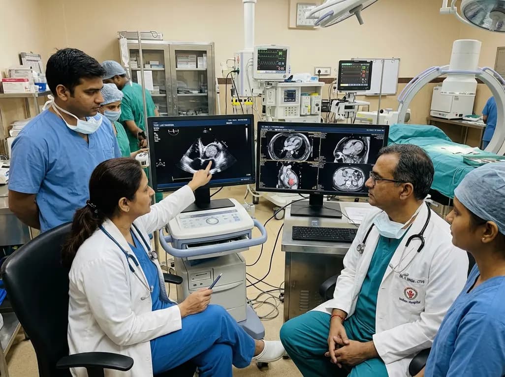
A rare case of an asymptomatic left ventricular hemangioma highlights the vital role of multimodal imaging in differentiating vascular tumors from thrombus. Complete surgical resection under transesophageal echocardiography guidance prevents thromboembolism and delivers optimal outcomes.
Today
A landmark study evaluates long-term quality of life in adolescents and adults with brachial plexus birth injury. Severe functional limitation and limited access to care significantly worsen outcomes, underscoring the urgent need for persistent, multidisciplinary management across adult healthcare transitions.
Today
An analysis of 1,000 posts by macro- and mega-influencers on Instagram and TikTok revealed that 78.5% contained nutrition misinformation, while only 24% of creators held valid credentials. This article examines the clinical risks of viral dietary fads and provides actionable guidance for healthcare professionals.
Today
