.webp&w=384&q=75)

Loading, please wait...
Loading, please wait...
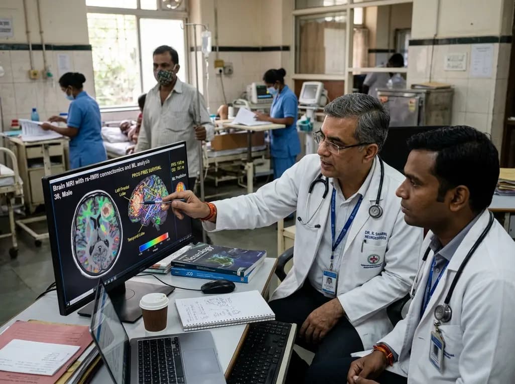
Glioblastoma represents the most aggressive and recurrent primary malignant brain tumor encountered in modern clinical practice. Clinicians constantly face significant prognostic uncertainty when designing personalized treatment pathways. Accurately estimating progression-free survival in glioblastoma prior to surgery or chemoradiotherapy offers an unprecedented opportunity to optimize surgical resection strategies, tailor therapeutic intensity, and guide patient counseling. Currently, conventional anatomical neuroimaging provides crucial structural details about tumor burden, mass effect, and perilesional edema. However, standard anatomical imaging often fails to capture the intricate, whole-brain functional disruptions that aggressive tumors inflict upon neural circuits.
To bridge this critical prognostic gap, recent advancements combine resting-state functional magnetic resonance imaging with sophisticated artificial intelligence algorithms. Resting-state functional connectivity measures spontaneous, synchronized blood-oxygen-level-dependent signal fluctuations across brain regions. Consequently, this non-invasive modality illuminates how malignant infiltration destabilizes large-scale neural networks beyond visible macroscopic tumor margins. By leveraging deep neural networks trained on pre-intervention functional connectivity metrics, researchers can uncover subtle neuroarchitectural signatures that correlate with rapid recurrence. This computational paradigm represents a substantial leap forward for modern neuro-oncology.
Malignant gliomas do not behave merely as isolated focal structural masses. Instead, these invasive neoplasms interact dynamically with host neural architecture and disrupt distributed connectomic pathways across both cerebral hemispheres. Specifically, high-grade glial tumors release neuroactive factors, alter synaptic microenvironments, and invade white matter tracts. These pathological actions fundamentally impair functional communication between distant cortical regions. Resting-state functional magnetic resonance imaging captures these complex disruptions by evaluating resting-state networks, including association networks that govern higher-order cognition and executive processing.
Recent neuroimaging findings demonstrate that glioblastoma induces widespread alterations in fundamental brain networks compared to healthy physiological baselines. In particular, dorsal attention, visual, frontal-parietal, and default mode networks frequently exhibit significant connectivity degradation when tumors encroach upon critical neuroanatomical hubs. Conversely, tumors situated within the right temporal lobe often present with distinct functional preservation patterns that correlate with comparatively favorable clinical trajectories. Therefore, evaluating pre-treatment whole-brain functional connectivity allows clinicians to appreciate the true global physiological impact of localized tumors. Understanding these extensive network alterations provides deeper physiological insight into tumor aggressiveness and disease dissemination.
While advanced neuroimaging delivers profound insights into neural connectomics, traditional clinical and molecular markers remain foundational in neuro-oncology prognostication. In comprehensive retrospective analyses, several baseline clinical variables show robust correlations with disease progression timelines. For example, patient sex significantly influences clinical trajectories, reflecting emerging evidence on sex-specific biological dimorphisms in glioma growth. Furthermore, established molecular biomarkers, most notably MGMT promoter methylation status, demonstrate powerful predictive value regarding progression intervals and temozolomide responsiveness.
Additionally, initial neurological presentations serve as meaningful clinical indicators of functional network vulnerability and disease severity. Patients presenting with motor weakness or memory impairment often experience significantly shorter progression-free survival in glioblastoma compared to those presenting with less disruptive focal symptoms. These neurological deficits directly reflect compromised motor pathways and association networks, emphasizing the connection between clinical symptomatology and connectomic disruption. Moreover, overall survival strongly correlates with progression-free intervals, highlighting that delays in early disease progression translate directly into extended survival. Consequently, integrating baseline neurological evaluations with molecular profiling and neuroimaging yields a comprehensive prognostic framework for practicing oncologists.
Applying deep learning to high-dimensional resting-state functional connectivity datasets requires robust computational pipelines to prevent overfitting and ensure clinical generalizability. Functional neuroimaging generates thousands of pairwise regional connectivity values for each patient. Therefore, employing rigorous algorithmic feature selection before training neural networks is essential. Modern investigations successfully apply maximum relevance minimum redundancy feature selection algorithms to isolate the most informative connectomic variables from noise.
Specifically, this algorithmic feature selection identifies thalamic and association network connections as the strongest independent predictors of progression timelines. Key networks identified include the somatomotor, ventral attention, dorsal attention, and default mode/parietal memory circuits. Deep neural network architectures trained on these selected connectivity features achieve remarkable predictive accuracy during leave-one-out cross-validation. For instance, validated models achieve a root mean square error of approximately 1.26 months, a mean absolute error of 1.08 months, and a coefficient of determination of 0.96. These statistical metrics underscore the exceptional precision with which pre-operative resting-state neuroimaging can forecast disease progression. As a result, computational analytics translate raw functional connectivity matrices into actionable, high-confidence prognostic timelines.
Integrating artificial intelligence predictions into preoperative workflows creates compelling opportunities for personalized patient management in neuro-oncology. Currently, neurosurgeons design surgical resection boundaries primarily based on structural contrast-enhancing lesions and functional motor or language mapping. However, preoperative knowledge of expected progression intervals could significantly refine aggressive surgical planning and adjuvant timing. When machine learning models predict rapid early recurrence, multidisciplinary tumor boards can proactively consider specialized clinical trials, novel targeted agents, or intensified local therapeutic strategies.
Furthermore, accurate prognostic modeling empowers clinicians to conduct more transparent and empathetic communication with patients and their families. Transparent counseling regarding expected progression-free timelines helps families make informed personal decisions, prioritize rehabilitation goals, and manage expectations realistically. Additionally, identifying vulnerable functional networks allows neurosurgeons to balance maximum cytoreductive resection against the preservation of essential cognitive networks like the default mode and attention systems. Preserving these critical association networks maintains postoperative functional independence and quality of life. Thus, combining connectomic analytics with clinical care models bridges the gap between technical neurosurgical precision and holistic, patient-centered oncological therapy.
Despite these promising computational breakthroughs, translating machine learning neuroimaging tools into everyday clinical practice presents several challenges. First, acquisition protocols for resting-state functional MRI must achieve standardization across diverse scanner manufacturers and magnetic field strengths. Variability in scan duration, patient movement, and preprocessing pipelines can introduce artifactual noise into connectivity matrices. Therefore, neuro-oncology consortia must establish reproducible imaging acquisition and post-processing protocols across institutional hospital networks.
Second, clinical validation requires expanding research cohorts across multi-center, multi-ethnic patient populations. While initial single-center studies show outstanding predictive accuracy, validating deep neural networks against larger external cohorts will confirm broad generalizability. Moreover, future algorithms should integrate multimodal inputs, uniting functional connectomics with genomic sequencing, radiomics, and dynamic perfusion imaging. Such multi-omic platforms could deliver continuous, real-time risk stratification throughout the disease course. Ultimately, collaborative efforts between computational data scientists, neuroradiologists, and neuro-oncologists will facilitate the deployment of validated artificial intelligence tools in routine hospital environments.
Resting-state functional MRI measures spontaneous neural activity across distributed brain networks. Advanced machine learning models analyze altered connectivity patterns in key association networks, such as attention and memory circuits, which correlate with glioblastoma invasiveness and reliably forecast progression timelines prior to surgery.
Significant clinical predictors include patient sex, MGMT promoter methylation status, baseline motor weakness, and memory impairment. In addition, localized network involvement and overall survival strongly correlate with progression intervals, demonstrating that molecular features and neurological presentation jointly determine clinical trajectories in high-grade gliomas.
Machine learning cannot replace histopathological and molecular diagnosis, but it serves as a powerful complementary tool. While tissue biopsy confirms tumor grade and genetic mutations, pre-intervention computational neuroimaging provides early prognostic stratification, non-invasive connectomic insights, and personalized risk profiling to guide comprehensive treatment planning.
Disclaimer: This content is for informational and educational purposes only and is not intended as medical advice. Healthcare professionals should exercise their independent clinical judgment. Refer to the latest local and national guidelines for clinical practice.
References

Read summarized clinical updates, watch expert medical content, and earn CME certifications right from your smartphone.
Open your smartphone camera to scan and install.





A novel machine learning model utilizing preoperative resting-state functional MRI accurately predicts progression-free survival in glioblastoma patients, linking whole-brain connectomic alterations with clinical outcomes to guide personalized treatment.
last month

A recent case highlights the rare coexistence of anti-AQP4 positive neuromyelitis optica spectrum disorder with long-standing rheumatoid arthritis. Recognizing atypical neurological signs in systemic autoimmunity is crucial to prevent irreversible cord damage and long-term disability.
Today

A ten-year cohort study of ABU case logs reveals that URPS fellowship training significantly drives higher annual case volumes, advanced surgical complexity, and increased female representation in pelvic floor reconstruction, underscoring the vital need for structured subspecialty training.
Today
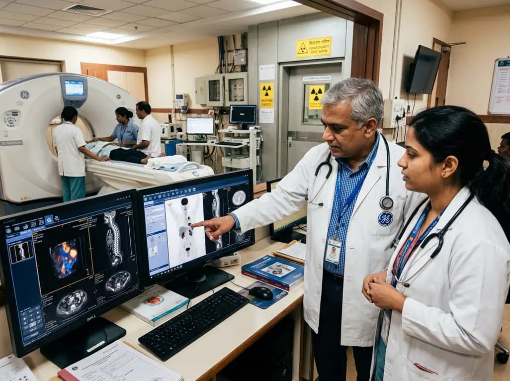
GE HealthCare has agreed to acquire Sofie Biosciences in a 945 million dollar all-cash transaction. The deal secures global rights to the investigational pan-cancer tracer FAPI-74 and expands cyclotron manufacturing infrastructure to accelerate precision theranostics and clinical molecular imaging workflows.
Today

Posttraumatic pisotriquetral instability is a rare wrist injury causing severe hypothenar pain and weakness. Explore clinical examination, imaging, and pisiformectomy.
Today

India's food safety authority has cracked down on high-caffeine beverages sold under stimulant branding, prompting legal challenges from major beverage corporations. This review examines FSSAI regulations, cardiac and metabolic complications, pediatric risks, and actionable counseling strategies for physicians.
Today
