.webp&w=384&q=75)

Loading, please wait...
Loading, please wait...

Coexisting autoimmune diseases frequently create complex clinical dilemmas for practicing physicians. In particular, neuromyelitis optica spectrum disorder represents a severe inflammatory demyelinating syndrome that rarely presents alongside rheumatoid arthritis. When a patient with longstanding rheumatoid disease develops progressive neurological deficits, clinicians often suspect secondary articular complications first. However, early recognition of independent central nervous system autoimmunity remains critical. Furthermore, prompt diagnostic identification prevents devastating axonal destruction and permanent neurological impairment in vulnerable individuals.
Longitudinally extensive transverse myelitis remains a classic hallmark of central demyelination. In this notable case, a woman with a ten-year history of rheumatoid arthritis experienced six years of progressive lower extremity weakness. Consequently, her functional mobility deteriorated significantly over time. Spinal magnetic resonance imaging demonstrated continuous cord inflammation extending from T4 to the filum terminale. Therefore, neuroimaging established a remarkably extensive lesion pattern. Clinicians frequently attribute gait difficulties in rheumatoid patients to compressive myelopathy from cervical subluxation. Additionally, peripheral neuropathy or muscular disuse atrophy can confound the clinical examination. In contrast, this extensive thoracic involvement pointed directly toward an aggressive demyelinating pathology. Consequently, clinicians must maintain vigilance whenever non-articular weakness emerges in systemic autoimmunity. Neurological examinations should assess sensory levels, deep tendon reflexes, and sphincter function promptly. Furthermore, thoracic and lumbar imaging effectively differentiates structural joint impingement from inflammatory cord demyelination. Consequently, comprehensive neuroimaging protocols safeguard patients against misdirected interventions.
The coexistence of systemic rheumatologic conditions and central demyelination indicates shared immunological dysregulation. Specifically, neuromyelitis optica spectrum disorder arises from humoral autoimmunity targeting the astrocytic aquaporin-4 water channel. In this patient, serum analysis revealed positive anti-aquaporin-4 antibodies alongside markedly elevated rheumatoid factor and anti-CCP titers. Consequently, both humoral pathways showed intense simultaneous activation. Shared pathogenic mechanisms involve hyperactive B-cell signaling and elevated interleukin-6 production. Interleukin-6 promotes plasma cell differentiation while compromising blood-brain barrier integrity. Furthermore, T-helper 17 cells contribute substantially to systemic tissue inflammation in both disorders. Therefore, common cytokine abnormalities may foster multiple parallel autoimmune attacks. Although rheumatoid arthritis primarily damages synovial membranes, circulating autoantibodies can trigger severe central pathology. Clinicians must recognize that spinal demyelination can occur regardless of peripheral joint stability. Thus, quiescent articular disease does not protect patients against active astrocytic destruction. Additionally, complement cascade activation exacerbates local tissue necrosis in the spinal cord parenchyma. Hence, targeting humoral autoimmunity remains essential for preventing recurrent central demyelination.
Establishing an accurate diagnosis demands meticulous differentiation between idiopathic demyelination and alternative inflammatory etiologies. In this patient, cerebrospinal fluid analysis demonstrated marked hyperproteinorachia without cellular pleocytosis. Additionally, laboratory testing showed negative oligoclonal bands. This distinct biochemical profile contrasts sharply with typical multiple sclerosis presentations, where oligoclonal bands occur consistently. Consequently, absent oligoclonal bands should direct diagnostic focus toward neuromyelitis optica spectrum disorder. Moreover, elevated protein levels reflect severe disruption of the blood-spinal cord barrier during extensive myelitis. Clinicians must also exclude infectious meningomyelitis, neurotuberculosis, and systemic vasculitis before initiating therapies. Furthermore, systemic lupus erythematosus and primary Sjögren syndrome frequently mimic these neuroimmunological manifestations. Therefore, obtaining comprehensive serological panels remains indispensable in complex clinical scenarios. Serological evaluation should always include aquaporin-4 antibodies and connective tissue markers. Ultimately, integrating laboratory findings with neuroimaging allows clinicians to establish timely diagnostic clarity. Additionally, routine repeat testing helps clarify antibody status in borderline presentations.
Acute management of severe transverse myelitis prioritizes immediate suppression of the inflammatory cascade. In this case, clinicians initiated high-dose intravenous methylprednisolone, which yielded meaningful motor recovery. However, corticosteroids represent merely the first step in acute care. When intravenous steroids fail to produce prompt functional gains, therapeutic plasma exchange should follow without delay. Furthermore, preventing catastrophic disease relapses requires long-term maintenance immunosuppression. Historically, clinicians utilized oral azathioprine or mycophenolate mofetil to prevent clinical recurrences. Nevertheless, modern neuroimmunology strongly favors targeted biologic therapies. B-cell depleting agents such as rituximab demonstrate exceptional efficacy across both rheumatologic and neurological manifestations. Additionally, interleukin-6 receptor antagonists and complement inhibitors offer powerful steroid-sparing alternatives. Consequently, selecting an agent that addresses both articular and neurological axes optimizes disease control. Clinicians must avoid multiple sclerosis therapies like interferon-beta, because these agents can severely aggravate myelitis attacks. Therefore, cross-specialty coordination ensures rapid implementation of tailored dual-purpose immunotherapies.
Long-term prognosis in central demyelinating disease depends heavily on sustained medical follow-up and treatment compliance. In this reported case, the patient voluntarily discontinued clinical monitoring, which prevented initiation of maintenance therapy. Consequently, she remains at imminent risk of devastating neurological relapses. Unlike multiple sclerosis, disability in aquaporin-4 seropositive demyelination accumulates through acute, destructive attacks rather than slow progressive neurodegeneration. Therefore, a single recurrent episode can cause permanent paraplegia, sensory loss, or respiratory failure. Clinicians must provide comprehensive patient education regarding the silent risk of future attacks. Furthermore, socioeconomic obstacles and lack of symptom awareness frequently hinder therapy adherence in clinical practice. Physicians should actively coordinate multidisciplinary support systems to maintain patient engagement. Moreover, serial neurological examinations ensure timely intervention before structural cord damage becomes permanent. Ultimately, rigorous treatment adherence serves as the primary determinant of long-term functional independence and survival. Consequently, sustained compliance prevents cumulative disability and preserves long-term quality of life.
Neuromyelitis optica spectrum disorder primarily causes longitudinally extensive transverse myelitis and severe optic neuritis, mediated by pathogenic anti-aquaporin-4 antibodies. In contrast, multiple sclerosis presents with shorter demyelinating lesions and exhibits positive cerebrospinal fluid oligoclonal bands. Furthermore, multiple sclerosis follows a progressive or relapsing course with partial spontaneous recovery, whereas neuromyelitis attacks cause permanent necrotic damage. Consequently, distinguishing these entities remains critical because multiple sclerosis therapies can exacerbate neuromyelitis attacks.
Yes, central nervous system demyelination can develop irrespective of peripheral joint inflammation. Systemic autoimmunity reflects widespread immune dysregulation, often involving shared cytokine hyperactivity such as interleukin-6 elevation. Therefore, stable articular manifestations do not preclude new central astrocytic injury. Clinicians must maintain a high index of clinical suspicion whenever rheumatoid arthritis patients present with new motor weakness, sensory changes, or bowel dysfunction, rather than attributing neurological signs to joint pathology.
Acute transverse myelitis requires immediate intervention with high-dose intravenous methylprednisolone, typically administered for three to five days. Furthermore, clinicians must promptly initiate therapeutic plasma exchange if corticosteroids fail to produce rapid neurological recovery. Intravenous immunoglobulin provides an effective alternative when plasma exchange is unavailable. Early anti-inflammatory therapy minimizes secondary axonal loss and supports motor restoration. Following acute stabilization, long-term immunosuppressive regimens prevent further attacks and protect functional mobility.
Disclaimer: This content is for informational and educational purposes only, should not be interpreted as medical advice, and is not a substitute for professional medical evaluation or treatment. Refer to the latest local and national guidelines for clinical practice.
References

Read summarized clinical updates, watch expert medical content, and earn CME certifications right from your smartphone.
Open your smartphone camera to scan and install.





A recent case highlights the rare coexistence of anti-AQP4 positive neuromyelitis optica spectrum disorder with long-standing rheumatoid arthritis. Recognizing atypical neurological signs in systemic autoimmunity is crucial to prevent irreversible cord damage and long-term disability.
Today

A ten-year cohort study of ABU case logs reveals that URPS fellowship training significantly drives higher annual case volumes, advanced surgical complexity, and increased female representation in pelvic floor reconstruction, underscoring the vital need for structured subspecialty training.
Today
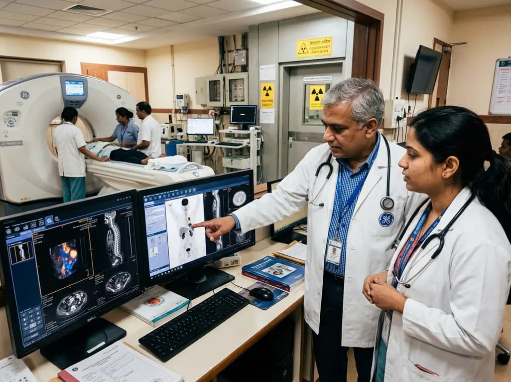
GE HealthCare has agreed to acquire Sofie Biosciences in a 945 million dollar all-cash transaction. The deal secures global rights to the investigational pan-cancer tracer FAPI-74 and expands cyclotron manufacturing infrastructure to accelerate precision theranostics and clinical molecular imaging workflows.
Today

Posttraumatic pisotriquetral instability is a rare wrist injury causing severe hypothenar pain and weakness. Explore clinical examination, imaging, and pisiformectomy.
Today

India's food safety authority has cracked down on high-caffeine beverages sold under stimulant branding, prompting legal challenges from major beverage corporations. This review examines FSSAI regulations, cardiac and metabolic complications, pediatric risks, and actionable counseling strategies for physicians.
Today
