.webp&w=384&q=75)

Loading, please wait...
Loading, please wait...

Ulnar-sided wrist trauma frequently challenges clinicians because complex anatomical structures converge within a compact space. Among these rare presentations, posttraumatic pisotriquetral instability remains an elusive and frequently overlooked condition that causes disabling pain and weakness. Although the sturdy surrounding ligamentous complex typically prevents joint disruption, severe mechanical trauma can dissociate the pisiform from the triquetrum. Consequently, early recognition and accurate management are vital to prevent long-term joint dysfunction and chronic neuropathy.
The pisotriquetral articulation functions as a unique, planar synovial joint positioned along the anterior ulnar wrist. The pisiform acts primarily as a sesamoid bone within the distal tendon of the flexor carpi ulnaris muscle. Therefore, this specialized bone elevates the tendon, significantly increasing the flexor moment arm across the wrist. Multiple robust fibrous structures stabilize the articulation. Specifically, the pisohamate and pisometacarpal ligaments provide primary distal restraint against proximal muscular traction forces. Additionally, the joint capsule and surrounding retinacular fibers resist excessive translation during active wrist movement. Because the ulnar neurovascular bundle traverses through Guyon's canal immediately adjacent to the pisiform, joint architecture directly influences adjacent neural physiology. When external forces disrupt these stabilizing tethers, the biomechanical equilibrium collapses rapidly. Furthermore, the triquetral facet experiences abnormal focal contact stresses during subsequent hand grasp. Consequently, patients develop marked functional disability during standard occupational or athletic activities.
Developing posttraumatic pisotriquetral instability generally requires substantial kinetic energy directed across the ulnar palm. Most cases stem from falls onto an outstretched hand with forced wrist hyperextension and ulnar deviation. During impact, intense sudden contraction of the flexor carpi ulnaris delivers powerful traction to the pisiform. Consequently, this immense tensile vector can avulse the distal ligamentous stabilizers or fracture the triquetral articular bed. In high-energy motor vehicle crashes or sports collisions, direct blunt impaction can also drive the pisiform out of alignment. Furthermore, concurrent shear forces frequently create intra-articular triquetrum avulsions. Once displaced, the unstable pisiform loses its anatomical congruence with the underlying triquetral facet. As a result, repetitive forearm muscle excursion produces pathologic subluxation and painful joint shearing. Moreover, persistent micro-instability can compress the adjacent deep motor branch of the ulnar nerve. Untreated cases therefore progress toward recalcitrant pisotriquetral chondromalacia, articular degeneration, and persistent hypothenar aching.
Patients suffering from pisotriquetral joint disruption typically present with persistent, localized ulnar-sided wrist pain. Symptoms intensify markedly during active wrist flexion, ulnar deviation, and heavy power grasping. Additionally, swelling, ecchymosis, and palpable tenderness often localize over the hypothenar eminence. Clinicians must conduct structured provocative physical examinations to distinguish this pathology from hook of hamate injuries or triangular fibrocartilage tears. Specifically, examiners perform the pisotriquetral shear test by grasping the pisiform firmly between thumb and forefinger while the wrist remains flexed. The clinician subsequently translates the pisiform laterally and medially against the triquetrum while applying gentle axial compression. A positive sign elicits sharp pain, instability, or coarse crepitus. Furthermore, clinicians must evaluate ulnar nerve motor and sensory function systematically. Because secondary compression within Guyon's canal occurs in displaced injuries, clinicians should look for hypothenar numbness, clawing, or intrinsic muscle weakness. Consequently, comprehensive physical assessment establishes clinical suspicion early.
Standard wrist radiography often obscures the pisotriquetral joint due to overlapping carpal anatomy on standard posteroanterior views. Therefore, physicians must obtain dedicated specialized radiographic projections whenever they suspect hypothenar injury. A lateral wrist radiograph in thirty degrees of supination cleanly profiles the pisotriquetral interface. Additionally, the semi-supine oblique view and carpal tunnel projection display pisiform alignment and reveal subtle avulsion fragments. However, plain radiographs may overlook non-displaced intra-articular fractures. Consequently, computed tomography serves as the gold standard for defining bony architecture and fragment displacement. High-resolution multiplanar computed tomography scans delineate articular congruity and identify concurrent triquetral fracture lines accurately. Moreover, magnetic resonance imaging provides unparalleled assessment of soft-tissue constraints, identifying tears in the pisohamate and pisometacarpal ligaments. Ultrasound also offers dynamic visualization of pisiform subluxation during active muscle contraction. Thus, multimodal imaging prevents delayed diagnosis and guides timely surgical decision-making.
While acute nondisplaced ligamentous sprains may occasionally respond to six weeks of cast immobilization, recurrent instability and displaced fracture-dislocations necessitate surgical treatment. Pisiformectomy, or complete surgical excision of the pisiform, represents the definitive treatment of choice for painful posttraumatic pisotriquetral derangement. Surgeons make an incision along the hypothenar crease, carefully exposing Guyon's canal. Next, they meticulously identify, mobilize, and protect the ulnar neurovascular structures throughout the procedure. The pisiform is then enucleated subperiosteally from the flexor carpi ulnaris tendon. Crucially, surgeons must repair and reconstitute the flexor carpi ulnaris continuity and distal ligamentous expansions to maintain grip strength. Following surgery, clinicians immobilize the wrist in a protective volar splint for two weeks. Subsequently, patients initiate guided passive and active range-of-motion exercises under physical therapy supervision. Strengthening protocols begin after six weeks, allowing gradual return to heavy manual labor and sports. Therefore, complete pisiform excision reliably relieves chronic pain while restoring optimal wrist function.
Traumatic dislocation occurs when high-energy blunt force impacts the palm or sudden hyperextension forces stress the wrist. A fall onto an outstretched hand causes forceful contraction of the flexor carpi ulnaris muscle. Consequently, this strong mechanical traction disrupts the stabilizing pisohamate and pisometacarpal ligaments. In severe incidents, associated intra-articular fractures of the triquetrum further destabilize the pisotriquetral articulation, leading to painful persistent subluxation or complete joint dislocation.
Complete excision of the pisiform generally causes minimal long-term functional loss if surgeons carefully preserve the flexor carpi ulnaris tendon continuity. Although the pisiform acts biomechanically as a sesamoid fulcrum to enhance flexion leverage, soft-tissue repair reconstructs the muscular insertion effectively. Consequently, patients typically regain excellent pain-free grip strength and range of motion. Rehabilitation ensures that manual laborers and competitive athletes return safely to high-demand activities without persistent mechanical deficit.
Clinicians differentiate pisotriquetral instability through precise anatomical palpation and provocative stress testing. Tenderness directly over the pisiform bone and a positive pisotriquetral shear test confirm local joint pathology. In contrast, triangular fibrocartilage complex tears cause pain within the foveal soft-tissue depression. Additionally, specialized radiographic projections, such as thirty-degree supinated oblique views and cross-sectional computed tomography, accurately distinguish pisotriquetral malalignment from hook of hamate fractures or distal radioulnar joint disease.
Disclaimer: This content is for informational and educational purposes only. It is not intended as medical advice, diagnosis, or treatment. Always consult a qualified healthcare professional regarding any medical condition or procedure. Refer to the latest local and national guidelines for clinical practice.
References

Read summarized clinical updates, watch expert medical content, and earn CME certifications right from your smartphone.
Open your smartphone camera to scan and install.





Posttraumatic pisotriquetral instability is a rare wrist injury causing severe hypothenar pain and weakness. Explore clinical examination, imaging, and pisiformectomy.
Today

Systemic amyloidosis management is undergoing a therapeutic revolution. This clinical review synthesizes updates from the International Society of Amyloidosis, detailing novel plasma cell therapies, TTR stabilizers, silencers, translational models, and artificial intelligence in drug development.
Today

A retrospective study of 824 patients undergoing over 2,200 intradetrusor onabotulinumtoxinA injections identifies sex- and etiology-specific predictors of 2-week post-void residual, highlighting the influence of baseline residual volumes, toxin dosage, and distinct urodynamic parameters on urinary retention risk.
Today
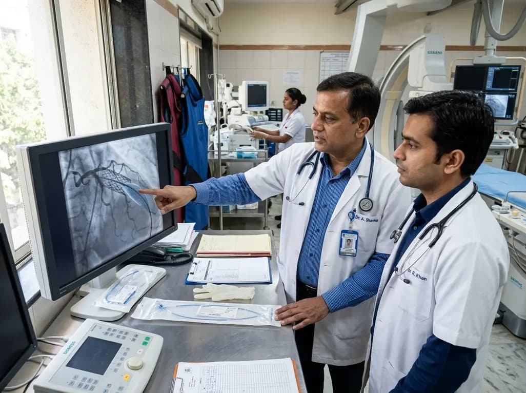
The US FDA has granted approval to the SELUTION SLR drug-eluting balloon for treating coronary in-stent restenosis. This milestone introduces a sustained-release sirolimus platform that eliminates the need for additional permanent metallic stents while maintaining robust vascular healing and luminal patency.
Today

A comprehensive review analyzes periconceptional GLP-1 receptor agonist exposure, revealing reassuring data on congenital anomalies and adverse perinatal outcomes while emphasizing the need for cautious patient counseling.
Today
