.webp&w=384&q=75)

Loading, please wait...
Loading, please wait...
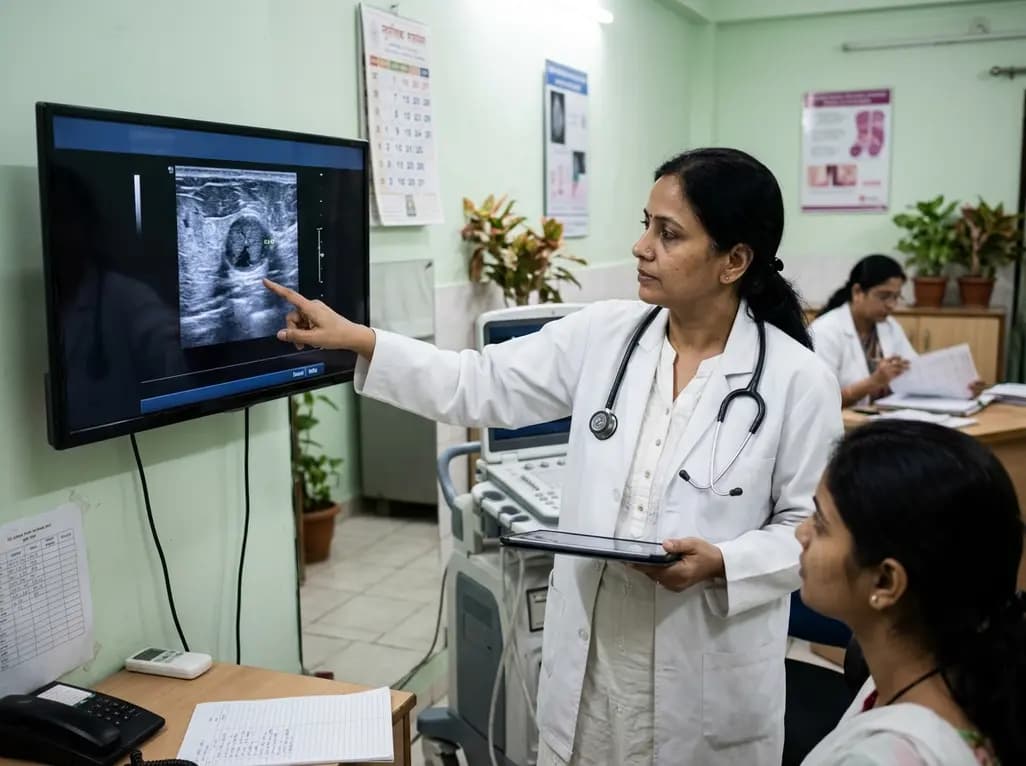
Managing ductal carcinoma in situ presents a clinical dilemma, as oncologists strive to prevent overtreatment while ensuring disease control. Accurately assessing DCIS upstaging risk is crucial during initial management, because core needle biopsies may overlook occult invasive carcinoma. When patients transition to definitive surgical excision, a notable proportion display microinvasion or invasive disease. Consequently, clinicians require dependable preoperative tools to distinguish low-risk lesions from progressive tumors. By integrating routine clinical findings with sonographic characteristics, modern predictive models offer a practical framework for risk estimation. These risk-stratification instruments help multidisciplinary teams evaluate disease biology safely and identify optimal candidates for active surveillance protocols.
Active surveillance trials seek to reduce unnecessary surgeries for indolent pre-invasive breast disease. However, the safety of non-operative approaches depends on precise initial staging. Standard core needle biopsies sample only a limited volume of tissue. As a result, approximately 20% to 40% of lesions diagnosed as pure DCIS show invasive components upon surgical removal. This substantial upgrade rate creates clinical concern, because invasive carcinoma requires systemic therapies, sentinel lymph node staging, or adjuvant radiation.
Therefore, occult malignancy represents the primary safety barrier for trials like COMET, LORIS, and LORD. If clinicians enroll patients with undetected invasive carcinoma into observation protocols, disease progression may occur before medical intervention. Consequently, objective predictive models are vital to mitigate these clinical risks. Multidisciplinary teams can use validated clinical and imaging metrics to estimate individual upstaging probabilities accurately. By establishing clear cutoffs, clinicians can confidently recommend active surveillance to low-risk patients while directing individuals with higher risks toward immediate surgical excision.
Multivariate analyses demonstrate that combining physical findings with ultrasound metrics provides robust predictive accuracy. Palpable breast masses serve as strong clinical indicators of underlying invasion. Because pure pre-invasive disease typically presents asymptomatically as mammographic calcifications, a palpable lump often indicates significant periductal reaction or extensive tumor volume. Therefore, physical examination provides essential baseline risk data during initial consultations.
Additionally, high-resolution sonography reveals critical morphological markers that signal invasive biology. Lesion dimension on ultrasound correlates directly with upstaging probability, as larger lesions present greater opportunities for sampling error. Furthermore, a non-parallel growth orientation—where lesion depth exceeds width—strongly predicts occult invasion. This vertical growth axis reflects aggressive stromal infiltration that disrupts normal tissue planes.
Moreover, secondary sonographic findings, including non-mass-like architecture, microlobulated margins, and suspicious axillary lymph nodes, significantly increase upstaging likelihood. Core biopsies of diffuse non-mass areas often miss small foci of basement membrane penetration. Consequently, detailed sonographic analysis bridges diagnostic gaps and refines preoperative risk stratification.
Predictive nomograms combine multiple clinical and sonographic variables into a single calibrated score. Multivariable logistic regression equations incorporate clinical palpability, sonographic dimensions, growth orientation, and biopsy nuclear grade. In multiple validation cohorts, these models achieve respectable discriminatory power, demonstrating area under the receiver operating characteristic curve (AUC) values between 0.70 and 0.86.
Furthermore, calibration assessments reveal strong concordance between predicted probabilities and observed surgical pathology. Decision curve analysis shows that these predictive tools deliver net clinical benefit across diverse decision thresholds. Unlike expensive genomic tests or contrast-enhanced MRI, conventional ultrasound is accessible, cost-effective, and safe for repeat examinations.
Therefore, mathematical models utilizing standard ultrasound metrics offer an economical and reliable solution for clinical practice. By converting complex imaging features into reproducible numerical probabilities, these tools help clinicians stratify patients into low-, intermediate-, and high-risk groups. This structured approach reduces subjective interpretation, optimizes diagnostic precision, and supports evidence-based surgical planning.
The primary application of predictive modeling involves refining patient selection for active surveillance trials. Existing de-escalation protocols rely heavily on biopsy pathology, such as estrogen receptor positivity and low nuclear grade. However, histological assessment alone cannot detect unsampled invasive components. Integrating sonographic parameters provides a vital safety check that enhances trial eligibility criteria.
When predictive models indicate minimal upstaging probability, clinicians and patients can choose active surveillance with enhanced reassurance. Conversely, when ultrasound reveals high-risk markers—such as large size or non-parallel orientation—clinicians should recommend primary surgical intervention. For these patients, non-operative management carries unacceptable risks.
Additionally, predictive models improve surgical and axillary staging decisions. If a patient with biopsy-proven DCIS faces high upstaging risk, surgeons can plan a concurrent sentinel lymph node biopsy during mastectomy. This proactive strategy avoids secondary operations, limits anesthesia exposure, and shortens treatment timelines. Thus, predictive tools successfully balance therapeutic de-escalation with rigorous oncological safety.
In Indian clinical settings, patient presentation and diagnostic resources create specific challenges where sonographic predictive models offer immense value. A significant proportion of Indian breast cancer patients present with dense breast tissue or palpable masses, which can complicate pure mammographic evaluation. Because high-frequency ultrasound is widely available and affordable across Indian healthcare centers, it represents an ideal modality for preoperative risk stratification.
Furthermore, sonographic models align seamlessly with resource-conscious oncology care. While advanced radiomic profiling and routine breast MRI may face accessibility or economic barriers in regional centers, conventional ultrasound requires no specialized infrastructure. Standardizing the evaluation of lesion orientation, margin characteristics, and axillary nodes enables clinicians across India to deliver uniform, high-quality care.
Moreover, as Indian academic centers participate in global discussions regarding cancer de-escalation, validated clinical-ultrasound tools can guide safe domestic clinical trials. Multidisciplinary collaboration among radiologists, oncologists, and surgeons ensures that patient care remains personalized and thorough. By adopting structured risk models, Indian clinicians can minimize overtreatment while preventing the underestimation of invasive disease.
Predicting DCIS upstaging risk is essential because core needle biopsies sample only small tumor areas. Approximately 20% to 40% of biopsy-proven DCIS cases harbor occult microinvasive or invasive components upon final excision. If clinicians place patients with undetected invasive cancer on active surveillance, disease progression may occur without timely systemic or local therapy. Accurate prediction ensures that only patients with truly non-invasive disease undergo conservative monitoring.
Key ultrasound features indicating elevated upstaging risk include larger lesion dimensions, a non-parallel growth orientation where depth exceeds width, and the presence of a palpable mass. Additionally, non-mass-like lesions, irregular margins, posterior acoustic shadowing, and suspicious axillary lymph nodes strongly correlate with invasive disease. Identifying these morphological markers alerts clinicians to potential biopsy underestimation and indicates the need for prompt surgical resection.
Multidisciplinary breast teams can integrate predictive models by standardizing ultrasound reporting and reviewing key imaging parameters alongside core biopsy pathology during tumor board meetings. Clinicians can calculate individual upstaging probabilities using validated equations that combine palpability, lesion size, growth orientation, and nuclear grade. This coordinated assessment helps surgeons, oncologists, and radiologists personalize treatment strategies, identify trial candidates, and plan appropriate axillary staging.
Disclaimer: This content is for informational and educational purposes only and does not constitute professional medical advice, diagnosis, or clinical guidance. Healthcare providers must exercise clinical judgment and evaluate individual patient circumstances. Refer to the latest local and national guidelines for clinical practice.
References

Read summarized clinical updates, watch expert medical content, and earn CME certifications right from your smartphone.
Open your smartphone camera to scan and install.





A clinical-ultrasound predictive model evaluates DCIS upstaging risk using lesion size, growth orientation, and palpability, aiding patient selection for active surveillance trials and safe surgical decision-making.
last month

Following 37,899 reported cases and 9 deaths, Odisha has scaled up active case detection, rapid diagnostic testing, and vector control across high-burden districts. Here is a comprehensive clinical breakdown of the state's aggressive containment protocols, diagnostic pathways, and targeted field management.
Today

A retrospective study of 86,012 pregnancies demonstrates that size-selective cfDNA enrichment boosts mean fetal fraction to 18.88% and achieves a 59.20% PPV for fetal copy number variants. However, 85.83% of CNVs showed size discrepancies, underscoring the mandatory role of confirmatory microarray testing.
Today

Radiofrequency catheter ablation of the cavotricuspid isthmus successfully terminates typical atrial flutter. This article reviews the mechanisms driving coronary sinus electrogram changes, wavefront activation pathways, and clinical criteria to verify bidirectional block during electrophysiology studies.
Today

A study comparing four knee flexion strength tests after ACL reconstruction using hamstring tendon autografts reveals marked variability in limb symmetry indices. Clinicians must recognize that different strength testing modalities yield divergent return-to-sport clearance rates.
Today
A cross-sectional study demonstrates that increased BMI in adolescents significantly enhances mandibular length and alters salivary adipokines like adiponectin and resistin, providing novel insights into craniofacial growth and orthodontic planning.
Today
