.webp&w=384&q=75)

Loading, please wait...
Loading, please wait...

Radiofrequency cavotricuspid isthmus ablation represents the definitive curative intervention for typical atrial flutter. During this procedure, operators target the anatomical corridor between the inferior vena cava and tricuspid valve annulus. Successful energy application terminates the reentrant circuit and restores sinus rhythm. However, clinicians frequently observe distinctive alterations in intracardiac recordings immediately following lesion completion. Specifically, changes in coronary sinus atrial electrograms provide critical insights into shifting myocardial activation pathways. Electrophysiologists must understand these signal modifications to distinguish true bidirectional conduction block from misleading pseudo-block patterns.
The cavotricuspid isthmus serves as an obligatory slow conduction pathway for typical counterclockwise atrial flutter. Anatomically, this region comprises three main segments: the anterior paraseptal zone, the central isthmus, and the inferolateral isthmus. Furthermore, the coronary sinus ostium lies immediately adjacent to the septal margin of this anatomical corridor. Consequently, radiofrequency energy delivery along this line directly influences regional atrial impulse propagation. In typical flutter, the electrical wavefront travels superiorly along the interatrial septum, activating the coronary sinus from proximal to distal poles. Moreover, the reentrant impulse constantly engages pectinate muscle bands and terminal crest ramifications. Therefore, creating a continuous transmural lesion eliminates this troublesome macroreentrant circuit. Subsequently, cardiac electrical activation must find alternative routes to reach the left atrium and coronary sinus musculature. When ablation disrupts the native pathway, wavefront propagation shifts entirely to superior interatrial bridges. As a result, catheter electrodes placed inside the coronary sinus capture markedly altered local electrograms. Electrophysiologists analyze these subtle signal morphology changes to verify the procedural efficacy of the ablation line.
Wavefront collision dynamics primarily explain the observed electrogram shifts after arrhythmia termination. Before ablation, wavefronts from the right atrial free wall traverse the isthmus rapidly to activate the coronary sinus musculature. However, an effective lesion line permanently blocks this direct pathway. Pacing from the low lateral right atrium forces the electrical wavefront to propagate cranially toward Bachmann bundle. Subsequently, the impulse descends along the interatrial septum and crosses the fossa ovalis to reach the coronary sinus. Because this alternative pathway covers a considerably longer anatomical distance, local activation delays emerge promptly. Therefore, electrophysiologists record a significant widening of the stimulus-to-electrogram interval along the multipolar coronary sinus catheter. In addition, the local bipole electrogram morphology frequently changes from narrow, sharp spikes into split or multicomponent signals. These double potentials indicate that local tissue activation occurs via two distinct, widely separated wavefronts. Thus, changes in coronary sinus electrogram characteristics directly reflect profound alterations in global atrial conduction pathways.
Persistent conduction across the cavotricuspid isthmus often involves complex anatomical connections within the coronary sinus sleeve. The coronary sinus possesses a distinct myocardial coat that extends several centimeters into the left atrioventricular groove. Moreover, this musculature connects directly with both the right atrial septum and the left atrial free wall. Consequently, electrical impulses can bypass endocardial ablation lines via epicardial muscular bridges. When epicardial bridging occurs, coronary sinus electrograms continue to show early activation despite extensive endocardial lesion delivery. Furthermore, the middle cardiac vein and the vein of Marshall can provide alternative conducting pathways around the lesion. Electrophysiologists often observe ambiguous coronary sinus electrogram intervals when these epicardial connections sustain partial propagation. Therefore, operators must carefully evaluate signal polarity and activation timing during differential pacing maneuvers. In addition, targeted radiofrequency delivery inside the proximal coronary sinus floor may become necessary to eliminate these resilient epicardial bridges. Consequently, precise electrogram interpretation prevents premature assumptions of complete line transection.
Achieving permanent clinical cure requires absolute proof of bidirectional conduction block across the isthmus. Electrophysiologists routinely verify block by pacing sequentially from the low lateral right atrium and proximal coronary sinus. However, clinicians must remain vigilant against pseudo-block, which creates an illusion of complete line interruption. For instance, slow conduction across a surviving muscle bundle can produce delayed signals that mimic complete block. In addition, pacing artifacts and far-field ventricular signals can contaminate proximal coronary sinus recordings. Therefore, operators employ differential pacing techniques to ascertain the true sequence of wavefront propagation. Pacing at increasing distances from the ablation line should produce predictable interval prolongations if complete block exists. Conversely, paradoxical interval shortening indicates persistent trans-isthmus conduction over a slow residual corridor. Furthermore, mapping catheters should reveal distinct double potentials with an isoelectric baseline along the entire line. Thus, rigorous scrutiny of coronary sinus electrograms protects patients from early flutter recurrence.
Accurate evaluation of coronary sinus electrogram changes directly correlates with long-term procedural success. When operators achieve verified bidirectional block, the recurrence rate of typical atrial flutter drops below five percent. In contrast, incomplete conduction block leads to arrhythmia relapse, which frequently manifests as clockwise flutter or atrial tachycardia. Moreover, prolonged procedural times and excessive energy delivery increase the risk of complications, such as cardiac perforation. Electrophysiologists in modern catheterization laboratories utilize high-density electroanatomical mapping to visualize coronary sinus activation patterns in real time. Consequently, 3D mapping systems integrate local activation times and voltage criteria with multipolar coronary sinus recordings. This multimodal approach allows clinicians to identify the exact site of conduction breakthrough along the ablation line quickly. Furthermore, post-ablation coronary sinus electrogram monitoring helps detect subtle shifts toward atypical atrial arrhythmias or fibrillation. Therefore, thorough electrophysiological assessment provides immediate assurance of therapeutic efficacy while enhancing long-term patient safety.
Coronary sinus electrograms change because the ablation line interrupts direct trans-isthmus conduction between the lateral right atrium and the interatrial septum. Consequently, electrical impulses must propagate superiorly over the crista terminalis and Bachmann bundle before descending toward the coronary sinus. This longer detour delays activation, widens stimulus-to-electrogram intervals, and alters local signal polarity, providing definitive evidence of altered wavefront pathways across the atrium.
Differential pacing verifies block by comparing conduction times from different pacing sites relative to the ablation line. When clinicians pace more medially within the coronary sinus, conduction to the lateral right atrium must travel further if complete block is present. Therefore, the measured activation time increases proportionately. If the interval paradoxical shortens, persistent trans-isthmus conduction remains, proving that bidirectional block has not been successfully achieved.
The myocardial sleeve of the coronary sinus frequently acts as an epicardial conduction bridge that circumvents endocardial ablation lesions. Consequently, impulses can bypass the isthmus line, leading to persistent conduction or early flutter recurrence. Electrophysiologists must recognize this anatomical variation by analyzing fractionated coronary sinus electrograms. Operators may need to deliver targeted ablation inside the proximal coronary sinus to eliminate these epicardial connections effectively.
Disclaimer: This content is for informational and educational purposes only and should not be considered medical advice. It is not intended to replace consultation with qualified healthcare professionals. Refer to the latest local and national guidelines for clinical practice.
References

Read summarized clinical updates, watch expert medical content, and earn CME certifications right from your smartphone.
Open your smartphone camera to scan and install.





Radiofrequency catheter ablation of the cavotricuspid isthmus successfully terminates typical atrial flutter. This article reviews the mechanisms driving coronary sinus electrogram changes, wavefront activation pathways, and clinical criteria to verify bidirectional block during electrophysiology studies.
Today
Psychometric validation of the WHO BeSD survey identifies vaccine importance and social responsibility as primary drivers of adult influenza vaccination uptake, offering clinicians actionable strategies to overcome hesitancy.
Today
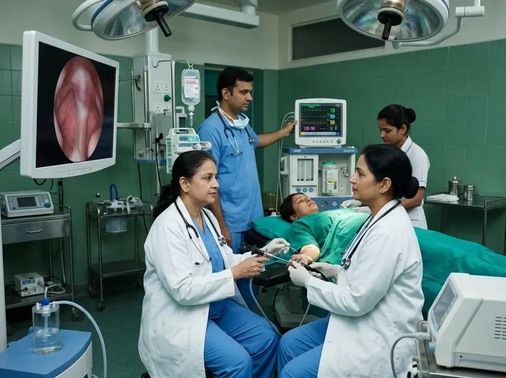
A meta-analysis of nine RCTs comparing remimazolam versus propofol in hysteroscopy reveals remimazolam significantly reduces injection pain, hypotension, and respiratory depression while maintaining comparable sedation success, recovery times, and procedural duration.
Today
A new study reveals that MRI-based meniscal coverage and cartilage thickness explain mechanical axis alignment in knee osteoarthritis comparably to joint line convergence angle. These soft-tissue insights improve preoperative planning for osteotomies and arthroplasty.
Today

A breakthrough study leverages natural language processing to accurately identify incident medication-related osteonecrosis of the jaw and track antiresorptive drug holidays from clinical narratives in electronic health records, advancing osteoporosis pharmacovigilance and dental safety.
Today
