

Loading, please wait...
Loading, please wait...

Upper limb motor impairment remains a debilitating consequence of cerebrovascular accidents, significantly reducing functional independence. In everyday life, human autonomy relies heavily on coordinated bilateral movements. Consequently, persistent deficits in post-stroke bimanual coordination severely compromise activities of daily living and long-term recovery. Although clinicians frequently treat these challenges, the underlying central-peripheral neurophysiological dynamics remain difficult to capture with conventional tools. Traditional clinical assessments rely primarily on subjective observations or gross motor scales, which overlook subtle neuromuscular communication. However, modern functional neuroimaging and electrophysiological tools offer deeper physiological insights. By synchronously acquiring functional near-infrared spectroscopy and surface electromyography signals, investigators can observe cortical activation alongside peripheral muscle execution. This multimodal approach clarifies how cerebral network disruption and impaired sensorimotor feedback undermine coordinated hand movements in chronic stroke survivors.
Coordinating both upper limbs requires continuous neural communication across cerebral hemispheres and peripheral nerves. Bilateral tasks depend on reciprocal transcallosal transfer alongside balanced interhemispheric inhibitory networks. Furthermore, precise motor execution demands intact corticomuscular coupling, linking cerebral commands with effector muscles. In healthy individuals, descending motor drives and ascending sensory feedback operate in synchronized harmony. However, subcortical ischemic lesions disrupt these pathways by severing internal capsule fibers and subcortical feedback loops. Consequently, the brain loses its capacity to synchronize bilateral muscle groups effectively. In response, cerebral motor networks undergo neuroplastic reorganization, frequently recruiting the contralesional hemisphere to assist the affected limb. While this reorganization provides compensatory motor drive, it often induces abnormal muscle synergies and impaired motor control. In addition, patients exhibit significantly weakened intermuscular coupling across upper limb muscle groups. Therefore, bilateral motor impairment reflects broader network dysfunction across the neuroaxis.
Modern neurorehabilitation research increasingly relies on multimodal diagnostic tools to evaluate neuromuscular integration. Functional near-infrared spectroscopy tracks cortical hemoglobin dynamics, capturing localized cerebral activation during motor tasks. Simultaneously, surface electromyography captures electrical activity generated by contracting forearm and arm muscles. Synchronizing fNIRS with sEMG allows investigators to measure corticomuscular coupling using transfer entropy analysis. Furthermore, transfer entropy quantifies directional information transfer between the cortex and peripheral muscles. This directional metric distinguishes efferent descending motor commands from afferent ascending sensory signals. Notably, research reveals that ascending muscle-to-cortex communication suffers selective damage following stroke. In contrast, descending motor pathways often preserve relative integrity during specific tasks. In addition, sEMG signals enable the detailed examination of intermuscular coupling and muscle synergies. Thus, multimodal fNIRS-sEMG analysis bridges central cortical dynamics with peripheral motor execution, delivering invaluable quantitative biomarkers for stroke recovery.
Neurological demands change markedly depending on whether patients execute symmetrical or asymmetrical arm movements. During symmetrical bimanual tasks, homologous muscle groups contract simultaneously in mirror-like coordination. Under these conditions, stroke patients exhibit a significant reduction in ascending muscle-to-cortex connectivity. Importantly, this reduced afferent coupling correlates positively with the Modified Barthel Index, indicating that sensory feedback deficits directly impair functional independence. Furthermore, patients display elevated cortical activation and strengthened functional connectivity, reflecting compensatory effort. Conversely, asymmetrical tasks require independent movements from each arm, demanding complex temporal coordination. During asymmetrical tasks, corticomuscular coupling shows fewer group differences, yet cortical functional connectivity remains heightened. Specifically, connectivity between the ipsilesional and contralesional supplementary motor areas correlates positively with clinical independence. In both tasks, however, patients demonstrate weakened intermuscular coupling and disorganized muscle synergies. Consequently, therapeutic interventions must address task-specific physiological demands during upper extremity rehabilitation.
Interpreting neurophysiological markers in chronic stroke requires careful differentiation from normal aging processes. As adults age, the brain experiences gradual structural changes, including white matter degeneration and reduced interhemispheric inhibition. Consequently, older individuals without neurological injury often exhibit altered cortical activation and decreased neuromuscular coherence. Attribution analysis helps researchers separate true stroke deficits from these physiological aging effects. Specifically, evidence confirms that cortical hyperactivation and disorganized muscle synergies stem predominantly from stroke damage. These findings represent pathological adaptations and compensatory recruitment following structural lesions. In contrast, sensory cortex activation changes and baseline corticomuscular coupling alterations reflect combined influences from aging and stroke. Therefore, neurorehabilitation studies must always incorporate age-matched control groups to avoid misinterpreting normal senescence as stroke pathology. By clearly isolating lesion-induced mechanisms from age-related decline, clinicians can establish precise diagnostic baselines and design targeted rehabilitation protocols.
Understanding task-dependent coupling dynamics offers meaningful opportunities to optimize upper extremity rehabilitation protocols. Because bimanual coordination deficits involve significant sensory feedback impairment, therapy must extend beyond simple repetitive motor training. Instead, therapists should integrate rich proprioceptive and tactile stimulation during bilateral arm exercises. For instance, combining bilateral movement therapy with neuromuscular electrical stimulation can enhance ascending afferent input into sensorimotor networks. Furthermore, non-invasive neuromodulation techniques can regulate imbalanced interhemispheric communication between bilateral supplementary motor areas. Utilizing robotic rehabilitation systems also provides real-time kinematic feedback, actively reshaping disordered muscle synergies. In addition, multimodal fNIRS-sEMG monitoring serves as a reliable objective tool to evaluate patient recovery over time. By tracking improvements in corticomuscular transfer entropy, clinicians can adapt rehabilitation intensity dynamically. Ultimately, translating these physiological insights into structured clinical practice will advance personalized stroke therapy and promote long-term functional recovery.
Impaired bimanual coordination after stroke arises primarily from disrupted ascending sensory feedback pathways and compromised interhemispheric communication. Although descending motor pathways may partially recover, the damaged brain struggles to integrate real-time muscle sensory inputs. Furthermore, lesions disconnect critical pathways linking the supplementary motor areas and corpus callosum. Consequently, patients develop abnormal muscle synergies, diminished intermuscular coupling, and maladaptive cortical hyperactivation, which collectively hinder the execution of coordinated bilateral motor tasks.
Multimodal fNIRS-sEMG combines hemodynamic cortical monitoring with electrophysiological muscle recordings, providing a comprehensive assessment of neuromuscular coordination. Traditional bedside scales provide qualitative scores, but they fail to capture directional neural dynamics. Synchronized fNIRS-sEMG measures corticomuscular transfer entropy, revealing whether motor failure stems from deficient brain drive or inadequate sensory feedback. Therefore, this non-invasive technology equips clinicians with quantifiable, objective biomarkers to track neuroplasticity, monitor motor recovery, and personalize stroke rehabilitation regimens.
Differentiating stroke pathology from chronological aging is essential because both processes independently affect sensorimotor performance. Normal physiological aging naturally degrades baseline corticomuscular coupling and sensory processing through cerebral atrophy and white matter changes. In contrast, abnormal muscle synergies and excessive cortical hyperactivation stem directly from stroke-induced brain injury. Therefore, incorporating age-matched controls ensures that rehabilitation specialists address actual stroke deficits rather than normal aging processes, avoiding inaccurate clinical prognostications and misdirected therapeutic protocols.
Disclaimer: This content is for informational and educational purposes only. It is not intended to be a substitute for professional medical advice, diagnosis, or treatment. Always seek the advice of your physician or other qualified health provider with any questions you may have regarding a medical condition. Refer to the latest local and national guidelines for clinical practice.
References

Read summarized clinical updates, watch expert medical content, and earn CME certifications right from your smartphone.
Open your smartphone camera to scan and install.





A multimodal fNIRS-sEMG analysis reveals that impaired post-stroke bimanual coordination stems from disrupted ascending sensory feedback and abnormal cortical reorganization, offering novel quantitative biomarkers for stroke recovery.
Today
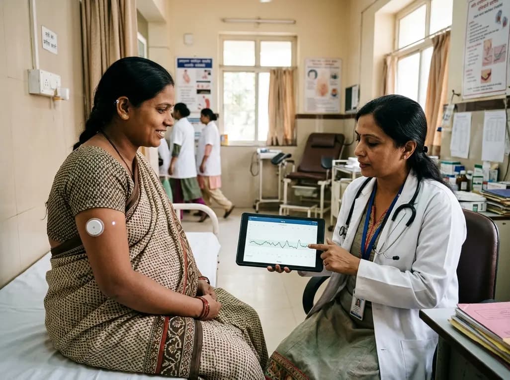
A prospective multicenter trial demonstrates that a 15-day factory-calibrated continuous glucose monitoring system provides high analytical accuracy (MARD ~7.7%) and safety in pregnant women with gestational diabetes mellitus.
Today
Recent research reveals distinct protein secretion profiles and morphometric shifts between euploid and aneuploid human blastocysts cultured in an advanced 3D endometrial platform, opening new avenues for non-invasive embryo assessment.
Today

A landmark study details how an enterprise pediatric remote patient management model expanded from a single cardiology pilot to 16 diverse specialty programs by combining centralized digital operations with specialty clinical leadership.
Yesterday
Peripheral nerve injuries present clinical challenges due to regenerative mismatches between sensory and motor neurons. This review explores cutting-edge tissue-engineered nerve grafts designed as central-peripheral signal relays to overcome denervation atrophy and restore functional motor-sensory recovery.
Today
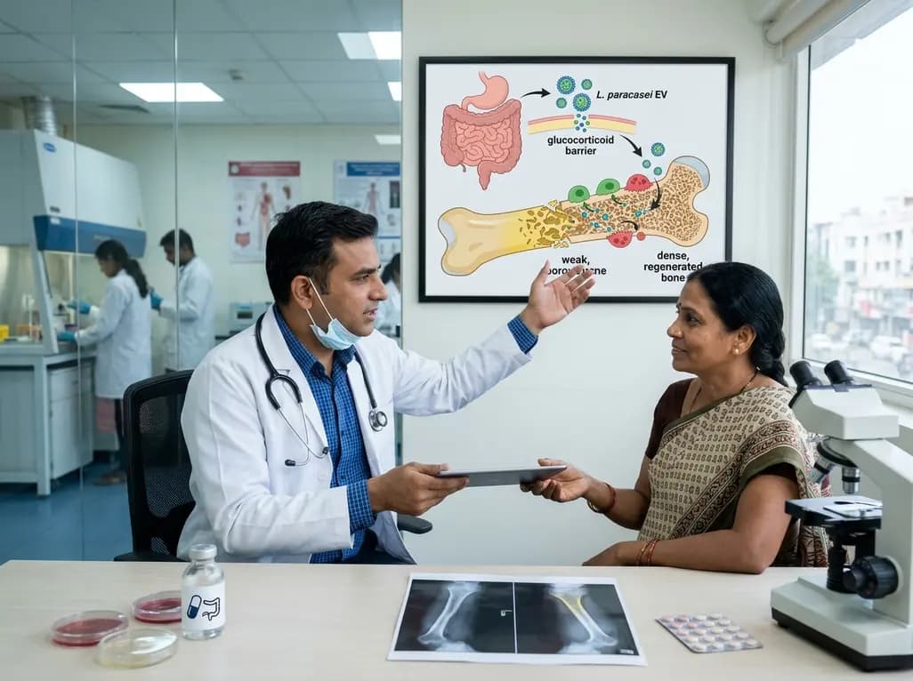
A novel oral nanoplatform using Lacticaseibacillus paracasei-derived extracellular vesicles delivers alendronate, reversing glucocorticoid-induced bone loss and restoring gut-immune-bone homeostasis.
Today
