

Loading, please wait...
Loading, please wait...

Implantation represents one of the most critical and complex bottlenecks in human assisted reproduction. During this window, dynamic biochemical dialogue occurs between the competent blastocyst and a receptive endometrium. Aneuploidy remains a leading cause of implantation failure and early spontaneous miscarriage. However, researchers have long struggled to model these peri-implantation events outside the maternal body. Standard laboratory protocols often evaluate pre-implantation blastocysts up to day 5 or 6, leaving post-implantation dynamics largely hidden. A groundbreaking study now provides essential insights into early developmental competence by characterizing the human embryo secretome and morphometric patterns during extended post-implantation culture.
Chromosomal abnormalities disrupt fundamental molecular pathways required for embryonic competence and tissue differentiation. Nevertheless, clinical embryologists currently rely primarily on pre-implantation genetic testing for aneuploidies (PGT-A), which requires an invasive trophectoderm biopsy. Investigators set out to determine whether euploid embryos, trisomy 21 embryos, and monosomy 21 embryos exhibit distinctive secretory and structural behaviors. They compared extended development in conventional culture media against an advanced biological environment. In doing so, the investigators tracked development up to 14 days post-fertilization to capture the critical peri-implantation transition.
Importantly, chromosomal errors alter early morphogenetic movements and disrupt hormonal signaling cascades. Previous research focused predominantly on pre-implantation embryonic morphology, which frequently fails to correlate with developmental trajectory. By examining post-blastocyst stages, this investigation clarifies how numerical chromosomal errors impede sustained attachment and tissue invasion. Consequently, analyzing these developmental checkpoints deepens our physiological understanding of early human pregnancy loss and recurrent implantation failure.
To overcome the biological limitations of standard plastic culture dishes, researchers deployed cell-engineered receptive endometrial scaffold technology, known as CREST. This advanced 3D culture system incorporates both endometrial epithelial and stromal cell compartments. Therefore, it closely mimics the natural architectural and biological cues of a receptive maternal endometrium. The study examined 69 supernumerary blastocysts donated for research, including 24 euploid, 21 trisomy 21, and 24 monosomy 21 embryos.
Embryos cultured without endometrial cellular support exhibited rapid developmental arrest beyond 10 days post-fertilization. Furthermore, these unassisted blastocysts demonstrated sharply diminished viability and negligible protein production. In contrast, the CREST platform extended developmental viability up to 14 days post-fertilization. The endometrial co-culture enhanced embryo survival and stimulated higher baseline protein secretion across all groups. This finding emphasizes that functional stromal-epithelial crosstalk is essential to sustain early embryonic organization and functional trophoblast differentiation.
The research team quantified key trophoblast and cellular stress markers at distinct time points, including days 7, 10, 12, and 14 post-fertilization. Specifically, they measured beta-human chorionic gonadotropin, human placental lactogen, pregnancy-associated plasma protein A, placental growth factor, and calreticulin. The resulting human embryo secretome revealed striking differences across karyotypes. Consequently, spent culture media reflected real-time embryonic functional competence.
Monosomy 21 blastocysts displayed significant secretory deficits almost immediately. These embryos demonstrated severely diminished levels of human placental lactogen, placental growth factor, and calreticulin as early as day 7 post-fertilization. By day 10, pregnancy-associated plasma protein A effectively segregated euploid embryos from both trisomy 21 and monosomy 21 cohorts. Furthermore, euploid embryos maintained a progressive, sustained rise in both pregnancy-associated plasma protein A and placental growth factor through day 14. In contrast, trisomic embryos failed to establish this upward trajectory, demonstrating clear biochemical divergence as differentiation progressed.
In addition to secretory signatures, the researchers evaluated quantitative morphometric indices across the 14-day culture window. They monitored total embryo surface area, degree of expansion, circularity, and axis ratio. Embryos cultured in cell-free control media failed to expand their blastocoelic cavities and maintained a static, rounded appearance throughout culture. In comparison, embryos supported by the receptive endometrial platform revealed marked morphological remodeling.
Euploid and trisomy 21 blastocysts both achieved significant implantation-like surface expansion and blastocoel collapse within the 3D scaffold. Moreover, both cohorts showed an increased axis ratio, signifying directional cell spreading and invasion. In sharp contrast, monosomy 21 embryos failed to execute these fundamental structural adaptations. Instead, monosomic embryos remained spherical, exhibited no axis elongation, and suffered early morphogenetic arrest. These data confirm that specific chromosomal deletions trigger severe structural failure well before definitive organogenesis begins.
These laboratory findings carry profound implications for modern reproductive medicine. Currently, clinicians evaluate embryo viability via static microscopic grading, time-lapse imaging, and invasive trophectoderm biopsy. Although PGT-A offers high chromosomal diagnostic accuracy, it raises procedural concerns regarding mosaicism and laboratory-induced cell trauma. The discovery that targeted secretomic panels differentiate chromosomal states supports the development of non-invasive spent culture media testing.
Furthermore, evaluating secretomic markers alongside morphometric parameters could refine non-invasive embryo prioritization algorithms. Reproductive endocrinologists could theoretically screen for impaired developmental trajectories prior to transfer. Additionally, the endometrial scaffold model provides a reliable drug testing platform. Clinicians and scientists can utilize this platform to evaluate targeted endometrial therapeutics designed to correct hostile maternal environments and reduce recurrent implantation failure rates.
While these insights advance reproductive science, clinicians must interpret the results within their biological context. First, the investigation examined a focused cohort restricted to chromosome 21 aneuploidies. Therefore, reproductive teams cannot immediately extrapolate these findings to other common autosomal trisomies or monosomies. Second, the current model lacks maternal vascular endothelial cells and uterine immune cell populations, such as uterine natural killer cells, which play decisive roles during clinical implantation.
Moreover, the authors evaluated a targeted panel of predetermined proteins rather than employing untargeted omics. Future research must combine high-throughput proteomics with extended organoid models across diverse chromosomal profiles. As in vitro microfluidic and biomimetic technologies mature, these platforms will likely bridge the gap between basic embryology and individual clinical care. Ultimately, refining these physiological models will help embryologists select the most competent blastocyst and improve live-birth outcomes.
Traditional in vitro systems culture embryos in synthetic liquid media on plastic surfaces, which lack biological signaling. In contrast, the CREST model utilizes a 3D architecture containing receptive endometrial epithelial and stromal cells. This physiological scaffold provides essential paracrine factors, supports natural structural remodeling, and extends embryo viability up to 14 days post-fertilization, allowing researchers to study post-implantation milestones accurately.
Pregnancy-associated plasma protein A served as a primary discriminator, distinguishing euploid embryos from both trisomy 21 and monosomy 21 embryos by day 10 post-fertilization. Additionally, placental growth factor rose progressively through day 14 exclusively in euploid embryos. In contrast, monosomy 21 blastocysts exhibited severely diminished secretion of calreticulin, human placental lactogen, and placental growth factor as early as day 7.
Currently, secretomic profiling cannot fully replace pre-implantation genetic testing. This investigation evaluated a targeted panel restricted to chromosome 21 variations during post-implantation culture. PGT-A remains the gold standard for comprehensive pre-implantation chromosomal screening. However, ongoing validation of non-invasive secretomic markers may eventually complement routine embryo assessment and reduce reliance on invasive biopsies in clinical assisted reproductive technology.
Disclaimer: This content is for informational and educational purposes only and should not be taken as professional medical advice. Always seek the advice of your physician or other qualified healthcare provider with any questions you may have regarding a medical condition. Refer to the latest local and national guidelines for clinical practice.
References
1. Yagüe-Serrano R et al. Mapping developmental and targeted secretory signatures of euploid and aneuploid human embryos using an advanced endometrial model. Hum Reprod. 2026 Oct 25. doi: undefined. PMID: 42927452.
2. Molè MA et al. Cell-engineered receptive endometrial scaffold technology (CREST) facilitates human embryo post-implantation development. Cell. 2025;188(4):890-907.
3. Teh WT, McBain J, Rogers P. What is the contribution of the endometrium to recurrent implantation failure? Human Reproduction. 2016;31(7):1381-1392.
4. Simon C et al. In vitro models of human embryo implantation: historical perspective and current advancements. Hum Reprod Update. 2020;26(6):773-788.

Read summarized clinical updates, watch expert medical content, and earn CME certifications right from your smartphone.
Open your smartphone camera to scan and install.





Recent research reveals distinct protein secretion profiles and morphometric shifts between euploid and aneuploid human blastocysts cultured in an advanced 3D endometrial platform, opening new avenues for non-invasive embryo assessment.
Today
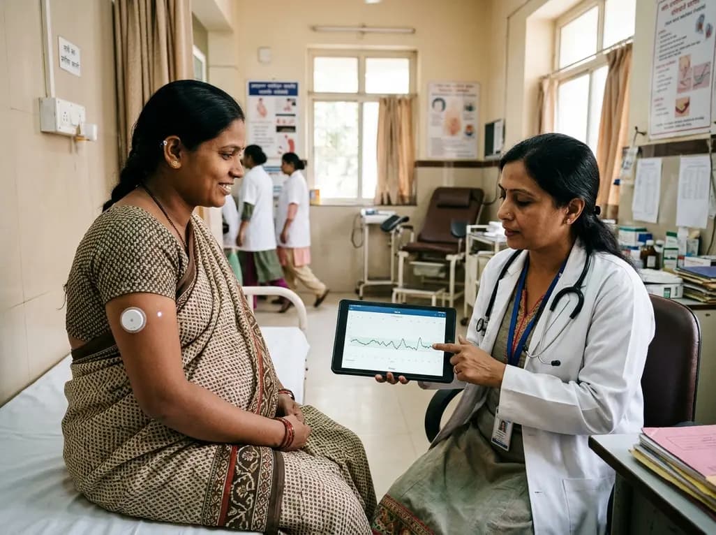
A prospective multicenter trial demonstrates that a 15-day factory-calibrated continuous glucose monitoring system provides high analytical accuracy (MARD ~7.7%) and safety in pregnant women with gestational diabetes mellitus.
Today

A landmark study details how an enterprise pediatric remote patient management model expanded from a single cardiology pilot to 16 diverse specialty programs by combining centralized digital operations with specialty clinical leadership.
Yesterday
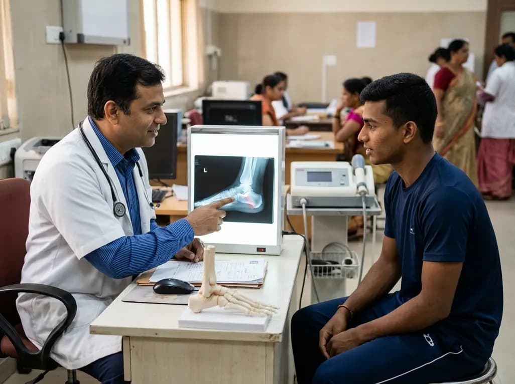
A collegiate hurdler with a 2-year neglected Degan type III anterior process calcaneus fracture achieved complete pain resolution and bony bridging using focused extracorporeal shock wave therapy, returning to competition without invasive surgery.
Today
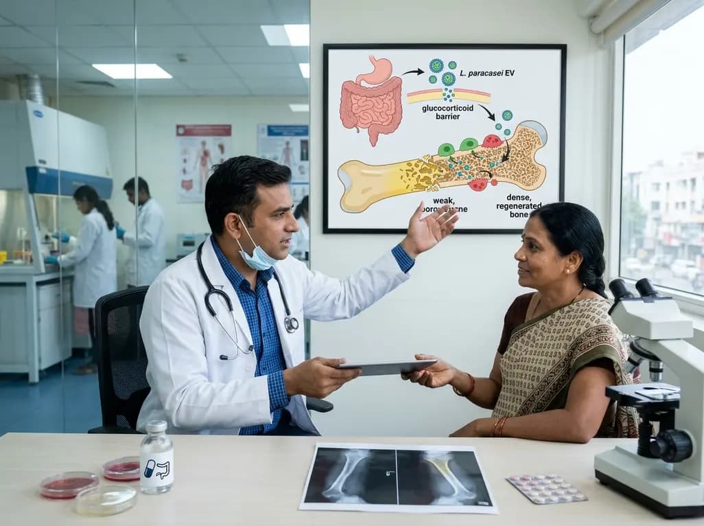
A novel oral nanoplatform using Lacticaseibacillus paracasei-derived extracellular vesicles delivers alendronate, reversing glucocorticoid-induced bone loss and restoring gut-immune-bone homeostasis.
Today
