

Loading, please wait...
Loading, please wait...

Peripheral nerve injuries present some of the most complex clinical challenges in modern restorative surgery. Severe physical trauma causes extensive axonal disruption, resulting in devastating loss of sensory and motor function. Consequently, accelerating peripheral nerve regeneration remains a pivotal goal for reconstructive surgeons, orthopedic teams, and clinical neuroscientists striving to prevent permanent physical impairment.
Surgical reconstruction of severed peripheral nerves often fails because of an unforgiving biological race against time. Following severe transection, injured axons regrow at a sluggish rate of approximately one millimeter each day. However, target skeletal muscles undergo rapid and relentless denervation atrophy long before advancing axonal growth cones can arrive. When gap defects exceed several centimeters, the required transit time guarantees prolonged distal denervation. Consequently, motor endplates degrade permanently and interstitial fibrosis replaces receptive myofibers.
Although microsurgeons achieve flawless microscopic coaptation, anatomical continuity rarely translates into meaningful functional movement. Therefore, clinicians must recognize that axonal elongation speed creates an intrinsic spatiotemporal mismatch with peripheral tissue survival. In addition, the chronic loss of neurotrophic support from target tissues accelerates the deterioration of distal Schwann cell columns. These bands of Büngner lose their pro-regenerative phenotype over extended intervals, gradually becoming unresponsive conduits of fibrous tissue. As a result, even robustly regenerating axons encounter a hostile, non-permissive distal pathway. Thus, overcoming this temporal bottleneck demands innovative strategies that accelerate elongation while preserving distant neuromuscular junctions.
A fundamental barrier to functional recovery lies in the divergent biological behavior of sensory and motor neurons. Sensory neurons, situated within dorsal root ganglia, possess an impressive cell-autonomous capacity for regenerative survival. In contrast, spinal motor neurons depend heavily on continuous retrograde trophic feedback from distal skeletal target fields. When axotomy interrupts this vital retrograde signal, motor neurons experience progressive metabolic and transcriptional exhaustion.
Furthermore, single-cell analyses reveal distinct differences in how these cell types reprogram their transcriptional machinery. While sensory neurons swiftly upregulate regeneration-associated genes independently, motor neurons suffer profound transcriptional silencing during protracted separation from muscle targets. Therefore, clinicians frequently observe satisfactory cutaneous sensory reinnervation while motor recovery remains profoundly defective. Moreover, contemporary reconstructive science still lacks comprehensive knowledge regarding subtype-specific transcriptional signatures within motor neuron pools. Consequently, therapeutic interventions that treat peripheral nerves as uniform cables inevitably fail to rescue vulnerable motor functional pathways. Clinicians must therefore advocate for therapies that selectively nourish motor phenotypes and sustain their intrinsic survival programs throughout prolonged recovery phases.
Surgeons currently regard autologous nerve transplantation as the clinical gold standard for bridging extensive nerve defects. Nevertheless, harvesting autografts causes permanent donor-site morbidity, localized numbness, and potential neuroma formation. In addition, limited donor supply restricts autograft utility in extensive polytrauma injuries with multiple transected nerves. These severe clinical constraints have driven intense commercial interest toward artificial nerve guidance conduits.
Unfortunately, first-generation synthetic conduits, fabricated from simple silicone or collagen tubes, exhibit mediocre efficacy in long-segment defects. While hollow tubes provide a physical barrier against encroaching scar tissue, they lack essential luminal guidance cues. Consequently, regenerating growth cones frequently become disorganized and misdirected across defect distances greater than two centimeters. Furthermore, simple hollow tubes cannot counteract the rapid degradation of distal Schwann cells. As a result, regenerating axons experience significant dispersion, leading to aberrant target reinnervation and persistent muscle weakness. Therefore, modern surgical research has abandoned bare mechanical conduits in favor of biologically responsive scaffolds. Clinicians now demand synthetic matrices that actively emulate the biochemical and topographical complexity of native tissue.
Modern tissue engineering seeks to overcome historical failures by reconstructing native neurovascular microenvironments within artificial grafts. Researchers now fabricate advanced conduits incorporating longitudinally aligned microchannels, nanofibrous textures, and conductive polymers. These aligned topographical elements mimic native bands of Büngner, directly steering regenerating axons across expansive structural gaps. Additionally, smart hydrogels allow for the sustained, localized delivery of essential neurotrophic factors, including nerve growth factor and glial-derived neurotrophic factor.
Moreover, cellularized biomaterials represent a major leap forward in peripheral nerve tissue engineering. Scientists harvest mesenchymal stem cells, adipose-derived progenitors, or differentiated Schwann-like cells to populate synthetic scaffolds prior to implantation. Consequently, these living cellular components secrete endogenous growth factors and continuously remodel the local extracellular matrix. Furthermore, vascularization strategies, such as incorporating endothelial cells or angiogenic peptides, prevent central core necrosis within thick artificial grafts. As a result, the regenerating nerve trunk maintains adequate oxygenation and nutrient perfusion throughout its reconstructive trajectory. Thus, combining directional mechanical guidance with targeted biochemical support transforms synthetic tubes into dynamic, living regenerative conduits.
To solve the sensory-motor mismatch permanently, bioengineers are introducing a revolutionary conceptual paradigm for next-generation nerve grafts. Instead of serving as passive physical bridges, advanced implants must function as active central-peripheral signal relay stations. These intelligent constructs do not simply guide elongating axonal tips; rather, they actively modulate somatic transcriptional programs within distant spinal cords.
Specifically, relay-style grafts incorporate targeted bioactive molecules and nanoparticle vectors that undergo active retrograde axonal transport. Once these molecular cargos reach spinal motor neuron somas, they trigger epigenetic modifications and reactivate suppressed pro-regenerative genetic pathways. Furthermore, subtype-targeted therapies deliver specialized cues designed specifically to sustain motor neuron viability while avoiding non-specific sensory misdirection. In addition, these smart systems release trophic signals directly to distal neuromuscular junctions, temporarily sustaining muscle viability despite absent physical innervation. Consequently, the graft effectively bridges the temporal gap between slow axonal elongation and rapid muscular denervation atrophy. Ultimately, this bidirectional signaling framework synchronizes central neuronal reprogramming with peripheral target preservation.
High-energy trauma from road traffic accidents and industrial injuries produces a massive peripheral nerve injury burden across India. Young individuals comprise the majority of these severe trauma cases, facing lifelong disability and loss of productivity. Therefore, Indian reconstructive surgeons, orthopedicians, and neurosurgeons must stay abreast of evolving tissue engineering paradigms.
Presently, autologous sural nerve grafting remains the primary resort in Indian surgical theaters despite its unavoidable donor morbidities. However, the imminent clinical translation of intelligent, biomimetic nerve conduits promises to democratize advanced nerve reconstruction nationwide. In addition, establishing specialized limb salvage and nerve repair centers will accelerate the integration of these emerging technologies into regular clinical practice. Multidisciplinary collaboration among microsurgeons, biomaterial scientists, and rehabilitation specialists is crucial to navigating cost-effective indigenous manufacturing. Furthermore, implementing intensive post-repair electrical stimulation and neurorehabilitation protocols will ensure that newly regenerated axons achieve meaningful end-organ control. Consequently, bridging technological innovation with accessible trauma care will dramatically improve functional independence and vocational rehabilitation for thousands of injured patients.
Sensory neurons exhibit cell-autonomous regenerative drive, maintaining transcriptional competence independently after peripheral transection. In contrast, spinal motor neurons depend strictly upon retrograde trophic feedback derived from target skeletal muscles. When trauma interrupts this retrograde signaling, motor neurons experience severe transcriptional silencing and metabolic exhaustion. Furthermore, prolonged target denervation degrades neuromuscular junctions, leaving regenerating motor axons without receptive functional targets even when anatomical continuity is restored.
Autologous nerve grafting remains the clinical gold standard, yet it presents major surgical drawbacks. Harvesting donor nerves inevitably causes permanent donor-site morbidity, sensory loss, and painful neuroma formation. In addition, severe polytrauma scenarios frequently exhaust available donor nerve supplies. Furthermore, structural and diameter mismatches between donor cables and recipient injured trunks can impair regeneration, leading to axonal dispersion and incomplete functional recovery despite meticulous microsurgical coaptation.
Conventional nerve guidance conduits provide only a passive physical pathway to shield regenerating fibers from surrounding fibrous scar tissue. Conversely, next-generation tissue-engineered grafts function as active signal relay stations. These advanced scaffolds incorporate aligned nanotopographical microchannels, living supportive cells, and controlled trophic factor release. Crucially, they deliver retrograde molecular signals directly to spinal motor neuron somas, remotely modulating genetic transcription and preventing denervation atrophy in distant target muscles.
Disclaimer: This content is for informational and educational purposes only... Refer to the latest local and national guidelines for clinical practice.
References

Read summarized clinical updates, watch expert medical content, and earn CME certifications right from your smartphone.
Open your smartphone camera to scan and install.





Peripheral nerve injuries present clinical challenges due to regenerative mismatches between sensory and motor neurons. This review explores cutting-edge tissue-engineered nerve grafts designed as central-peripheral signal relays to overcome denervation atrophy and restore functional motor-sensory recovery.
Today

A prospective multicenter trial demonstrates that a 15-day factory-calibrated continuous glucose monitoring system provides high analytical accuracy (MARD ~7.7%) and safety in pregnant women with gestational diabetes mellitus.
Today
Recent research reveals distinct protein secretion profiles and morphometric shifts between euploid and aneuploid human blastocysts cultured in an advanced 3D endometrial platform, opening new avenues for non-invasive embryo assessment.
Today

A landmark study details how an enterprise pediatric remote patient management model expanded from a single cardiology pilot to 16 diverse specialty programs by combining centralized digital operations with specialty clinical leadership.
Yesterday
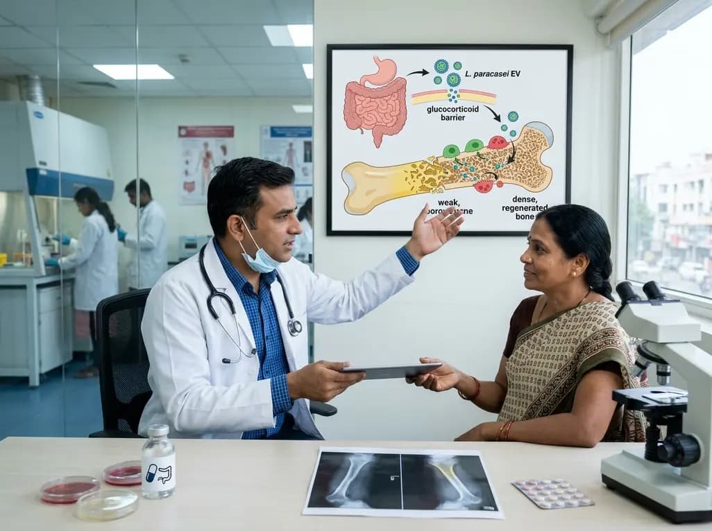
A novel oral nanoplatform using Lacticaseibacillus paracasei-derived extracellular vesicles delivers alendronate, reversing glucocorticoid-induced bone loss and restoring gut-immune-bone homeostasis.
Today
