

Loading, please wait...
Loading, please wait...

Clinical differentiation between cerebral amyloid angiopathy and Alzheimer disease remains a persistent challenge in neurodegenerative medicine. Both conditions frequently present in elderly populations and share overlapping pathophysiological features, most notably vascular and parenchymal amyloid-beta accumulation. However, therapeutic pathways and hemorrhage risks diverge dramatically between these entities. Therefore, clinicians urgently require precise molecular biomarkers to distinguish them in vivo. Recent breakthrough evidence demonstrates that evaluating pericentral tau distribution on positron emission tomography provides an unprecedented imaging distinction. While Alzheimer disease exhibits hierarchical tau accumulation that spares primary cortices until terminal stages, cerebral amyloid angiopathy displays a distinct cortical topography. Consequently, by assessing relative tracer retention across specific brain regions, clinicians can gain vital diagnostic clarity. In addition, novel neuroimaging analyses now focus on regional topographic ratios rather than absolute tracer uptake. As a result, this innovative approach promises to refine therapeutic strategies and reduce hemorrhage complications.
Differentiating cerebral amyloid angiopathy from Alzheimer disease represents a common dilemma in everyday cognitive neurology. Both disorders feature amyloid beta peptides that disrupt central nervous system homeostasis. In Alzheimer disease, amyloid accumulates primarily within the parenchymal neuropil as senile plaques. In contrast, cerebral amyloid angiopathy involves amyloid deposition within the walls of cortical and leptomeningeal arterioles. Furthermore, these two conditions frequently coexist in older individuals, confusing the clinical picture. Patients presenting with cognitive decline or mild behavioral changes might harbor severe cerebral vascular fragility. Misclassifying these patients carries profound clinical consequences. Specifically, modern anti-amyloid immunotherapies carry substantial risks of amyloid-related imaging abnormalities. When clinicians administer these therapies to patients with unrecognized severe angiopathy, patients face a heightened risk of microhemorrhages and lobar hematomas. Unfortunately, conventional structural magnetic resonance imaging sequences only identify downstream hemorrhagic footprints rather than early pathophysiology. Therefore, specialists require reliable molecular neuroimaging markers to differentiate both conditions before catastrophic vascular complications occur. Tau-targeted positron emission tomography offers a highly promising pathway to resolve this diagnostic dilemma.
Tau neurofibrillary tangles follow a predictable anatomical progression in typical Alzheimer disease, famously categorized by Braak staging. The pathology originates in the transentorhinal cortex, progresses into the hippocampus, and then gradually advances into multimodal association cortices. Crucially, Alzheimer pathology spares primary sensory and motor areas, including the pericentral cortex, until the most advanced stages of disease. In contrast, cerebral amyloid angiopathy displays an entirely different spatial pattern of tau deposition. Pathologists observe that vascular-associated tau aggregates appear early and distribute more diffusely across superficial cortical layers. Therefore, investigating pericentral tau distribution serves as an anatomical indicator of true disease identity. Rather than merely assessing absolute tracer uptake, analyzing the ratio of pericentral-to-global tau retention exposes key disease differences. Standardized uptake value ratios in the pericentral cortex, when normalized to global cortical retention, highlight this divergence. While Alzheimer disease yields relatively low pericentral-to-global ratios due to severe associative cortical binding, angiopathy displays elevated ratios. Consequently, this relative pericentral tau distribution metric transforms routine tau-PET scans into highly specific diagnostic instruments for clinicians worldwide.
A multicenter observational study recently established the definitive diagnostic accuracy of this imaging biomarker. Investigators first enrolled a derivation cohort comprising 22 healthy controls, 80 patients with cerebral amyloid angiopathy, and 64 individuals with Alzheimer disease. All participants underwent amyloid-PET alongside 18F-florzolotau tau-PET scanning. Patients with angiopathy exhibited higher pericentral and global tau retention than healthy controls, yet lower absolute uptake than Alzheimer patients. However, the pericentral-to-global tau ratio was significantly higher in angiopathy than in Alzheimer disease (0.93 ± 0.07 versus 0.82 ± 0.08). The researchers identified an optimal cutoff value of 0.90 for this ratio. This cutoff generated a sensitivity of 68.9% and a specificity of 87.1%, achieving an area under the curve of 0.836. Subsequently, the researchers confirmed these diagnostic parameters in an external validation cohort of 65 angiopathy and 19 Alzheimer patients using 18F-flortaucipir PET. In this validation group, the ratio delivered 63.1% sensitivity and an impressive 94.7% specificity, yielding an area under the curve of 0.844. Furthermore, multivariable regression demonstrated that this spatial distribution remained independent of chronological age and total amyloid burden.
The stark anatomical disparity between these conditions reflects contrasting biological mechanisms of tau seeding and clearance. In classic Alzheimer disease, hyperphosphorylated tau spreads trans-synaptically along interconnected functional neuronal networks. Primary sensory and motor regions possess distinct cytoarchitectonic properties and lower synaptic connectivity with early-affected limbic hubs, protecting them from early tau pathology. Conversely, cerebral amyloid angiopathy triggers tau deposition through prominent microvascular dysfunction. Severe vascular amyloid deposition damages the basement membrane, impairs perivascular glymphatic clearance, and promotes localized neuroinflammation. Furthermore, histological examinations reveal perivascular tau-positive neurites wrapping around damaged cortical arterioles. Because cerebral amyloid angiopathy primarily targets leptomeningeal and superficial cortical microvessels across the brain convexities, it induces diffuse cortical clearance failure. Consequently, primary motor and sensory gyri along the central sulcus experience early tau accumulation alongside associative regions. Conditional probability models in the study confirmed that pericentral tau involvement occurs far earlier in angiopathy trajectories than in Alzheimer disease progressions. Thus, vascular breakdown generates a unique, widespread cortical tau signature that distinctly defies classic Braak staging rules.
These imaging discoveries provide actionable clinical utility for specialists navigating complicated dementia diagnoses. In clinical settings, differentiating pure Alzheimer disease from mixed pathology or primary amyloid angiopathy heavily influences therapeutic management. Currently, neurologists exercise tremendous caution when prescribing monoclonal anti-amyloid therapies due to severe vascular hemorrhage risks. By incorporating pericentral tau distribution assessments into PET interpretation workflows, radiologists can deliver clearer risk stratification. When a scan reveals an elevated pericentral-to-global ratio approaching or exceeding 0.90, clinicians should maintain a high suspicion for underlying amyloid angiopathy. Additionally, identifying this angiopathy-predominant signature guides blood pressure optimization and antiplatelet selection. Clinicians frequently hesitate to discontinue anticoagulation or antiplatelet agents without objective proof of vascular amyloid fragility. Moreover, this biomarker provides clinical trials with an enrichment tool to screen prospective candidates cleanly. Ultimately, implementing relative pericentral tau ratios bridges molecular imaging and clinical bedside decisions. As tau-PET tracers become more accessible across global medical centers, quantitative regional analyses will ensure safer, personalized therapies for patients with complex neurodegenerative diseases.
In Alzheimer disease, tau deposition spares primary sensorimotor cortices until late stages, creating a lower pericentral-to-global ratio. Conversely, cerebral amyloid angiopathy features diffuse microvascular dysfunction that causes early tau deposition in primary cortices. Consequently, a pericentral-to-global tau ratio of 0.90 or higher reliably identifies angiopathy over Alzheimer disease.
Differentiating these disorders is essential because treatment strategies differ significantly. Emerging anti-amyloid immunotherapies carry major risks of brain swelling and hemorrhage in patients with severe cerebral amyloid angiopathy. Accurately identifying angiopathy prevents catastrophic microvascular bleeding, optimizes strict blood pressure management, and guides appropriate decisions regarding antithrombotic and anticoagulant therapies.
Yes, researchers validated this discriminative method across multiple distinct radiotracers, including 18F-florzolotau and 18F-flortaucipir. Rather than relying on absolute uptake values, calculating regional-to-global retention ratios normalizes tracer kinetics across different imaging platforms. This standardized metric ensures reproducible results across diverse nuclear medicine departments and clinical imaging settings.
Disclaimer: This content is for informational and educational purposes only and does not constitute medical advice, diagnosis, or treatment recommendations. Clinical decisions must always be made by qualified healthcare professionals based on individual patient assessments and established diagnostic criteria. Refer to the latest local and national guidelines for clinical practice.
References

Read summarized clinical updates, watch expert medical content, and earn CME certifications right from your smartphone.
Open your smartphone camera to scan and install.





A study in Neurology reveals that the relative pericentral tau distribution on tau-PET differentiates cerebral amyloid angiopathy from Alzheimer disease with high specificity, offering clinicians a valuable tool to guide treatment decisions and reduce vascular complications.
Today

A validated natural language processing pipeline leverages clinical notes and intermediate annotations to identify risky alcohol use across surgical patients, offering automated preoperative risk stratification.
Today

The GESS-AEEM consensus establishes a clear clinical implementation framework for testosterone therapy in postmenopausal women with hypoactive sexual desire disorder, focusing on transdermal dosing, precise laboratory monitoring, and safety communication.
Yesterday
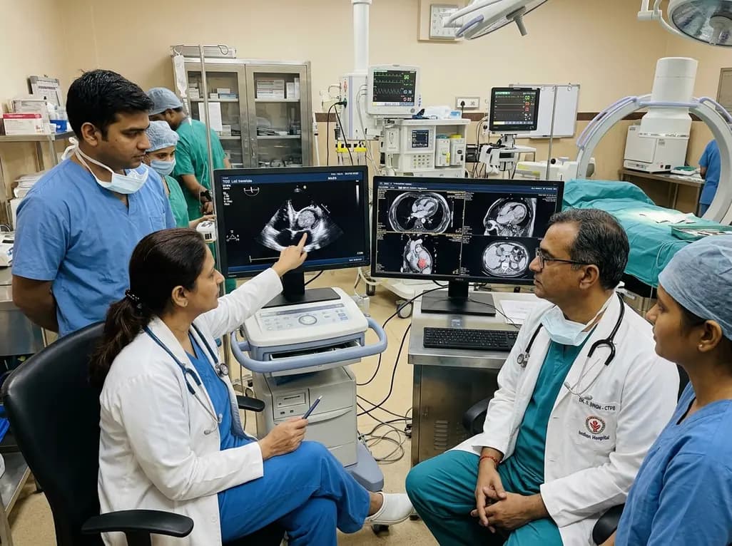
A rare case of an asymptomatic left ventricular hemangioma highlights the vital role of multimodal imaging in differentiating vascular tumors from thrombus. Complete surgical resection under transesophageal echocardiography guidance prevents thromboembolism and delivers optimal outcomes.
Today

A landmark study evaluates long-term quality of life in adolescents and adults with brachial plexus birth injury. Severe functional limitation and limited access to care significantly worsen outcomes, underscoring the urgent need for persistent, multidisciplinary management across adult healthcare transitions.
Today

An analysis of 1,000 posts by macro- and mega-influencers on Instagram and TikTok revealed that 78.5% contained nutrition misinformation, while only 24% of creators held valid credentials. This article examines the clinical risks of viral dietary fads and provides actionable guidance for healthcare professionals.
Today
