.webp&w=384&q=75)

Loading, please wait...
Loading, please wait...
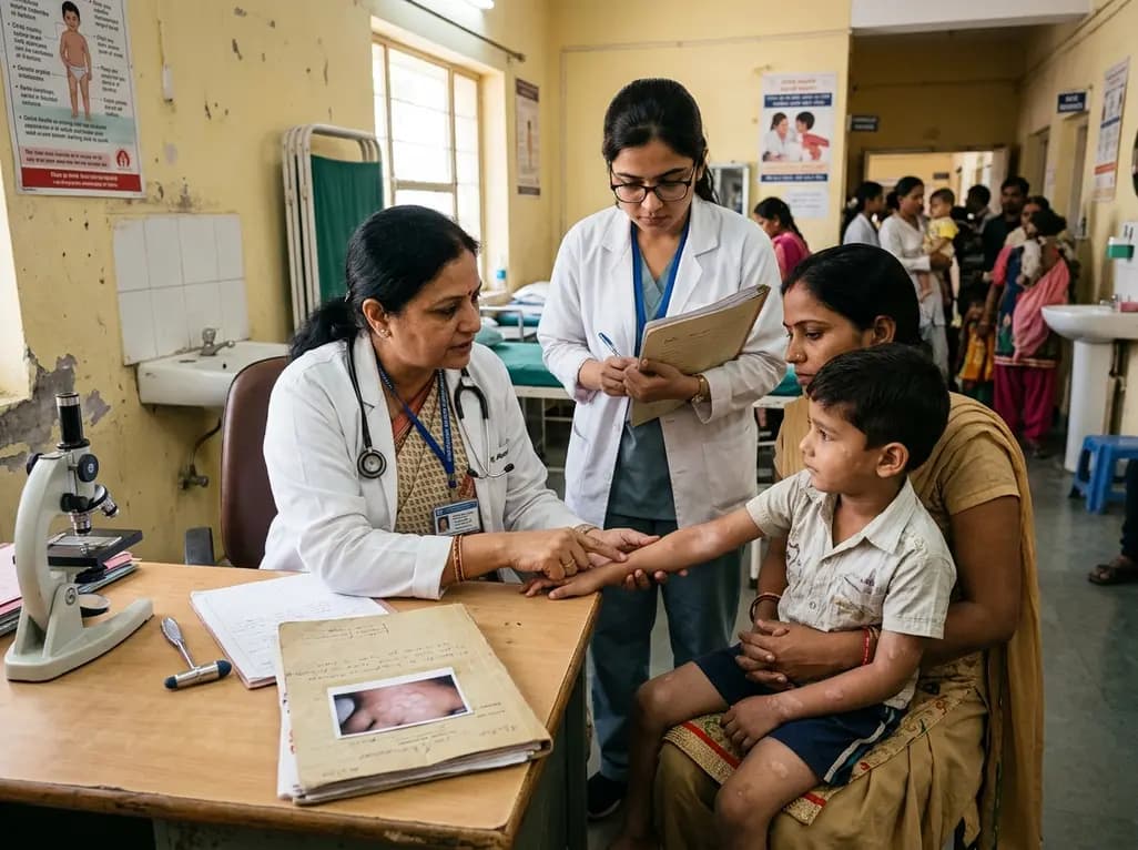
Cutaneous T-cell lymphomas rarely present in children and adolescents, creating significant diagnostic dilemmas across clinical practices. Among these neoplasms, pediatric mycosis fungoides represents the predominant variant encountered in juvenile populations. Clinicians often confuse its initial lesions with common inflammatory conditions like atopic eczema, pityriasis alba, or leprosy. However, early detection remains critical to avoid disease progression and establish targeted skin-directed management. A comprehensive nationwide multicenter study recently examined the clinicopathologic characteristics, treatment patterns, and response rates among young patients.
The multicenter Turkish cohort evaluated 133 pediatric patients who received a confirmed diagnosis before eighteen years of age. Male patients constituted 60.9% of the overall study cohort, showing a slight male predominance. Furthermore, the median age at diagnosis stood at thirteen years, with an interquartile range spanning nine to fifteen years. Most children presented with early-stage disease, and investigators identified stage IA illness in 59.4% of the participants. Consequently, extensive tumor burden or nodal spread remained uncommon at the initial clinical encounter.
Moreover, the initial clinical features often resembled benign chronic dermatoses. Classic erythematous patches and plaques appeared in 78.2% of the cases. Meanwhile, hypopigmented lesions occurred in 36.1% of patients. Interestingly, 42.1% of children presented with overlapping clinical morphologies simultaneously. Thus, dermatologists must recognize that childhood cases rarely manifest with uniform cutaneous plaques alone. Instead, varied patch patterns frequently coexist on identical patients. Because young skin demonstrates high immunological plasticity, atypical clinical phenotypes frequently emerge. Recognizing these diverse clinical signs ensures timely specialist referral.
A major revelation from this multicenter cohort involved the prolonged median diagnostic delay of eighteen months. Furthermore, the interquartile range reached up to forty-eight months before definitive histopathological verification. This notable delay occurs because pediatric lesions mimic common childhood rashes. For instance, clinicians routinely treat hypopigmented patches as eczema or pityriasis lichenoides chronica for months. Therefore, pediatricians and dermatologists must maintain a high index of suspicion for persistent scaly macules.
Additionally, overlapping phenotypes further complicate prompt identification. When hypopigmented macules accompany classical erythematous patches, clinicians might overlook underlying malignancy. In contrast, adult mycosis fungoides typically presents with more distinct erythematous plaques. Consequently, physicians often exhaust several courses of topical corticosteroids or moisturizers before considering cutaneous lymphoma. Nevertheless, early skin biopsies provide the only reliable method to curtail this delay. When children exhibit non-responsive hypopigmented patches, specialists must perform repeat punch biopsies promptly. Early clinical suspicion prevents prolonged parental anxiety and avoids unnecessary diagnostic detours.
Accurate confirmation demands meticulous histopathological evaluation and immunohistochemical assessment. Typically, the dermal-epidermal junction reveals atypical epidermotropic T lymphocytes aligning along the basal layer. In addition, juvenile cases frequently demonstrate a cytotoxic CD8-positive phenotype, especially within hypopigmented lesions. In contrast, adult classical forms predominantly display CD4-positive helper T lymphocytes. Pathologists frequently report haloed intraepidermal lymphocytes, mild spongiosis, and papillary dermal fibrosis. Therefore, distinguishing early lesions from benign inflammatory dermatoses requires extensive clinicopathological correlation.
Furthermore, the Turkish cohort confirmed that 59.4% of children harbored stage IA disease at diagnosis. This finding confirms that pediatric cases predominantly present at early cutaneous stages. Systemic dissemination and visceral involvement remain exceedingly rare in juvenile patients. However, pathologists must carefully rule out secondary cutaneous lymphomas or aggressive cytotoxic presentations. Consequently, repeated tissue biopsies and molecular T-cell receptor gene rearrangement studies occasionally become necessary. Close collaboration between pediatric dermatologists and expert dermatopathologists ensures accurate diagnosis and prevents overstaging.
Therapeutic management for childhood cutaneous T-cell lymphoma favors conservative skin-directed treatments. In the national cohort, dermatologists administered phototherapy to 60.9% of the young patients. Specifically, narrowband ultraviolet B and psoralen combined with ultraviolet A represented the primary phototherapeutic modalities. Overall, the cohort demonstrated remarkable therapeutic responsiveness, achieving an early response rate of 91.0% between three and six months. Thus, skin-directed therapies clear skin lesions effectively in most children.
Moreover, multivariable logistic regression evaluated predictors of early therapeutic response in this large pediatric cohort. Patients presenting with limited body surface area involvement achieved faster and more complete remission. Additionally, clinicians successfully utilized high-potency topical corticosteroids as primary or adjuvant treatments for localized patches. In contrast, systemic chemotherapy remained reserved exclusively for refractory or advanced cases. Because pediatric patients face decades of potential life years, clinicians avoid aggressive cytotoxic drugs whenever possible. Consequently, phototherapy provides a safe, highly effective first-line standard that minimizes long-term organ toxicities.
The findings from this large cohort offer crucial clinical insights for Indian clinical practice. In India, physicians frequently evaluate children presenting with solitary or generalized hypopigmented macules. Common differentials include pityriasis alba, post-inflammatory hypopigmentation, indeterminate leprosy, and early vitiligo. Therefore, Indian practitioners must include pediatric mycosis fungoides in their differential diagnosis for persistent hypopigmented lesions. When typical treatments fail, physicians must avoid prolonged, empirical steroid administration without histological confirmation.
Furthermore, phototherapy infrastructure exists widely across secondary and tertiary medical centers in India. Narrowband UVB therapy offers a practical, affordable, and accessible option for Indian children with extensive patches. However, compliance requires structured counseling regarding photoprotection, treatment duration, and regular follow-up visits. Because recurrence remains common after stopping phototherapy, dermatologists must maintain long-term surveillance schedules. Additionally, training general practitioners to recognize overlapping morphologies can significantly shorten diagnostic delay across urban and rural centers. Ultimately, heightened clinical awareness ensures prompt diagnosis and favorable prognosis for affected children.
Pediatric mycosis fungoides presents much more frequently with hypopigmented macules and overlapping clinical morphologies than the adult form. While adult patients typically exhibit erythematous scaling plaques or progressive tumors, children commonly present with indolent, pale patches that mimic benign rashes. Furthermore, children frequently show a predominant CD8-positive immunophenotype on biopsy. Despite having higher rates of unusual morphological variants, young patients generally achieve superior early treatment responses and face lower rates of disease progression.
Diagnostic delay occurs primarily because early lesions closely mimic common benign conditions, including pityriasis alba, atopic dermatitis, eczema, and leprosy. Clinicians frequently prescribe empirical topical steroids or emollients for many months without conducting tissue biopsies. Additionally, primary care physicians rarely suspect cutaneous malignancies in otherwise healthy children. Consequently, the median time from symptom onset to definitive histopathological diagnosis often extends beyond eighteen months, highlighting the necessity of biopsy for persistent, treatment-resistant childhood macules.
Skin-directed therapy serves as the preferred first-line intervention for pediatric mycosis fungoides. Specifically, narrowband ultraviolet B phototherapy provides exceptional efficacy, achieving favorable early responses in more than ninety percent of pediatric patients. For localized early-stage patches, clinicians also prescribe potent topical corticosteroids with excellent clinical results. Clinicians actively avoid systemic chemotherapeutic agents because juvenile patients demonstrate favorable prognoses with minimal progression risks, ensuring effective disease control while avoiding severe medication toxicities.
Disclaimer: This content is for informational and educational purposes only... Refer to the latest local and national guidelines for clinical practice.
References

Read summarized clinical updates, watch expert medical content, and earn CME certifications right from your smartphone.
Open your smartphone camera to scan and install.





A Turkish multicenter cohort of 133 pediatric mycosis fungoides patients revealed an 18-month diagnostic delay, high rates of hypopigmented (36.1%) and overlapping (42.1%) lesions, and a 91% early response rate, mainly driven by phototherapy.
4 weeks back

Elevated serum HIF-1α levels correlate significantly with MELD scores and independent risk of hepatic decompensation, highlighting the biomarker's emerging diagnostic and prognostic utility in chronic liver disease.
Today

A ten-year cohort study of ABU case logs reveals that URPS fellowship training significantly drives higher annual case volumes, advanced surgical complexity, and increased female representation in pelvic floor reconstruction, underscoring the vital need for structured subspecialty training.
Today
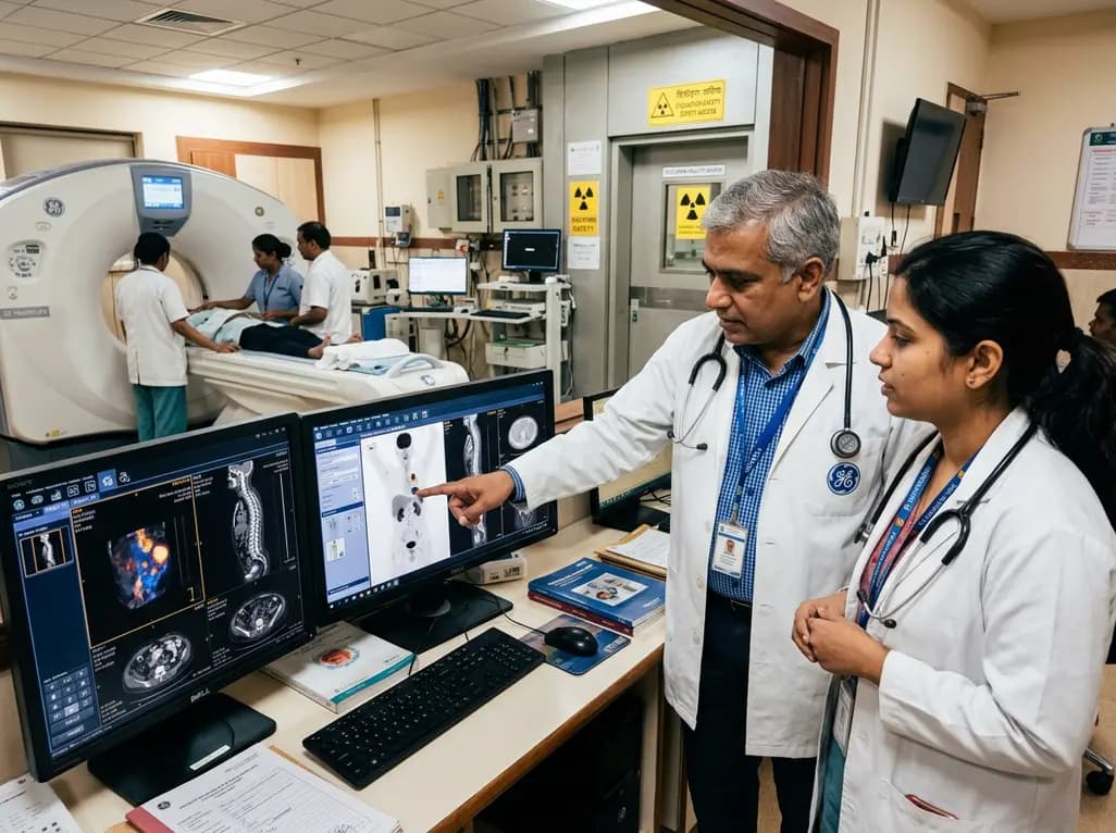
GE HealthCare has agreed to acquire Sofie Biosciences in a 945 million dollar all-cash transaction. The deal secures global rights to the investigational pan-cancer tracer FAPI-74 and expands cyclotron manufacturing infrastructure to accelerate precision theranostics and clinical molecular imaging workflows.
Today

Posttraumatic pisotriquetral instability is a rare wrist injury causing severe hypothenar pain and weakness. Explore clinical examination, imaging, and pisiformectomy.
Today

India's food safety authority has cracked down on high-caffeine beverages sold under stimulant branding, prompting legal challenges from major beverage corporations. This review examines FSSAI regulations, cardiac and metabolic complications, pediatric risks, and actionable counseling strategies for physicians.
Today
