.webp&w=384&q=75)

Loading, please wait...
Loading, please wait...

Pediatric movement disorders present substantial diagnostic challenges for clinicians worldwide. In particular, pediatric lesion-induced dystonia arises after diverse early cerebral insults, creating heterogeneous clinical phenotypes that often confound classical localization principles. Historically, clinicians attributed these hyperkinetic postures strictly to focal basal ganglia injury. However, recent neuroimaging evidence demonstrates that acquired childhood dystonia reflects widespread disruption across interconnected motor circuits rather than isolated subcortical damage. Understanding these complex network mechanisms allows pediatricians and neurologists to evaluate neurological vulnerability with greater precision.
Pediatric brain injuries arise from multiple distinct clinical insults, yet they often yield strikingly similar hyperkinetic movement phenotypes. Perinatal hypoxic-ischemic encephalopathy, kernicterus from hyperbilirubinemia, and arterial ischemic stroke represent distinct vascular and metabolic pathologies. Historically, clinicians struggled to explain why these disparate disease processes cause identical twisting postures and sustained muscular contractions.
Recent investigations resolve this clinical paradox. Although primary lesions occur in distinct structures depending on the etiology, these anatomical injuries converge upon common neural circuits. In neonatal hypoxia, bilateral putaminal necrosis frequently predominates due to elevated metabolic demands during perinatal asphyxia. Conversely, severe hyperbilirubinemia selectively damages the globus pallidus through neurotoxic bilirubin deposition. Meanwhile, pediatric stroke causes focal cortical or subcortical ischemic infarction.
Despite these clear etiological distinctions, lesion-network mapping reveals a shared topographical vulnerability. Researchers demonstrate that these focal disruptions affect identical functional pathways within the developing brain. Consequently, pediatric lesion-induced dystonia represents an integrated network disturbance rather than an isolated nuclear injury. This conceptual shift helps clinicians correlate variable magnetic resonance imaging findings with predictable movement patterns.
Advanced neuroimaging techniques now clarify how specific structural injuries correlate with hyperkinetic motor signs. Multivariate lesion-symptom mapping in pediatric cohorts has identified the putamen and globus pallidus as critical anatomical drivers of secondary dystonia. Specifically, ischemic stroke and hypoxic-ischemic encephalopathy demonstrate robust statistical associations with putaminal injury. In contrast, kernicterus consistently maps to bilateral globus pallidus damage.
These findings confirm classical clinico-anatomical observations while refining quantitative precision. The putamen functions as the primary input gateway for corticostriatal motor projections. Therefore, structural damage in this area disrupts sensorimotor integration and impairs voluntary motor execution. At the same time, pallidal injury compromises inhibitory basal ganglia output, which releases excessive motor drive to cortical targets.
Furthermore, the timing of brain injury markedly alters these structural outcomes. The immature neonatal nervous system undergoes rapid synaptic remodeling and myelination. As a result, early focal damage triggers maladaptive neuroplasticity throughout developing subcortical nuclei. Clinicians observing delayed post-injury dystonia frequently witness the consequences of this abnormal developmental reorganization. Recognizing these distinct anatomic hubs enables neurologists to anticipate dystonia risks in neonates surviving severe asphyxia or bilirubin encephalopathy.
Beyond focal nuclear damage, lesion network mapping using normative pediatric connectomes reveals a shared whole-brain circuit. Intriguingly, lesions causing dystonia across all etiologies connect directly to the somato-cognitive-action network and the cingulo-opercular action-mode network. This finding establishes a crucial link between traditional motor pathways and higher-order cortical control systems.
The somato-cognitive-action network integrates body movement with executive action planning, autonomic regulation, and cognitive processing. Traditionally, medical education taught that the primary motor cortex forms a continuous homunculus controlling discrete muscles. However, contemporary connectomics shows that effector-specific motor regions alternate with integrative action centers coordinating complex goal-directed behaviors. When focal subcortical lesions disconnect these integrative nodes, motor output loses normal inhibitory control.
Additionally, network mapping demonstrates connections to the anterior and medial cerebellum. The cerebellum maintains precise timing, sensory prediction, and motor error correction. When basal ganglia lesions disrupt cerebello-thalamo-cortical loops, the motor cortex receives corrupted feedforward signals. Consequently, voluntary movements trigger involuntary, overflow muscle contractions and prolonged twisting postures. This unified circuit perspective explains why patients with diverse lesion sites exhibit similar dystonic posturing.
Unraveling this unified brain network carries profound implications for pediatric dystonia therapy. Standard pharmacotherapies like baclofen, trihexyphenidyl, and carbidopa-levodopa often provide modest symptom relief in acquired childhood dystonia. However, identifying precise circuit hubs opens innovative avenues for targeted neuromodulation and surgical intervention.
Currently, deep brain stimulation primarily targets the internal globus pallidus or subthalamic nucleus. Yet, clinical responses among children with secondary dystonia remain notoriously variable compared to primary genetic dystonias. Network localization explains this variability with remarkable clarity. Because the functional lesion network encompasses cerebellar and cingulo-opercular pathways, alternative stimulation targets may provide superior therapeutic relief. For instance, neuromodulation targeting specific cerebellar outflow tracts or thalamic relay nodes might bypass irreversibly damaged basal ganglia.
In addition, these network insights provide a scientific rationale for customized neurorehabilitation programs. Physical and occupational therapists can design intensive sensorimotor training regimens that leverage preserved cortical regions within the action network. By driving adaptive neuroplasticity across intact nodes, rehabilitation specialists can promote motor compensation. Ultimately, integrating connectomic mapping into clinical decision-making will help pediatric movement disorder teams personalize multidisciplinary care.
These network insights hold immense clinical relevance for healthcare systems in developing countries like India. Despite significant improvements in perinatal care, conditions such as birth asphyxia, neonatal hyperbilirubinemia, and pediatric CNS infections continue to pose substantial public health burdens. Consequently, secondary dyskinetic cerebral palsy and acquired dystonias remain prevalent causes of childhood physical disability in Indian tertiary centers.
Early recognition of motor network disruption allows clinicians to initiate proactive monitoring rather than waiting for fixed postural deformities. In resource-limited settings lacking functional neuroimaging, understanding structural lesion correlations guides clinical prognosis. For example, identifying putaminal hyperechogenicity on cranial ultrasound or pallidal hyperintensity on MRI alerts pediatricians to dystonia risks. Consequently, clinicians can initiate timely motor rehabilitation, optimize nutritional support, and avoid unwarranted antiepileptic medications.
Furthermore, preventing neonatal jaundice toxicity through prompt phototherapy and exchange transfusion directly protects the globus pallidus, preserving critical motor circuits. Improving primary obstetric emergency management similarly reduces hypoxic insults to the striatum. By bridging cutting-edge network neuroscience with frontline pediatric care, Indian physicians can enhance both preventive strategies and long-term neurodevelopmental outcomes.
Pediatric lesion-induced dystonia occurs when early brain insults damage developing motor pathways. Common causes include perinatal hypoxic-ischemic encephalopathy, neonatal hyperbilirubinemia resulting in kernicterus, arterial ischemic stroke, trauma, and intracranial infections. Although primary injuries occur in different anatomical sites, they disrupt a shared brain-wide circuit governing motor coordination.
The shared pediatric dystonia network encompasses the basal ganglia, specifically the putamen and globus pallidus, alongside the anterior and medial cerebellum. Additionally, it connects to cortical areas overlapping the cingulo-opercular action-mode and somato-cognitive-action networks, which coordinate complex motor planning, cognitive integration, and voluntary movement execution.
Deep brain stimulation shows variable results in secondary dystonia because underlying structural lesions permanently destroy targeted basal ganglia nuclei or interrupt critical axonal pathways. Furthermore, because dystonia involves a widespread network including cerebellar and cortical regions, focal pallidal stimulation may not sufficiently normalize activity across all dysfunctional nodes.
Disclaimer: This content is for informational and educational purposes only and is not intended as medical advice. Healthcare professionals should exercise their independent clinical judgment. Refer to the latest local and national guidelines for clinical practice.
References

Read summarized clinical updates, watch expert medical content, and earn CME certifications right from your smartphone.
Open your smartphone camera to scan and install.





A landmark study reveals that pediatric lesion-induced dystonia from hypoxia, kernicterus, and stroke converges on a shared brain network involving the basal ganglia, cerebellum, and somato-cognitive-action network, offering new insights for targeted therapies.
Today

A crossover study demonstrates that locally deployed large language models with exact-match knowledge augmentation significantly elevate outpatient prescription review accuracy to 97.2%, curbing hallucinations and securing patient data without requiring complex cloud infrastructure.
Today
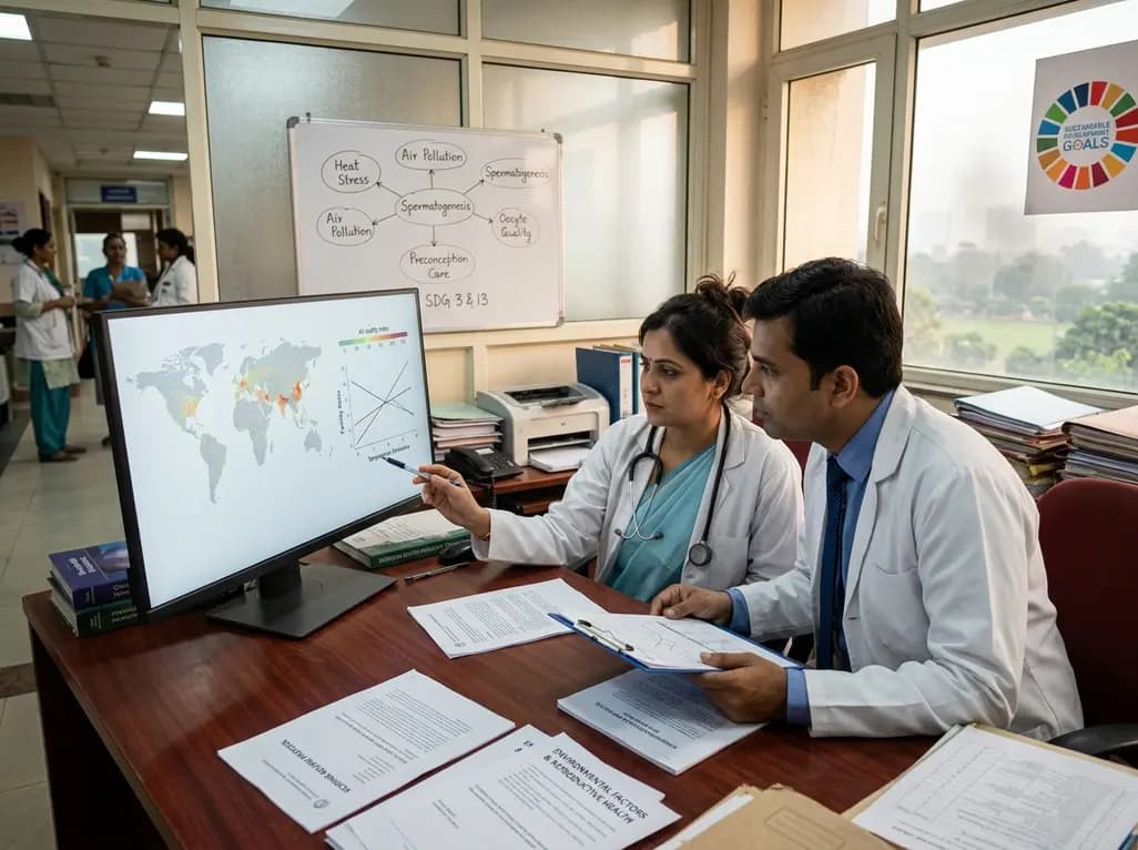
A new scoping review protocol systematically maps global evidence on how climatic shifts, ambient heat, and pollution impact human fertility. Discover key physiological mechanisms, clinical preconception strategies, and critical public health alignments under Sustainable Development Goals 3 and 13.
Today

Pharmacogenomics and artificial intelligence are revolutionizing cardiology by tailoring therapies to individual genetic profiles and clinical data, reducing adverse drug events, and improving cardiovascular patient outcomes.
Today
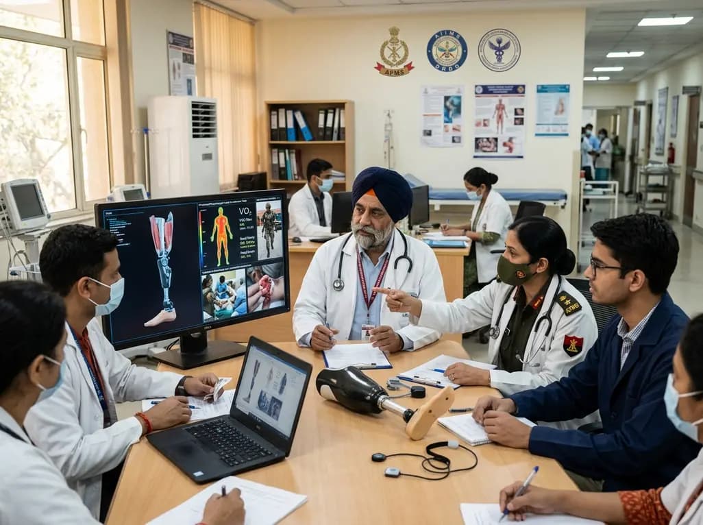
India's premier scientific bodies have joined the Armed Forces Medical Services to solve unique physiological and operational challenges faced by soldiers. The national collaboration covers combat casualty care, artificial intelligence diagnostics, bionic prosthetics, and extreme-environment physiological resilience.
Today

A retrospective cohort study evaluates the short- and long-term impacts of SARS-CoV-2 infection on patients with Graves' disease receiving antithyroid drug therapy, highlighting thyroid status destabilization, clinical symptom exacerbations, and post-viral sequelae.
Today
