

Loading, please wait...
Loading, please wait...
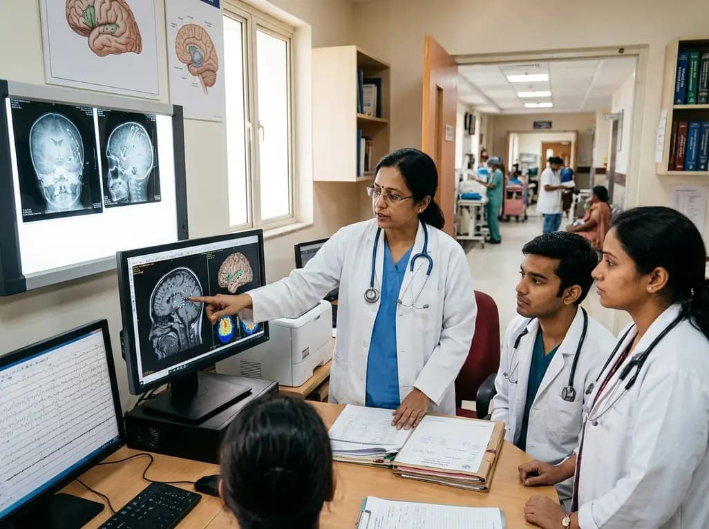
Medically refractory focal epilepsy presents profound neurodevelopmental challenges for affected children and their families. Fortunately, modern neurosurgical innovations now offer viable therapeutic options for previously inaccessible epileptogenic zones. Recent clinical evidence demonstrates that pediatric insular epilepsy surgery provides meaningful seizure control for patients who fail antiepileptic drug therapies. Historically, neurosurgeons hesitated to operate within the insular cortex due to dense surrounding vascular networks and critical functional pathways. However, emerging meta-analytic data clarify safety benchmarks, prognostic indicators, and diagnostic strategies that optimize pediatric care and long-term developmental recovery.
Diagnosing insular seizure foci in young patients remains a formidable clinical challenge. Because the insular cortex connects extensively with the frontal, temporal, and parietal lobes, seizure semiology often mimics other focal epilepsies. For example, children frequently exhibit complex hypermotor behaviors, visceral somatosensory auras, autonomic alterations, or asymmetric tonic posturing. Consequently, scalp electroencephalography often fails to localize the true epileptogenic focus accurately. Additionally, routine magnetic resonance imaging may show subtle abnormalities or appear entirely normal in many refractory cases. Therefore, clinicians frequently misclassify insular seizures as frontal or temporal lobe epilepsy. This diagnostic confusion often delays appropriate surgical evaluation and prolongs ineffective antiepileptic medication trials. Furthermore, persistent unmanaged seizures severely impair cognitive development, academic performance, and overall quality of life. Pediatric epileptologists must maintain a high index of suspicion when semiology reveals rapid propagation or early autonomic signs. Ultimately, establishing accurate localization requires comprehensive multimodal neuroimaging and advanced electrophysiological investigations before surgical planning.
Stereoelectroencephalography has revolutionized invasive presurgical evaluation for deep epileptogenic zones in pediatric patients. Specifically, stereoelectroencephalography enables neurosurgeons to insert robotic depth electrodes directly into the insular gyri with exceptional precision. Individual participant data demonstrate that children who undergo preoperative stereoelectroencephalography experience significantly lower odds of seizure recurrence. In fact, invasive stereoelectroencephalography evaluation reduces recurrence odds by approximately seventy-five percent compared to non-invasive evaluations. Moreover, depth electrodes map the exact spatial boundaries of the epileptogenic network without requiring large craniotomies. Surgeons can also assess whether seizure onsets involve adjacent opercular structures or deeper subcortical pathways. Consequently, surgical teams can tailor resections or ablative margins specifically to the active seizure focus. Furthermore, invasive recordings minimize unnecessary tissue destruction, preserving nearby functional parenchyma. Stereoelectroencephalography also allows clinicians to perform functional stimulation mapping at the bedside. Therefore, neurosurgeons can delineate language and motor pathways before executing irreversible interventions, solidifying the procedure as a critical pillar in pediatric insular epilepsy surgery.
Surgical treatment options for refractory insular lesions primarily include microsurgical resection and magnetic resonance-guided thermal ablation. Traditionally, neurosurgeons favored open microsurgical resection via transsylvian or transcortical approaches. Microsurgical techniques permit direct lesion removal and thorough tissue acquisition for histological examination. However, open resection carries notable risks, particularly vascular injury to middle cerebral artery branches and lenticulostriate perforators. In contrast, magnetic resonance-guided laser interstitial thermal therapy offers a minimally invasive alternative. Surgeons introduce thin laser fibers through tiny burr holes to ablate the epileptogenic zone under real-time thermal monitoring. Interestingly, recent meta-analytic data reveal no statistically significant difference in seizure freedom between resective and ablative modalities. Procedure type did not dictate long-term seizure control in pediatric patients. In addition, extending the resection beyond the insula did not independently improve seizure-free outcomes. Thus, neurosurgical teams can select either modality based on lesion architecture, anatomical accessibility, and center experience, providing flexible therapeutic opportunities.
Achieving complete seizure control represents the paramount goal in pediatric epilepsy surgery. In systematic pooled data, approximately fifty-seven percent of children achieved seizure freedom at a median follow-up of thirty-seven months. Researchers defined seizure freedom using Engel Class I or International League Against Epilepsy Class 1 and 2 criteria. Crucially, statistical analyses identified stereoelectroencephalography as the primary independent predictor of postoperative seizure freedom. Conversely, demographic factors like age at surgery did not alter the likelihood of seizure control. Similarly, pathology subtype and surgical extent did not produce statistically significant differences in seizure remission. These observations suggest that precise electrophysiological definition of the epileptogenic zone matters more than raw tissue volume removal. Additionally, achieving early seizure freedom halts progressive developmental impairment in children with epileptic encephalopathy. Long-term seizure cessation also allows gradual reductions in antiepileptic medications, decreasing drug-related behavioral side effects. Consequently, pediatric patients experience meaningful cognitive catch-up, enhanced social engagement, and improved family wellbeing.
Surgical intervention near the insular cortex inevitably raises legitimate concerns regarding permanent neurological impairment. The insula borders critical eloquent structures, including the internal capsule, corona radiata, and middle cerebral vasculature. According to pooled meta-analytic findings, permanent neurological deficits occurred in only eleven percent of operated children. Furthermore, motor deficits represented the most common post-surgical complication due to capsular proximity. Interestingly, statistical regression demonstrated that younger age at surgery correlated with higher odds of motor deficit. Younger infants possess smaller anatomical corridors and developing vascular systems, which increases vulnerability during surgical manipulation. In contrast, speech impairments and sensory deficits occurred far less frequently. Importantly, clinicians documented that over two-thirds of all observed postoperative deficits remained transient. Most children regained functional mobility within several months of targeted physical rehabilitation. Therefore, comprehensive preoperative counseling should explain both transient risks and potential long-term benefits to caregivers, while surgical teams employ continuous intraoperative monitoring.
Current systematic evidence shows that surgery achieves seizure freedom in approximately 57 percent of pediatric patients with refractory insular epilepsy. Both open microsurgical resection and thermal laser ablation produce comparable rates of seizure control over long-term follow-up. Furthermore, utilizing preoperative stereoelectroencephalography significantly enhances clinical outcomes, reducing the odds of seizure recurrence by seventy-five percent. Consequently, surgery represents an effective therapeutic option when antiepileptic medications fail to control pediatric seizures.
Postoperative neurological complications occur in a minority of pediatric cases, with permanent deficits affecting approximately 11 percent of patients. Motor deficits, such as hemiparesis, represent the most frequent neurological complication due to the close proximity of the internal capsule. Importantly, more than two-thirds of observed deficits are transient, resolving with comprehensive rehabilitation. Younger children face slightly higher risks of motor injury, emphasizing the need for meticulous intraoperative monitoring and surgical precision.
Stereoelectroencephalography provides accurate three-dimensional electrophysiological mapping of deep insular cortex networks. Because insular seizures frequently propagate rapidly to surrounding frontal and temporal regions, surface electroencephalography often misidentifies the true epileptogenic focus. Stereotactically placed depth electrodes confirm ictal onset zones and protect eloquent cortex during subsequent interventions. Multivariable analyses confirm that children assessed with stereoelectroencephalography achieve significantly higher rates of seizure freedom compared to those undergoing surgery without invasive depth recording.
Disclaimer: This content is for informational and educational purposes only and does not constitute medical advice. While based on recent research, clinical approaches must be individualized. Always consult qualified healthcare professionals before initiating or modifying any medical treatment. Refer to the latest local and national guidelines for clinical practice.
References

Read summarized clinical updates, watch expert medical content, and earn CME certifications right from your smartphone.
Open your smartphone camera to scan and install.





A landmark individual participant data meta-analysis demonstrates that pediatric insular epilepsy surgery offers significant seizure freedom, particularly with preoperative stereoelectroencephalography (SEEG), while revealing critical insights into postoperative neurological safety and transient motor deficits.
Today

A national cross-sectional survey reveals marked disparities in infectious disease diagnostic capacity across hospital tiers, underscoring the urgent need for integrated referral networks and enhanced point-of-care testing to improve outbreak detection and patient outcomes.
Today

The GESS-AEEM consensus establishes a clear clinical implementation framework for testosterone therapy in postmenopausal women with hypoactive sexual desire disorder, focusing on transdermal dosing, precise laboratory monitoring, and safety communication.
Yesterday
A rare case of an asymptomatic left ventricular hemangioma highlights the vital role of multimodal imaging in differentiating vascular tumors from thrombus. Complete surgical resection under transesophageal echocardiography guidance prevents thromboembolism and delivers optimal outcomes.
Today
A landmark study evaluates long-term quality of life in adolescents and adults with brachial plexus birth injury. Severe functional limitation and limited access to care significantly worsen outcomes, underscoring the urgent need for persistent, multidisciplinary management across adult healthcare transitions.
Today
An analysis of 1,000 posts by macro- and mega-influencers on Instagram and TikTok revealed that 78.5% contained nutrition misinformation, while only 24% of creators held valid credentials. This article examines the clinical risks of viral dietary fads and provides actionable guidance for healthcare professionals.
Today
