

Loading, please wait...
Loading, please wait...
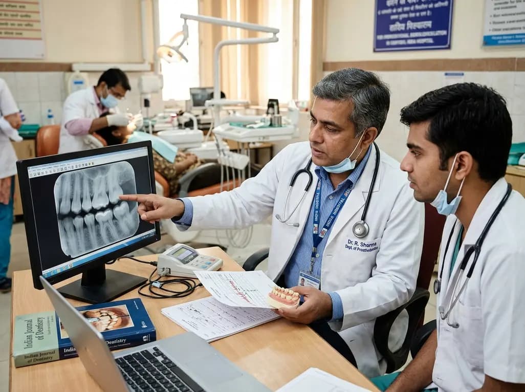
Occlusal tooth wear represents a significant clinical challenge for dental practitioners worldwide. This multifactorial condition causes loss of mineralized tooth structure, leading to compromised aesthetics and severe functional deterioration. Clinicians frequently encounter patients who present with accelerated dental attrition and erosion. However, determining the precise biomechanical drivers behind this degradation remains difficult. Masticatory function depends heavily on continuous neuromuscular coordination between the jaw elevator muscles and periodontal mechanoreceptors. When mechanical stresses exceed normal physiologic tolerances, tooth surfaces undergo irreversible attrition. Furthermore, age-related tissue remodeling alters dental anatomy and masticatory efficiency over time. Dental professionals traditionally assess surface degradation through visual grading systems, yet these tools often overlook functional loading. Therefore, clinicians must investigate how individual bite strength and sensory feedback mechanisms interact during daily mastication. Understanding these biomechanical dynamics helps practitioners identify high-risk individuals before severe coronal destruction occurs. Consequently, comprehensive diagnostic protocols should incorporate objective physical measurements alongside traditional evaluations. Ultimately, dental teams can design far more durable restorative treatment plans by addressing root functional causes.
Maximum voluntary bite force provides an objective measure of the functional capacity of the masticatory system. Specifically, elevator muscles like the masseter and temporalis dictate the magnitude of compressive forces during chewing. In clinical investigations, researchers quantify this parameter using specialized transducers positioned between opposing teeth. High bite force generates substantial mechanical stress across enamel and restorative interfaces. Consequently, patients with elevated muscular output repeatedly subject their dentition to intense microtrauma. In particular, a recent clinical study examined 51 adult patients with a mean age of 63 years. The investigators recorded maximum voluntary bite force and analyzed its relationship with cumulative dental attrition. The statistical analysis demonstrated a moderate positive correlation between bite force and overall wear severity. Moreover, multivariable regression models confirmed that this strong association persisted after controlling for patient age and sex. These findings emphasize that elevated muscular force serves as an independent driver of progressive tooth destruction. Therefore, evaluating absolute bite force offers critical diagnostic insight into patient vulnerability. Identifying hyperfunctional masticatory muscles early allows clinicians to implement protective strategies promptly.
Occlusal tactile acuity reflects the remarkable ability of the human brain to detect minute interocclusal thicknesses. Mechanoreceptors embedded within the periodontal ligament convey delicate tactile signals through trigeminal afferent fibers. Consequently, this sensory feedback loop protects teeth from excessive occlusal loads by modulating jaw muscle activity. Clinicians assess tactile acuity by placing calibrated aluminum foils between teeth and noting detection thresholds. Interestingly, the latest clinical data revealed a small negative correlation between tactile acuity and posterior tooth wear. Patients with diminished tactile acuity exhibited more severe structural loss in their posterior molars. Because impaired sensory acuity delays inhibitory neuromuscular reflexes, patients may inadvertently apply damaging forces during functional contacts. In contrast, patients with acute sensory perception detect prematurities quickly and adjust their chewing strokes accordingly. However, sensory acuity does not completely counterbalance excessive voluntary bite force during parafunctional episodes. Furthermore, age-related sensory declines might exacerbate wear progression in elderly cohorts. Clinicians must therefore recognize that reduced tactile sensitivity poses a hidden risk for progressive posterior wear.
Robust clinical assessment requires validated scoring methodologies to document dental surface loss accurately. For example, researchers utilized the Tooth Wear Evaluation System 2.0 to quantify anterior, canine, and posterior wear patterns. Additionally, they applied the California Dental Association criteria to evaluate wear across existing prosthetic restorations. The research findings demonstrated that mechanical load plays a primary role in structural degradation across dental arches. While anterior teeth often display pronounced aesthetic wear, posterior teeth absorb the greatest vertical masticatory stresses. Consequently, higher maximum bite forces correlate directly with widespread coronal deterioration across the entire dental arch. Multivariable regression analyses further established that bite force remains a significant predictor of wear regardless of age. Nevertheless, age independently contributes to cumulative tissue loss through decades of repetitive functional friction. Thus, practitioners must differentiate between normal physiological aging and accelerated mechanical destruction. When excessive bite force coexists with diminished tactile acuity, destructive forces amplify dramatically. Clinicians who understand these complex physiological interactions can accurately diagnose mechanical etiology and develop targeted interventions.
Managing advanced dental wear requires comprehensive treatment planning that addresses biological, sensory, and biomechanical factors. Dentists often reconstruct severely worn dentitions using ceramics, composite resins, or fixed implant-supported prostheses. However, placing expensive restorations without managing elevated bite force frequently leads to catastrophic material fractures. Therefore, clinicians must routinely assess occlusal force distribution before initiating extensive restorative therapy. In addition, digital occlusal analysis devices and pressure sensors help practitioners map occlusal contacts during dynamic excursion. Clinicians can prescribe custom occlusal splints to protect vulnerable restorations from nocturnal bruxism and diurnal clenching forces. Moreover, patient education regarding dietary acids and parafunctional habits reinforces mechanical protection across vulnerable tooth structures. Dentists should also recognize that patients with reduced tactile acuity cannot reliably sense minor occlusal interferences. As a result, practitioners must meticulously calibrate articulating contacts to prevent localized hyper-occlusion and mechanical overload. Furthermore, regular clinical monitoring enables dental teams to detect early signs of material fatigue or biological breakdown. Ultimately, combining protective nightguards, precision occlusal balancing, and durable restorative materials ensures long-term clinical success and preserves remaining dentition.
Maximum voluntary bite force determines the magnitude of mechanical stress delivered to opposing dental arches during mastication and clenching. When elevator muscles exert intense compressive forces, enamel and restorative surfaces experience severe friction and shear stress. Consequently, microcracks form and enamel prisms fracture over time. Clinical research demonstrates that patients who generate higher bite forces exhibit significantly greater cumulative structural wear, even after controlling for age and anatomical variations.
Occlusal tactile acuity relies on periodontal mechanoreceptors that detect minute interocclusal thicknesses and prematurities during jaw movements. This neurosensory mechanism triggers protective inhibitory reflexes in the trigeminal motor system, preventing muscular overload. When patients suffer from diminished tactile acuity, their ability to modulate chewing force decreases substantially. Consequently, unattenuated biting forces strike posterior teeth repeatedly, accelerating localized surface loss and predisposing restored teeth to early biomechanical failure.
Clinicians can incorporate bite force evaluation by utilizing digital occlusal sensors, clinical electromyography, or mechanical force gauges before placing restorations. Identifying patients with excessive bite force enables practitioners to select high-strength restorative materials like monolithic zirconia rather than weaker ceramics. Furthermore, dentists can fabricate protective occlusal splints and eliminate premature occlusal contacts during dynamic excursion. This proactive biomechanical approach minimizes restoration chipping, prevents abutment loosening, and ensures long-term restorative stability.
Disclaimer: This content is for informational and educational purposes only... Refer to the latest local and national guidelines for clinical practice.
References
Stanisic N et al. The Relationship Between Occlusal Tactile Acuity, Bite Force and Tooth Wear. J Oral Rehabil. 2026 Nov 03. doi: 10.1111/joor.70365. PMID: 42973391.
Gupta A et al. To Evaluate the Severity, Distribution of Occlusal Tooth Wear and its Correlation with Bite Force in Young North Indian Adults. Open Dent J. 2018;12:735-742.
Gu Y, Bai Y, Xie X. Bite Force Transducers and Measurement Devices. Front Bioeng Biotechnol. 2021;9:665081.

Read summarized clinical updates, watch expert medical content, and earn CME certifications right from your smartphone.
Open your smartphone camera to scan and install.





A clinical study demonstrates that maximum voluntary bite force strongly correlates with occlusal tooth wear, whereas reduced occlusal tactile acuity relates to posterior wear. Clinicians must incorporate biomechanical load evaluations and sensory factors into comprehensive restorative and prosthodontic workflows.
Today

The PhenX Toolkit offers over 1,000 consensus-based protocols to standardize phenotypes and environmental exposures. This open-access resource enhances scientific rigor, supports cross-study data pooling, and integrates with platforms like REDCap to empower biomedical researchers worldwide.
Today

A major ABCD study analysis reveals that prenatal co-exposure to cannabis and tobacco significantly impairs fluid cognitive abilities in middle childhood, whereas crystallized intelligence remains unaffected. The findings highlight urgent needs for targeted antenatal screening and intervention.
Today
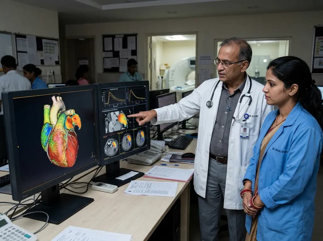
Dynamic contrast 4DCT enables precise mapping of left atrial appendage blood transport in atrial fibrillation. By quantifying regional stasis, wash-in kinetics, and residence time, this imaging advance offers clinician-tailored thromboembolic risk stratification beyond standard clinical scores.
Today
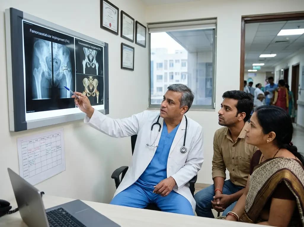
Deciding the optimal interval for staged bilateral hip arthroscopy in femoroacetabular impingement syndrome requires weighing the risks of rapid intervention against delayed joint degeneration. Shared decision-making balances functional rehabilitation milestones with bilateral symptom progression.
Today

India is poised to transform chronic disease management through wearable health technology. Discover how continuous remote monitoring and predictive algorithms empower clinicians to detect early cardiovascular signals, personalize metabolic interventions, and deliver equitable preventive care across populations.
Today
