

Loading, please wait...
Loading, please wait...

Emerging imaging protocols using dynamic contrast 4DCT provide unprecedented insights into intracardiac hemodynamics among patients with cardiac arrhythmias. Clinicians recognize that non-valvular atrial fibrillation substantially increases the risk of cardioembolic stroke. In addition, stagnant blood within the left atrial appendage serves as the primary nidus for intracardiac thrombus formation. Traditional assessment strategies rely on static anatomic evaluation or conventional transesophageal echocardiography. However, these conventional modalities cannot map three-dimensional contrast dispersion over consecutive cardiac cycles. Dynamic contrast 4DCT resolves this critical diagnostic limitation by tracking the wash-in and wash-out kinetics of iodinated contrast across the entire atrial volume. Consequently, cardiologists can now quantify regional blood stasis with exceptional spatial and temporal fidelity. Radiologists acquire volumetric image series across multiple heartbeats during contrast administration. Furthermore, advanced computational post-processing fits voxel-wise time-attenuation data to extract precise physiological flow parameters. As a result, clinicians gain an objective window into blood residence time and micro-mixing patterns within complex appendage anatomies. This functional imaging paradigm moves cardiovascular medicine beyond purely geometrical risk indicators. Ultimately, dynamic contrast 4DCT illuminates patient-specific pathophysiology and promises to refine stroke prevention strategies in high-risk electrophysiology cohorts.
Contemporary cardiology relies heavily on empirical clinical risk scores to guide anticoagulation therapy. For instance, clinicians routinely calculate the CHA2DS2-VASc score to estimate thromboembolic risk in atrial fibrillation. While these clinical rubrics offer valuable population-level guidance, they exhibit notable limitations for individual patients. Specifically, clinical risk scores overlook individual anatomical variations and patient-specific flow disturbances within the left atrium. Many patients with low clinical scores still develop debilitating cardioembolic strokes. Conversely, other individuals maintain elevated clinical scores yet demonstrate vigorous appendage emptying without demonstrable thrombogenesis. Transesophageal echocardiography provides valuable qualitative Doppler flow velocities at the appendage ostium. Nevertheless, echocardiography remains semi-invasive and fails to capture deep apex flow recirculations. Additionally, single-phase computed tomography often generates false-positive findings because sluggish contrast mixing mimics mural thrombus. Therefore, electrophysiologists require objective, reproducible tools that directly measure blood stasis throughout the appendage cavity. By resolving both temporal dynamics and structural geometry, dynamic cardiovascular imaging fills this diagnostic void. Clinicians can thereby identify localized areas of circulatory stasis that standard clinical indices inevitably overlook. Consequently, hemodynamically guided evaluations provide a stronger foundation for prophylactic anticoagulation decisions.
Dynamic imaging protocols characterize blood transport by mapping contrast attenuation changes over time. During contrast transit, radiologists collect sequential volumetric datasets across numerous cardiac cycles. Mathematical algorithms subsequently fit Hounsfield unit values across each voxel using gamma-variate models. This robust analytical framework yields three vital transport metrics. First, researchers calculate the delta residence time, which represents the delay in contrast arrival between the distal appendage and the orifice. A prolonged residence time indicates severe circulatory sluggishness within deep trabeculated lobes. Second, investigators measure the delta peak enhancement ratio. This parameter compares the maximum contrast enhancement within distal voxels against the peak enhancement at the appendage ostium. Markedly reduced ratios reveal poor contrast replenishment and persistent fluid stagnation. Third, the processing software computes the total volume of delayed filling within the appendage chamber. This volumetric metric quantifies the precise anatomical burden of stagnant blood. Furthermore, investigators consolidate these individual parameters into a composite Left Atrial Appendage Transport Index through principal component analysis. Therefore, this multidimensional score synthesizes temporal delay, wash-in magnitude, and stagnant blood volume into a single reproducible descriptor. Such quantitative rigor allows clinicians to distinguish subtle physiologic differences across diverse patient cohorts.
Clinical investigations demonstrate substantial heterogeneity in left atrial appendage transport across patients with atrial fibrillation. Even when patients present with similar baseline clinical characteristics, their internal hemodynamic profiles vary dramatically. For example, some individuals exhibit rapid contrast exchange that closely resembles sinus rhythm hemodynamics. In contrast, other patients demonstrate pronounced regional stasis, marked by extensive volumes of delayed filling and extended residence times. The composite transport index effectively captures this wide biological spectrum. Consequently, clinicians can identify individuals who harbor elevated thrombogenic risk despite harboring moderate clinical risk scores. Furthermore, the index illuminates the hemodynamic consequences of distinct morphological appendage phenotypes. Complex branching anatomies, such as cauliflower or windswept morphologies, frequently generate localized flow separation and stagnant recirculation zones. Conversely, simpler single-lobe structures often maintain more efficient blood clearance. Additionally, dynamic measurements correlate well with invasive biomarkers of endothelial dysfunction and local platelet activation. Therefore, integrating dynamic contrast transport data enhances mechanistic understanding of cardioembolic pathogenesis. By pinpointing the exact spatial distribution of stasis, this methodology provides cardiologists with an actionable physiological target. Ultimately, objective transport quantification supports more personalized, precision-oriented clinical management in electrophysiology clinics.
Dynamic transport quantification provides essential guidance for procedural planning in structural heart disease and electrophysiology. During catheter ablation for atrial fibrillation, extensive atrial scarring or electrical isolation of the appendage can impair regional contractility. Consequently, post-ablation stunning often exacerbates blood stasis, thereby increasing early thromboembolic vulnerability. Pre-procedural dynamic computed tomography enables interventional teams to anticipate post-ablation hemodynamics and tailor peri-procedural anticoagulation regimens accordingly. Similarly, left atrial appendage closure procedures benefit substantially from dynamic contrast mapping. Interventional cardiologists must verify complete ostial occlusion and accurately exclude pre-existing thrombi before device deployment. Traditional imaging sometimes misidentifies sluggish apical flow as an intracardiac clot, leading to unnecessary procedural cancellations. In contrast, dynamic contrast tracking reliably differentiates low-flow blood clearance from fixed filling defects. Moreover, post-implantation imaging can detect persistent peridevice leaks and characterize residual flow dynamics behind the occluder. Thus, operators gain precise feedback regarding device endothelialization and potential device-related thrombus formation. As transcatheter solutions expand globally, hemodynamic imaging will play a pivotal role in refining device selection. Ultimately, these functional assessments ensure safer procedural outcomes and optimize long-term thromboprophylaxis for complex cardiac patients.
Dynamic contrast 4DCT tracks contrast attenuation across sequential cardiac cycles rather than evaluating a single static phase. Stagnant blood demonstrates delayed wash-in and gradual, progressive contrast opacification over several heartbeats. In contrast, an organized intracardiac thrombus represents a persistent filling defect that never enhances with iodinated contrast. By capturing these temporal wash-in kinetics, clinicians can confidently distinguish low-velocity blood clearance from true thrombi, eliminating false-positive diagnoses and avoiding unnecessary procedure delays.
The Left Atrial Appendage Transport Index currently complements rather than replaces established clinical scoring tools like CHA2DS2-VASc. While clinical scores identify broad population-level risk factors, they fail to reflect local hemodynamic stasis and individual appendage anatomy. The transport index adds objective, patient-specific physiologic data that uncovers occult thrombogenic risk in low-risk individuals or confirms safety in borderline cases. As ongoing prospective clinical validation continues, integrating hemodynamic metrics with clinical scores will enable personalized anticoagulation management.
Dynamic cardiac 4DCT requires wide-detector computed tomography scanners capable of imaging the entire left atrium during contrast injection without table translation. While multi-phase dynamic acquisitions deliver higher cumulative radiation doses than single-phase examinations, modern dose-reduction technologies significantly mitigate this risk. Clinicians employ prospective ECG-triggering, low tube voltages, and advanced iterative reconstruction algorithms to keep radiation doses within clinically acceptable ranges. Careful patient selection ensures that diagnostic benefits regarding stroke prevention substantially outweigh potential radiation and iodinated contrast exposures.
Disclaimer: This content is for informational and educational purposes only and does not constitute medical advice, diagnosis, or treatment. Healthcare professionals should exercise independent clinical judgment. Refer to the latest local and national guidelines for clinical practice.
References

Read summarized clinical updates, watch expert medical content, and earn CME certifications right from your smartphone.
Open your smartphone camera to scan and install.





Dynamic contrast 4DCT enables precise mapping of left atrial appendage blood transport in atrial fibrillation. By quantifying regional stasis, wash-in kinetics, and residence time, this imaging advance offers clinician-tailored thromboembolic risk stratification beyond standard clinical scores.
Today

Maharashtra has launched a comprehensive initiative to halve anaemia prevalence across high-risk groups by 2030. The public health strategy integrates aggressive screening, point-of-care diagnostics, parenteral iron therapies, and targeted interventions for nutritional deficits and underlying hemoglobinopathies.
Today

A rare presentation of late myelomeningocele sac rupture devoid of a neural placode demonstrates excellent 2-year functional outcomes, shedding light on the mechanical, chemical, and hydrostatic dynamics of congenital spinal dysraphism.
Today
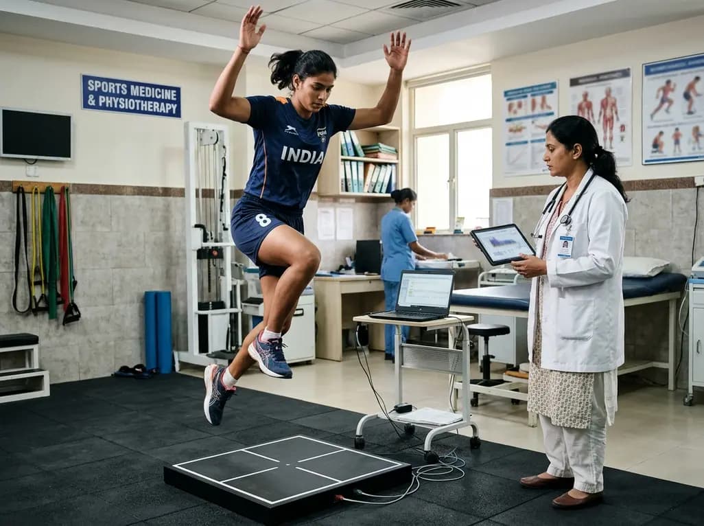
A systematic review and meta-analysis highlights the substantial impact of plyometric jump training on jump performance, reactive strength, and landing kinematics in female team-sport athletes, providing evidence-based programming guidelines for sports medicine and orthopedics.
Today

India is poised to transform chronic disease management through wearable health technology. Discover how continuous remote monitoring and predictive algorithms empower clinicians to detect early cardiovascular signals, personalize metabolic interventions, and deliver equitable preventive care across populations.
Today
