.webp&w=384&q=75)

Loading, please wait...
Loading, please wait...

Alzheimer's disease presents a substantial diagnostic challenge for clinicians worldwide, particularly in developing healthcare systems. Currently, definitive biological confirmation relies heavily on positron emission tomography and cerebrospinal fluid analysis. However, both modalities remain invasive, cost-prohibitive, and largely inaccessible across resource-limited settings. Consequently, clinicians urgently need reliable, noninvasive screening tools to detect early neurodegeneration. Routine magnetic resonance imaging provides a practical, widely available alternative. Traditional structural assessment evaluates regional atrophy and global cortical thinning. Nevertheless, subtle microstructural alterations often emerge long before overt tissue loss becomes detectable on standard scans. To address this critical gap, researchers have developed multiscale structural mapping to extract rich morphological information from standard clinical imaging. By evaluating multidimensional cortical metrics from conventional T1-weighted acquisitions, advanced imaging pipelines can detect subtle pathological shifts earlier. Therefore, refining structural analysis enhances timely diagnosis and patient risk stratification. Moreover, early identification enables prompt interventions, lifestyle modifications, and appropriate referral for disease-modifying therapies. As global dementia cases rise, scalable neuroimaging methods become indispensable for clinical practice. Furthermore, noninvasive imaging pipelines reduce patient anxiety and eliminate the complications associated with lumbar puncture. Consequently, computational neuroimaging offers an equitable diagnostic solution.
For decades, clinicians and neuroradiologists have utilized cortical thickness as a primary structural imaging biomarker. Thinning in the entorhinal cortex and temporal lobes reflects progressive neurodegenerative pathology. However, cortical thickness alone captures only late-stage structural loss and exhibits substantial biological overlap with normal aging. To enhance diagnostic precision, investigators recently introduced multiscale structural mapping as an integrated diagnostic approach. This framework combines cortical thickness measurements with gray-white matter contrast derived from a single T1-weighted scan. Gray-white matter contrast reflects underlying myelin integrity and dendritic density near the cortical interface. As Alzheimer's pathology progresses, microstructural breakdown blurs the boundary between cortical gray matter and subcortical white matter. Consequently, evaluating contrast alongside cortical thickness yields deeper insights into preclinical microarchitectural degradation. Furthermore, this combined analysis captures localized microstructural disruptions before macroscopic tissue loss manifests. Therefore, multiscale structural mapping provides a sensitive morphological footprint of neurodegeneration. Clinicians gain access to richer quantitative data without subjecting patients to prolonged scanner acquisition times. Ultimately, multiparametric structural evaluation bridges the gap between conventional neuroimaging and molecular biomarker sensitivity. In addition, this strategy optimizes standard clinical MRI sequences, maximizing diagnostic yield without increasing institutional expenses.
Building upon the multiscale concept, Baek and colleagues engineered an upgraded analytical pipeline termed MSSM+. This advanced framework expands beyond cortical thickness and gray-white matter contrasts by integrating geometric surface features. Specifically, MSSM+ incorporates vertex-level sulcal depth and cortical curvature measurements into the analytical model. Sulcal anatomy and curvature undergo subtle mechanical deformation during progressive cerebral atrophy. By merging these geometric parameters with contrast and thickness, MSSM+ constructs an extensive multiparametric surface representation. However, modeling complex vertex-level cortical geometries across whole-brain meshes creates significant computational challenges. To solve this problem, the authors introduced surface supervertex mapping to partition the cortical surface. This innovative method clusters adjacent vertices into coherent, anatomically informed surface patches called supervertices. Consequently, surface supervertex mapping preserves critical inter-regional and intra-regional spatial relationships while reducing computational dimensionality. In contrast to arbitrary voxel grids, these patches naturally respect human cortical folding patterns. Furthermore, the grouping maintains high biological fidelity across diverse brain regions. As a result, the pipeline efficiently captures intricate structural alterations across both sulcal depths and gyral crowns without data dilution. Thus, MSSM+ establishes a robust foundation for sophisticated deep learning architectures.
To analyze these partitioned surface patches, researchers introduced the Supervertex Vision Transformer architecture. Traditional convolutional neural networks struggle with irregularly curved non-Euclidean cortical meshes. In contrast, the Supervertex Vision Transformer applies self-attention mechanisms directly across supervertices, learning global structural interdependencies. This design enables anatomically informed feature extraction across disparate cortical zones. When evaluated on clinical datasets, MSSM+ demonstrated substantial diagnostic advantages over earlier methods. Specifically, the framework identified significantly more extensive and statistically robust group differences between Alzheimer's disease patients and cognitively normal controls. Furthermore, in binary diagnostic classification tasks, MSSM+ paired with the Supervertex Vision Transformer demonstrated superior discriminative power. The model achieved a notable 3 percentage point increase in the area under the precision-recall curve compared to standard multiscale mapping. This metric confirms that the architecture maintains high diagnostic precision even when classifying imbalanced clinical populations. Moreover, the self-attention layers highlight disease-affected areas, offering clinicians intuitive visual interpretability. Therefore, the integrated model successfully translates subtle structural alterations into actionable diagnostic predictions, supporting clinical decision-making during initial patient evaluations. Additionally, this high precision minimizes false-positive findings, reducing unnecessary downstream testing and alleviating severe patient psychological stress.
A persistent barrier to deploying artificial intelligence in clinical neuroimaging involves scanner heterogeneity. In routine practice, hospitals utilize scanners from various manufacturers with varying field strengths and acquisition protocols. These technical discrepancies frequently cause severe inter-vendor signal variability, degrading model generalization across institutions. Crucially, the researchers conducted vendor-specific analyses to assess model resilience across different magnetic resonance imaging manufacturers. The results revealed that MSSM+ significantly reduced signal variability compared to isolated cortical thickness, gray-white matter contrasts, or standard multiscale mapping. Furthermore, the Supervertex Vision Transformer maintained consistently superior classification performance across all major hardware vendors. This multi-vendor stability holds immense clinical importance for Indian healthcare settings. Many regional diagnostic centers operate diverse scanning equipment ranging from older systems to advanced platforms. Consequently, a robust biomarker that resists scanner bias ensures equitable diagnostic accuracy across tertiary referral centers and community hospitals. Ultimately, adopting vendor-resilient tools like MSSM+ and the Supervertex Vision Transformer bridges infrastructural gaps. By providing accessible, high-performing screening directly from standard T1-weighted images, this pipeline can streamline clinical triaging before invasive confirmatory testing. In summary, this technological advance moves computational neuroimaging closer to routine, real-world neurological practice.
Conventional cortical thickness assesses only macrostructural tissue loss across brain surfaces. In contrast, MSSM+ integrates cortical thickness with gray-white matter contrast, vertex-level sulcal depth, and curvature from standard T1-weighted MRI. This multiparametric approach captures subtle microstructural and geometric degradation, enabling earlier and more accurate detection of Alzheimer's disease neurodegeneration.
Cortical surfaces possess complex, non-Euclidean geometry that traditional convolutional neural networks struggle to process efficiently. The Supervertex Vision Transformer resolves this limitation by partitioning surfaces into anatomically coherent patches called supervertices. Applying self-attention mechanisms across these patches allows the model to learn long-range spatial relationships while preserving critical local anatomical details.
Scanner variability between different manufacturers often introduces technical noise that distorts automated neuroimaging metrics. MSSM+ significantly suppresses inter-vendor signal variability while sustaining high classification accuracy across diverse scanner models. Consequently, clinicians in community and tertiary centers can deploy this automated tool without needing vendor-specific model recalibration or expensive proprietary scanning sequences.
Disclaimer: This content is for informational and educational purposes only and does not constitute medical advice, diagnosis, or treatment. Healthcare professionals should exercise independent clinical judgment. Refer to the latest local and national guidelines for clinical practice.
References

Read summarized clinical updates, watch expert medical content, and earn CME certifications right from your smartphone.
Open your smartphone camera to scan and install.





A novel neuroimaging framework combining multiscale structural mapping (MSSM+) and Supervertex Vision Transformers enhances early Alzheimer's disease detection on T1 MRI scans while minimizing scanner variability across manufacturers.
Today

A crossover study demonstrates that locally deployed large language models with exact-match knowledge augmentation significantly elevate outpatient prescription review accuracy to 97.2%, curbing hallucinations and securing patient data without requiring complex cloud infrastructure.
Today

A new scoping review protocol systematically maps global evidence on how climatic shifts, ambient heat, and pollution impact human fertility. Discover key physiological mechanisms, clinical preconception strategies, and critical public health alignments under Sustainable Development Goals 3 and 13.
Today

Pharmacogenomics and artificial intelligence are revolutionizing cardiology by tailoring therapies to individual genetic profiles and clinical data, reducing adverse drug events, and improving cardiovascular patient outcomes.
Today
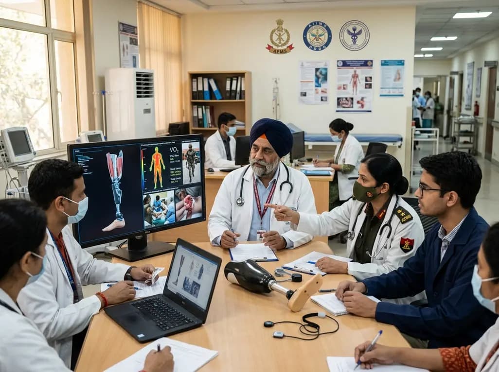
India's premier scientific bodies have joined the Armed Forces Medical Services to solve unique physiological and operational challenges faced by soldiers. The national collaboration covers combat casualty care, artificial intelligence diagnostics, bionic prosthetics, and extreme-environment physiological resilience.
Today

A retrospective cohort study evaluates the short- and long-term impacts of SARS-CoV-2 infection on patients with Graves' disease receiving antithyroid drug therapy, highlighting thyroid status destabilization, clinical symptom exacerbations, and post-viral sequelae.
Today
