.webp&w=384&q=75)

Loading, please wait...
Loading, please wait...

Ischemic stroke triggers widespread pathophysiological alterations that extend far beyond the focal vascular territory. Clinicians frequently observe delayed cognitive decline and neurological deterioration that cannot be explained solely by primary infarct volume. Emerging translational research demonstrates that evaluating MRI R2* stroke inflammation provides critical non-invasive insights into secondary neurodegeneration within remote, structurally disconnected subcortical regions. Understanding these secondary changes allows clinicians to appreciate the network-level devastation caused by focal cerebral ischemia.
Ischemic strokes directly destroy cerebral tissue, but they also interrupt structural axonal tracts connecting distant hubs. Consequently, secondary neurodegeneration develops along damaged pathways through anterograde and retrograde diaschisis. The thalamus represents a primary target for this progressive damage because it serves as an integrative relay station linking cortical networks. When cortical infarcts sever corticothalamic or thalamocortical projections, the affected thalamic nuclei endure prolonged metabolic stress and cellular degradation.
Clinicians in neurology and radiology encounter significant hurdles when attempting to identify remote post-stroke injury early. Routine anatomical imaging modalities, such as T1-weighted or T2-weighted MRI, often fail to detect early secondary degenerative processes before macroscopic tissue loss occurs. Therefore, secondary injury frequently progresses silently for months before presenting as overt thalamic atrophy or delayed functional impairment. Clinicians require robust imaging tools to track these early biological events non-invasively. Tracking microstructural alterations along disconnected pathways provides a window of opportunity to intervene before irreversible atrophy occurs. Advanced neuroimaging biomarkers capable of detecting subclinical axonal degeneration and accompanying glial reactions are therefore essential for improving post-stroke monitoring and long-term prognosis.
To investigate whether relaxometry can detect remote damage, researchers evaluated a prospective cohort of 156 ischemic stroke patients undergoing longitudinal neuroimaging. Investigators acquired MRI scans at baseline and at one-year follow-up, mapping fiber tracts disconnected by acute infarcts. Specifically, researchers measured quantitative R2* relaxation rates within distinct medial and lateral thalamic nuclei groups to evaluate local tissue composition changes over twelve months.
Multivariate linear regression analyses demonstrated that baseline structural disconnectivity status independently predicted significant R2* increases at one year. Importantly, thalamic nuclei with preserved axonal connections exhibited no significant R2* changes over the same period. This finding confirmed a direct and independent relationship between initial axonal disconnection and subsequent remote tissue alterations. Furthermore, traditional clinical confounders, such as patient age, vascular comorbidities, and initial lesion volume, did not diminish this association. By isolating the impact of tract disconnection, the clinical study established that R2* relaxometry reliably reflects progressive microstructural reorganization. These robust patient observations indicate that remote subcortical changes follow specific topological network disruptions rather than diffuse, generalized aging or systemic vascular pathology. Consequently, quantitative R2* mapping offers clinicians an objective method to quantify delayed, remote tissue damage across cerebral networks.
To elucidate the cellular mechanisms driving remote relaxometry changes, researchers conducted parallel experiments in a photothrombotic stroke mouse model. Investigators performed longitudinal MRI, histological characterization, quantitative gene expression analysis, and mass spectrometry across an eight-week timeframe. In vivo imaging in mice mirrored the clinical findings, showing significant R2* elevations specifically restricted to disconnected thalamic nuclei.
Focal R2* increases peaked at two weeks post-stroke, preceding significant thalamic atrophy measured at eight weeks. Mass spectrometry demonstrated that this signal elevation corresponded spatially and temporally with elevated non-heme iron concentrations. Furthermore, immunohistochemical analysis revealed that the accumulated iron was bound to ferritin within activated microglial cells rather than free in the parenchyma. Microglia responded to degenerating axon terminals by phagocytosing cellular debris and sequestering metabolically active iron. Consequently, the heightened R2* relaxation rate directly reflects ferritin-bound iron storage within neuroinflammatory cells. This translational validation confirms that early R2* elevations represent active microglial neuroinflammation rather than passive, non-specific edema. Because microglial activation precedes structural tissue loss, the peak in R2* signal provides a precise temporal marker of the inflammatory cascade occurring during secondary neurodegeneration.
Recognizing remote secondary neurodegeneration as an active, inflammatory process carries substantial implications for stroke neurology practice. Stroke survivors often experience post-stroke cognitive impairment, emotional dysregulation, and delayed motor decline months after index hospitalization. In many instances, these chronic symptoms stem from secondary thalamic damage rather than the primary cortical lesion. Therefore, clinicians must look beyond the initial ischemic core when assessing long-term functional prognosis.
Quantitative MRI techniques that track tissue iron and inflammation offer a valuable roadmap for patient stratification. By identifying patients who exhibit marked thalamic disconnectivity and elevated R2* values, neurologists can anticipate individuals at high risk for accelerated secondary neurodegeneration. In addition, these imaging biomarkers enable targeted rehabilitation strategies aimed at reinforcing alternative functional pathways. Clinicians can also tailor cognitive interventions before extensive thalamic atrophy becomes established. Furthermore, understanding the precise timeline of secondary microglial activation provides an objective framework for therapeutic trials. Interventions aimed at dampening chronic neuroinflammation could potentially preserve critical thalamic nuclei if administered during the subacute post-stroke window. Integrating quantitative relaxometry into follow-up neuroimaging protocols enhances clinical surveillance and supports comprehensive neurovascular care.
The identification of R2* relaxometry as an in vivo marker of remote neuroinflammation opens exciting therapeutic horizons. Past neuroprotective clinical trials overwhelmingly focused on salvaging the acute ischemic penumbra within hours of stroke onset, yielding disappointing translational results. However, secondary neurodegeneration unfolds over weeks to months, providing a significantly wider therapeutic window for neuroprotective interventions. Consequently, targeting secondary neuroinflammation represents an attractive strategy to preserve connected brain hubs.
Future clinical trials can leverage quantitative R2* imaging as an objective pharmacodynamic biomarker to evaluate anti-inflammatory or iron-chelating agents. Researchers can non-invasively measure whether experimental therapeutics successfully suppress microglial iron accumulation within disconnected thalamic nuclei. Moreover, combining R2* relaxometry with quantitative susceptibility mapping and advanced diffusion tractography will refine our anatomical understanding of post-stroke network degeneration. As high-field MRI scanners become more prevalent in clinical practice, automated relaxometry processing pipelines could soon enter standard radiology workflows. Ultimately, translating these advanced imaging techniques into routine post-stroke monitoring will empower physicians to implement personalized neuroprotective regimens, protect vulnerable structural networks, and significantly improve long-term functional outcomes for stroke survivors worldwide.
MRI R2* relaxometry measures the effective transverse relaxation rate of tissue water protons. In post-stroke secondary neurodegeneration, R2* is highly sensitive to paramagnetic susceptibility effects. Elevated values specifically reflect the focal accumulation of iron bound to ferritin within activated microglial cells in disconnected structures.
The thalamus functions as a critical integrative hub maintaining dense bidirectional axonal connections with the cerebral cortex. When cortical strokes sever these white matter projections, the associated thalamic nuclei lose trophic input. Consequently, retrograde and anterograde axonal degeneration triggers delayed microglial activation, iron deposition, and progressive neuronal atrophy.
Standard MRI sequences like T1 and T2 provide qualitative anatomical contrast, which only reveals macroscopic volume loss months after stroke. In contrast, quantitative R2* relaxometry detects subtle, subacute microstructural alterations. It captures active cellular neuroinflammation and iron deposition weeks before structural tissue shrinkage becomes visible on conventional imaging.
Disclaimer: This content is for informational and educational purposes only and is not intended as a substitute for professional medical advice, diagnosis, or treatment. Always seek the advice of your physician or other qualified health provider with any questions you may have regarding a medical condition. Refer to the latest local and national guidelines for clinical practice.
References

Read summarized clinical updates, watch expert medical content, and earn CME certifications right from your smartphone.
Open your smartphone camera to scan and install.





A translational study demonstrates that MRI R2* relaxometry effectively tracks remote secondary neuroinflammation and ferritin-bound iron accumulation within disconnected thalamic nuclei following ischemic stroke, providing a non-invasive imaging biomarker for long-term post-stroke neurodegeneration.
Today

A crossover study demonstrates that locally deployed large language models with exact-match knowledge augmentation significantly elevate outpatient prescription review accuracy to 97.2%, curbing hallucinations and securing patient data without requiring complex cloud infrastructure.
Today

A new scoping review protocol systematically maps global evidence on how climatic shifts, ambient heat, and pollution impact human fertility. Discover key physiological mechanisms, clinical preconception strategies, and critical public health alignments under Sustainable Development Goals 3 and 13.
Today

Pharmacogenomics and artificial intelligence are revolutionizing cardiology by tailoring therapies to individual genetic profiles and clinical data, reducing adverse drug events, and improving cardiovascular patient outcomes.
Today
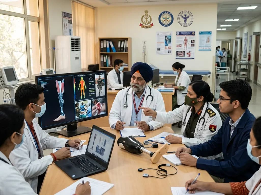
India's premier scientific bodies have joined the Armed Forces Medical Services to solve unique physiological and operational challenges faced by soldiers. The national collaboration covers combat casualty care, artificial intelligence diagnostics, bionic prosthetics, and extreme-environment physiological resilience.
Today

A retrospective cohort study evaluates the short- and long-term impacts of SARS-CoV-2 infection on patients with Graves' disease receiving antithyroid drug therapy, highlighting thyroid status destabilization, clinical symptom exacerbations, and post-viral sequelae.
Today
