.webp&w=384&q=75)

Loading, please wait...
Loading, please wait...

Gastric carcinoma remains a formidable global health challenge, demonstrating high disease-related mortality across several geographical regions, including East and South Asia. Recent oncological investigations have uncovered crucial molecular drivers that sustain tumor resilience and promote aggressive cellular invasion. Among these newly identified oncogenic factors, the expression of MAGEA6 in gastric cancer has emerged as a significant determinant of adverse pathological behavior. Researchers continue to explore how cancer cells rewire metabolic and organellar processes to escape apoptosis. Consequently, deciphering the intracellular pathways regulated by cancer-testis antigens provides valuable insights into tumor survival mechanisms.
Melanoma-associated antigen A6, commonly abbreviated as MAGEA6, belongs to the cancer-testis antigen family. Typically, normal human tissues restrict the physiological expression of these proteins primarily to germ cells within the testis. However, malignant transformations frequently reactivate MAGEA6 across diverse human carcinomas, including gastric malignancies. Analyses of large-scale genomic datasets, such as The Cancer Genome Atlas, reveal marked upregulation of MAGEA6 transcripts in malignant gastric tissues compared to adjacent benign specimens. Moreover, clinical correlations demonstrate that elevated levels correspond to advanced clinical stage, greater lymph node dissemination, and reduced overall survival. In addition, experimental models validate that this aberrant expression directly augments cell motility and proliferation. When oncologists evaluate gastric tumor specimens, they frequently note that elevated MAGEA6 correlates with a significantly more aggressive disease course. Thus, these findings highlight MAGEA6 not merely as an incidental marker of transformation, but rather as an active driver of tumor advancement. Consequently, understanding its molecular interactions offers vital opportunities to identify novel therapeutic targets. Scientists emphasize that unraveling MAGEA6-mediated downstream signaling could illuminate mechanisms underlying drug resistance and invasive disease progression in gastrointestinal oncology.
Mitochondria govern cellular bioenergetics, oxidative balance, and the execution of intrinsic apoptotic cascades in normal and malignant cells. However, rapidly proliferating neoplasms experience significant metabolic stress, leading to accumulated mitochondrial structural damage. To survive these adverse microenvironmental stresses, malignant cells utilize mitophagy, a specialized form of macroautophagy dedicated to clearing dysfunctional mitochondria. Recent experimental investigations indicate that MAGEA6 directly enhances mitochondrial autophagy within gastric carcinoma cells. By utilizing sensitive mito-Keima fluorescence assays and transmission electron microscopy, investigators observed that MAGEA6 expression markedly increases autophagic clearance of damaged organelles. Furthermore, JC-1 mitochondrial membrane potential assays confirmed that MAGEA6 prevents fatal mitochondrial depolarisation and collapse under conditions of cellular stress. As a result, malignant cells maintain robust energy production while circumventing cytochrome c release and subsequent apoptotic cell death. Therefore, MAGEA6 effectively preserves mitochondrial pool integrity, allowing tumor cells to sustain aggressive growth under nutrient-deprived microenvironmental conditions. Additionally, sustained mitochondrial quality control shields neoplastic tissues against oxidative stress and metabolic crisis. Consequently, this elevated mitophagic flux provides gastric carcinoma cells with a distinct survival advantage over surrounding non-malignant tissues.
To delineate the exact molecular cascade facilitating organelle clearance, researchers evaluated downstream mediators linked to MAGEA6 overexpression. Mechanistic analyses revealed that MAGEA6 upregulates the calcium-binding protein S100A9 within gastric cancer cells. Subsequently, elevated S100A9 expression triggers the activation of the canonical PINK1 and PRKN mitophagy signaling axis. Under basal conditions, healthy mitochondria rapidly degrade the serine/threonine kinase PINK1 to maintain homeostasis. However, when S100A9 levels rise, PINK1 stabilizes on the outer mitochondrial membrane, where it recruits and activates the E3 ubiquitin ligase PRKN. Furthermore, activated PRKN ubiquitinates key outer mitochondrial membrane proteins, recruiting autophagic cargo receptors and initiating autophagosome formation. Knockdown assays demonstrated that silencing either S100A9 or PRKN effectively abrogated the pro-survival advantages conferred by MAGEA6. Thus, the MAGEA6/S100A9/PINK1/PRKN cascade forms a tightly coordinated regulatory circuit driving mitochondrial quality control. In contrast to normal cells, which maintain tightly restricted mitophagy, gastric cancer cells exploit this specific signaling pathway to accelerate malignant turnover. Accordingly, these findings clarify how aberrant cancer-testis antigen expression directly interfaces with core mitochondrial quality control machinery to dictate cancer cell fate.
The functional outcomes of MAGEA6 upregulation extend far beyond baseline cytoprotection in primary gastric lesions. In vitro functional assays, including Transwell migration assays and 5-ethynyl-2'-deoxyuridine cell proliferation measurements, show enhanced invasiveness in MAGEA6-positive lines. Because active mitophagy maintains energetic homeostasis, malignant cells acquire superior mechanical flexibility and migration potential. Consequently, elevated MAGEA6 expression strongly correlates with lymphovascular invasion, deep muscularis infiltration, and peritoneal dissemination in clinical cohorts. Moreover, these migratory capabilities enable gastric cancer cells to survive detachment-induced apoptosis, commonly known as anoikis, during lymphatic and vascular transit. Therefore, patients displaying elevated MAGEA6 expression often experience earlier disease recurrence and diminished progression-free intervals following radical surgical resection. In addition, this mitophagic resilience shields metastatic clones against ischemic stress and hypoxic insults in distant organ niches. Pathologists and clinical oncologists should recognize that this phenotype reflects heightened metastatic potential rather than indolent disease. Thus, identifying MAGEA6 expression in endoscopic biopsy specimens may aid in stratifying patients who require more aggressive neoadjuvant or adjuvant systemic therapeutic protocols.
Given the vital role of the MAGEA6/S100A9/PINK1/PRKN pathway in driving malignancy, targeted therapeutic interventions represent an attractive oncological avenue. Because MAGEA6 exhibits restricted expression in adult somatic tissues, selective therapies could achieve profound antitumor efficacy while sparing normal gastric mucosa. For example, therapeutic vaccines and chimeric antigen receptor T-cell approaches targeting cancer-testis antigens are currently undergoing rigorous translational evaluation. Additionally, small-molecule inhibitors disrupting S100A9 function or inhibiting PRKN-mediated mitophagy could resensitize resistant gastric cancer cells to conventional cytotoxic regimens. Furthermore, standard chemotherapy regimens, such as fluoropyrimidine and platinum-based combinations, frequently fail due to cytoprotective autophagic responses in refractory tumors. Consequently, combining systemic chemotherapy with mitophagy inhibitors could disrupt mitochondrial stability and trigger robust apoptotic cell death in MAGEA6-overexpressing tumors. However, clinical implementation requires validated immunohistochemical biomarkers to select appropriate patient cohorts. Clinical trials must systematically investigate these combination regimens to evaluate safety, bioavailability, and overall therapeutic efficacy. Ultimately, targeting MAGEA6 and its downstream mitophagy pathway could improve clinical outcomes for patients suffering from advanced or metastatic gastric cancer.
Clinical data indicate that elevated MAGEA6 expression strongly correlates with poor prognosis in gastric cancer patients. Because MAGEA6 promotes cellular proliferation, invasiveness, and mitophagy, tumors harboring high expression levels demonstrate aggressive biological behavior. Consequently, these patients frequently present with advanced disease stages, increased lymph node metastasis, and higher rates of post-surgical recurrence. Therefore, evaluating MAGEA6 expression may serve as a valuable prognostic biomarker to identify high-risk individuals requiring intensified clinical monitoring and therapy.
Mitophagy functions as a critical organelle-specific quality control mechanism that eliminates damaged or depolarized mitochondria. In gastric carcinoma, rapidly dividing cells experience severe oxidative and metabolic stress that threatens organellar stability. By clearing dysfunctional mitochondria, mitophagy prevents cellular collapse and inhibits the release of pro-apoptotic factors such as cytochrome c. Furthermore, this process recycles vital metabolic intermediates, enabling cancer cells to sustain energy generation, resist cytotoxic therapy, and promote continuous tumor survival under hypoxic microenvironments.
This molecular pathway represents a promising target because MAGEA6 exhibits restricted expression in healthy adult somatic tissues, reducing off-target toxicities. Additionally, the downstream axis directly controls mitochondrial resilience, which cancer cells depend on to evade apoptosis. Inhibiting S100A9 or PRKN disrupts mitophagic flux, rendering malignant cells vulnerable to oxidative damage and metabolic failure. Therefore, pharmacological inhibitors targeting this pathway could synergize with conventional chemotherapy to overcome treatment resistance and impede metastatic progression effectively.
Disclaimer: This content is for informational and educational purposes only... Refer to the latest local and national guidelines for clinical practice.
References

Read summarized clinical updates, watch expert medical content, and earn CME certifications right from your smartphone.
Open your smartphone camera to scan and install.





A recent study demonstrates that MAGEA6 drives gastric cancer progression by activating mitophagy via the S100A9/PINK1/PRKN signaling pathway. Overexpression enhances tumor migration and preserves mitochondrial integrity, highlighting this pathway as an attractive target for therapeutic intervention.
Today
The phase II DAPA-AIC trial demonstrates that dapagliflozin 10 mg daily significantly preserves left ventricular ejection fraction compared to placebo over four months in adults undergoing anthracycline chemotherapy, highlighting a potential preventive strategy in cardio-oncology.
Today

India's national digital health service achieved a historic milestone of fifty crore virtual consultations. Notably, female patients account for over fifty-seven percent of utilisation, demonstrating improved healthcare accessibility for remote communities, chronic disease management, and primary triage.
Today
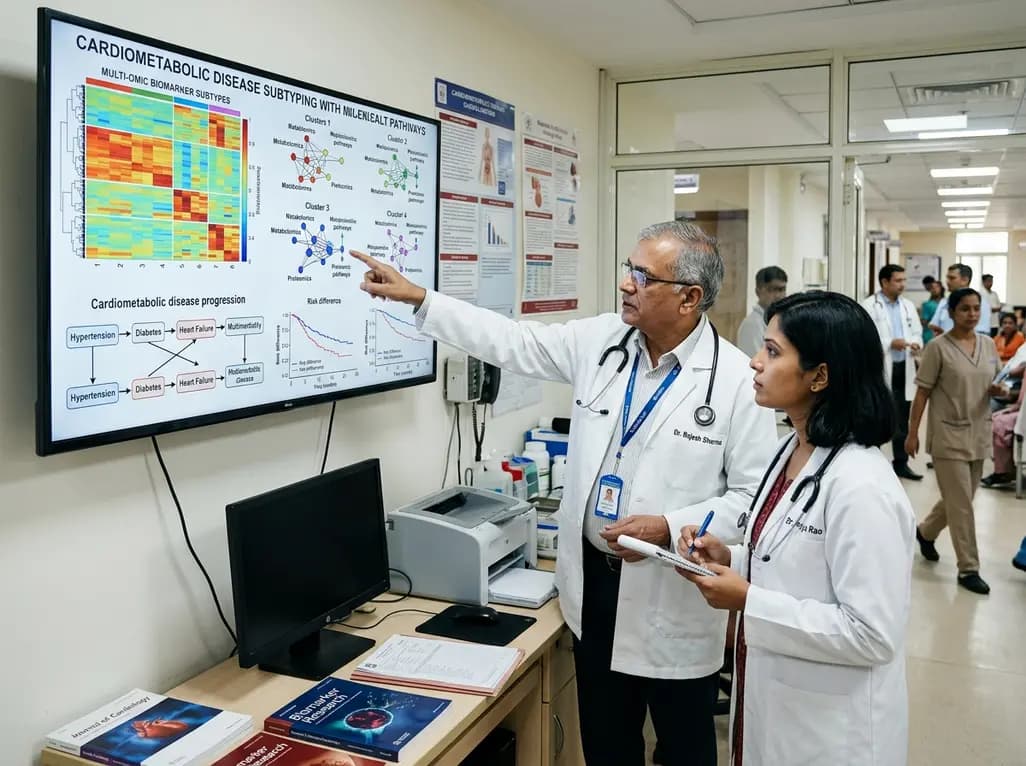
A large UK Biobank study identified four routine biomarker subtypes that predict cardiometabolic disease progression to multimorbidity. Integrating multi-state modelling, metabolomics, and proteomics revealed distinct biological pathways, offering new avenues for tailored clinical risk stratification.
Today
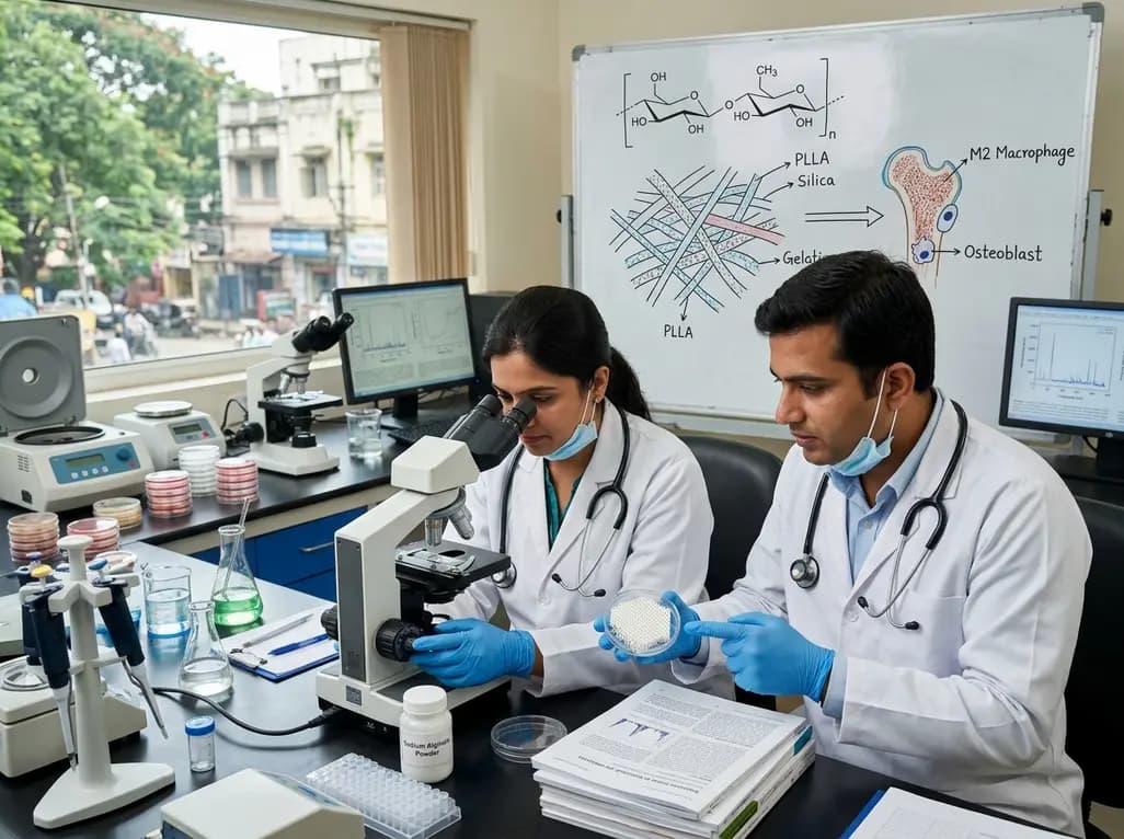
Dual-nanofiber interpenetrating network scaffolds combining silica and PLLA/gelatin nanofibers with sodium alginate achieve complementary mechanical stability, immunomodulatory M2 macrophage polarization, and enhanced osteogenesis for advanced bone repair.
Today

Researchers at IISER Berhampur have engineered a covalent organic framework immunosensor that detects urinary NGAL. This novel nanotechnology platform distinguishes diabetic nephropathy from other renal conditions, enabling rapid, minimally invasive stratification and earlier clinical intervention for patients.
Today
