

Loading, please wait...
Loading, please wait...

Kidney transplantation represents the gold standard definitive therapy for patients with end-stage renal disease. However, unexpected post-transplant surgical complications can rapidly jeopardize both graft function and patient survival. Among these emergencies, kidney allograft rupture stands out as a rare, life-threatening complication that demands immediate clinical recognition and decisive surgical management. Recent comprehensive analyses synthesize decades of evidence to clarify the real-world epidemiology, clinical triggers, and salvage outcomes of this condition. Therefore, nephrologists and transplant surgeons must understand current incidence trends and salvage pathways to optimize recipient care.
Historically, kidney allograft rupture posed a frequent and catastrophic hazard in early renal transplant programs. However, contemporary data demonstrate a striking reduction in its occurrence over recent decades. A comprehensive scoping review evaluated 86 articles comprising 34 consecutive series and 102 individual cases. Across single-center series, the pooled incidence of rupture reached 1.84%, spanning a wide range between 0.25% and 10.75%. Notably, the incidence fell dramatically from 4.7% in studies published before 1990 to 1.5% in modern cohorts.
This three-fold decrease reflects major advances in histocompatibility testing, modern immunosuppressive protocols, and refined surgical techniques. Furthermore, enhanced postoperative surveillance enables clinicians to detect graft dysfunction before extreme parenchymal swelling occurs. In addition, prompt hemodynamic monitoring and prophylactic measures mitigate early vascular complications. Consequently, while kidney allograft rupture remains an acute emergency, transplant centers encounter this event far less often today than in previous eras. Nevertheless, clinicians must maintain vigilance because timely intervention dictates survival.
Understanding the underlying triggers of rupture is essential for timely clinical prevention. Specifically, acute allograft rejection represents the dominant precipitating factor, causing 58% of documented ruptures. During severe acute rejection, intense inflammatory infiltration leads to profound interstitial edema. Because the rigid renal capsule restricts expansion, elevated internal pressure causes the parenchyma to tear along its convex border.
Additionally, transplant renal vein thrombosis accounts for approximately 14% of rupture cases. This venous outflow occlusion produces rapid intrarenal congestion, acute capsular distension, and subsequent mechanical disruption. Furthermore, acute tubular necrosis contributes to roughly 13% of ruptures through severe tissue ischemia and interstitial swelling. Chronic glomerulonephritis and systemic hypertension represented the primary native kidney diseases among affected recipients, whose mean age was 35 years. Moreover, male recipients accounted for nearly two-thirds of reported cases. Therefore, acute vascular congestion and severe immunologic injury remain the central pathophysiological drivers of this complication.
A classic triad of symptoms often heralds the onset of graft rupture during the early postoperative period. Specifically, graft pain or localized tenderness occurs in 88% of patients as the expanding capsule stretches and tears. Furthermore, acute intra-abdominal hemorrhage induces a sudden fall in hemoglobin levels in 62% of individuals. Consequently, hemodynamic instability develops rapidly, manifesting as systemic hypotension in 57% of affected recipients.
In addition to systemic shock, patients frequently experience sudden oliguria, abdominal distension, and visible swelling over the graft site. Clinicians must immediately deploy emergency bedside Doppler ultrasonography when these signs emerge. Ultrasonography typically demonstrates substantial perigraft fluid collections, compressive hematomas, and disrupted cortical continuity. Moreover, Doppler interrogation assesses renal arterial and venous flows to identify underlying vascular occlusion. Laboratory testing concurrently reveals worsening graft function and sudden metabolic acidosis. Therefore, rapid bedside clinical assessment and imaging confirmation remain crucial steps before moving the patient to the operating theatre.
Immediate surgical exploration is mandatory when clinicians suspect acute rupture accompanied by internal hemorrhage. Historically, transplant surgeons routinely performed graft nephrectomy to control bleeding and protect patient life. However, modern surgical practice increasingly emphasizes organ salvage whenever parenchymal integrity permits. Across the individual cases reviewed, surgical teams preserved 48% of ruptured allografts, while 52% required transplant nephrectomy.
Salvage techniques rely on meticulous hemostatic control and parenchymal repair. Specifically, surgeons utilize deep absorbable sutures buttressed with surgical felt, gelatin sponges, or oxidized cellulose. Furthermore, teams apply topical hemostatic sealants, argon beam coagulation, and external mesh wrapping to arrest diffuse bleeding. In selected settings, autologous rectus sheath grafts provide robust mechanical support over large lacerations. Concurrently, nephrologists intensify immunosuppressive therapy with high-dose intravenous corticosteroids or lymphocyte-depleting antibodies to treat underlying rejection. Consequently, conservative surgical salvage effectively stabilizes graft function without compromising recipient safety in appropriately selected candidates.
Patient survival following allograft rupture remains remarkably high despite the severity of the acute hemorrhage. Across analyzed series, patient survival reached 94%, demonstrating the efficacy of rapid resuscitation and emergency surgical intervention. However, long-term allograft survival exhibits significant variability depending on the underlying etiology and the severity of tissue destruction.
Interestingly, the scoping review revealed a critical discrepancy between isolated case reports and systematic cohort data. Standalone case reports documented a graft preservation rate of 62%, creating an overly optimistic impression of salvage success. In contrast, consecutive clinical series reported graft preservation in only 39% of cases. Thus, publication bias in standalone case literature materially overstates the actual likelihood of successful salvage. Overall, aggregate series indicate that roughly 55% of all ruptured grafts are preserved across various institutional settings. Additionally, early graft salvage prevents immediate resumption of dialysis and improves quality of life. Ultimately, transparent reporting from consecutive cohorts provides transplant clinicians with realistic expectations regarding graft recovery and helps guide definitive surgical decisions.
Acute cellular or vascular rejection represents the primary precipitating cause of graft rupture, accounting for nearly sixty percent of cases. Additionally, transplant renal vein thrombosis induces severe venous congestion, which significantly increases intracapsular pressure and causes tissue disruption. Acute tubular necrosis also contributes to graft edema and capsular tearing in early postoperative recovery. Less frequently, severe ischemic injury, trauma, or biopsy site lacerations trigger this critical surgical complication.
Clinicians suspect allograft rupture when patients present with sudden graft tenderness, hypotension, and rapid drops in hemoglobin levels. Consequently, urgent ultrasonography serves as the frontline modality to detect perigraft hematomas, active bleeding, and parenchymal tears. Furthermore, Doppler evaluation assesses renal vascular patency and rules out acute venous thrombosis. Ultimately, exploratory laparotomy provides definitive visual confirmation of capsular rupture while simultaneously allowing surgical control of life-threatening hemorrhage.
Surgeons can successfully salvage ruptured kidney grafts in selected patients who maintain hemodynamic stability. Specifically, operative repair involves direct capsular suturing, hemostatic biomaterials, and compression techniques alongside aggressive antirejection immunosuppressive therapy. However, consecutive cohort studies demonstrate that roughly half of all cases ultimately require transplant nephrectomy. Therefore, prompt surgical decision-making balances organ preservation against the recipient's life, especially when severe tissue destruction or uncontrollable hemorrhage occurs.
Disclaimer: This content is for informational and educational purposes only and should not be taken as professional medical advice. Always seek the guidance of a qualified healthcare provider with any questions you may have regarding a medical condition or treatment. Refer to the latest local and national guidelines for clinical practice.
References

Read summarized clinical updates, watch expert medical content, and earn CME certifications right from your smartphone.
Open your smartphone camera to scan and install.





A scoping review of kidney allograft rupture reveals an incidence drop to 1.5% since 1990. Acute rejection is the primary cause, while approximately half of affected grafts are preserved through surgical salvage.
Today

A Stony Brook University study published in npj Digital Medicine shows physicians preferred AI-generated discharge summaries over human drafts in 95% of complex cases. While large language models enhance accuracy, readability, and completeness, expert clinician verification remains crucial to catch documentation errors.
Today

Delhi is undergoing a major healthcare overhaul with enhanced Ayushman Bharat insurance up to ₹10 lakh, increased MBBS and PG medical training seats, expanded primary network mandirs, new tertiary neuro-ICUs, and public-private reproductive technology initiatives to build a resilient, preventative healthcare model.
Today
Aon forecasts India's healthcare inflation will ease to 10.1% in 2027 from 11.5% in 2026. Despite moderation, healthcare expenses continue to outpace general inflation, propelled by non-communicable diseases, advanced medical therapeutics, and expanding utilization patterns across diverse patient populations.
Today
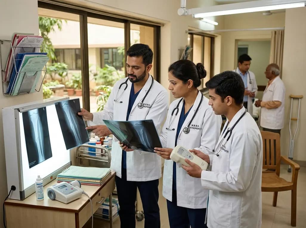
Non-union fractures occur in 2-5% of cases, causing significant disability and cost. Conservative modalities such as LIPUS, PEMF, and focused ESWT offer non-surgical healing options for defects up to 5-10 mm when adequate mechanical stability exists without infection.
Today
Explore how novel oxygen-releasing PVA hydrogels prevent early post-transplant hypoxic damage in pancreatic beta cells, sustaining viability and insulin secretory function.
Today
