

Loading, please wait...
Loading, please wait...

Modern orthopedic care has advanced significantly over recent decades. However, impaired fracture healing remains a major clinical hurdle. Non-union complicates approximately 2% to 5% of all skeletal fractures worldwide. Consequently, patients suffer prolonged pain, functional impairment, and extensive work absence. Although revision surgery represents the traditional standard of care, operative intervention carries inherent risks. Clinicians now evaluate the conservative treatment of non-unions as a viable, limb-preserving alternative that avoids surgical trauma.
Fracture non-union creates substantial biological and socio-economic burdens. When normal fracture healing arrests, physiological repair mechanisms fail to bridge the osseous gap. Most trauma classifications define non-union as persistent fracture mobility and absent radiological progression after several months. In addition, healthcare expenditures escalate rapidly because patients require extended medical rehabilitation and specialized assistive devices.
Biologically, non-unions divide into hypertrophic, oligotrophic, and atrophic variations. Hypertrophic non-unions display abundant callus formation because vascularity remains preserved, yet inadequate mechanical stability prevents terminal consolidation. Conversely, atrophic non-unions lack vital biological activity and cellular ingrowth. In these compromised tissues, microvascular perfusion declines and local mesenchymal stem cells become quiescent. Therefore, restoring osteogenic activity without invasive dissection offers tremendous physiological benefits. Non-operative biophysical therapies address these deficits by reactivating dormant regenerative pathways. Ultimately, targeted conservative interventions stimulate cellular proliferation, encourage local neoangiogenesis, and trigger physiological bone remodeling without introducing surgical morbidity.
Successful conservative management requires rigorous patient triage. Clinicians must confirm that the fracture microenvironment satisfies fundamental mechanical and biological prerequisites. Specifically, non-surgical modalities succeed primarily when bony defect gaps measure between 5 and 10 mm or less. If defect sizes exceed this critical threshold, osteoprogenitor cells cannot bridge the structural void conservatively.
Furthermore, mechanical stability remains an absolute requirement. Existing hardware, intramedullary nails, external casts, or functional braces must stabilize the fracture fragments adequately. Persistent pathological motion shears fragile microvascular sprouts and prevents primary mineral deposition. Moreover, surgeons must definitively exclude active infection before initiating conservative therapies. Septic non-unions consistently require formal debridement, antibiotic eradication, and revision fixation. Clinicians also review patient-specific metabolic factors, including vitamin D status, thyroid function, and glycemic control. Similarly, addressing tobacco use and non-steroidal anti-inflammatory medications enhances the overall probability of non-operative success.
Biophysical stimulation provides non-invasive energy to stimulate dormant bone healing. Low-intensity pulsed ultrasound, commonly known as LIPUS, transmits low-frequency acoustic pressure waves across soft tissue envelopes directly into fracture sites. These mechanical forces stimulate cell surface integrin receptors through mechanotransduction. Consequently, osteoblasts and chondrocytes upregulate vital healing factors, including vascular endothelial growth factor and bone morphogenetic proteins. Patients self-administer LIPUS at home once daily for twenty minutes. The treatment is completely painless and produces negligible soft-tissue heating.
Similarly, pulsed electromagnetic fields (PEMF) deliver low-energy magnetic waves across the fracture gap. These electromagnetic pulses induce physiological electrical currents within extracellular bone matrices. As a result, the therapy activates intracellular calcium-calmodulin pathways and accelerates osteogenic differentiation. Patients wear lightweight PEMF coils over limb casts or braces for several hours each day. Because both LIPUS and PEMF allow convenient home application, patient compliance remains exceptionally high. These non-invasive modalities reliably rescue delayed unions without disrupting daily patient routines.
Focused extracorporeal shockwave therapy (ESWT) delivers high-energy acoustic pressure pulses to recalcitrant bone interfaces. Unlike low-intensity ultrasound, focused shockwaves generate significant focal energy densities that penetrate deep skeletal structures. Consequently, administration requires direct physician supervision within a hospital or clinic setting. Clinicians typically perform the procedure under general anesthesia, regional nerve block, or conscious sedation to prevent patient discomfort.
During the session, the physician utilizes precise fluoroscopic or sonographic guidance to align the shockwave applicator with the non-union cleft. The applied energy produces focused acoustic cavitation and micro-disruptions within sclerotic fracture margins. In response, local tissues release angiogenic signaling cascades and mobilize circulating endothelial progenitor cells. Furthermore, shockwaves convert indolent pseudarthroses into active osteogenic hematomas, effectively restarting the biological repair sequence. Numerous clinical trials demonstrate that focused shockwave therapy achieves radiographic union rates between 70% and 80% in well-selected long bone non-unions. Thus, this single-session or short-course outpatient intervention serves as an exceptionally powerful alternative to complex revision osteosynthesis.
Although conservative therapies provide significant advantages, clinicians must acknowledge their clear limitations. Non-surgical modalities cannot overcome severe mechanical failure or structural instability. If broken implants, loose screws, or profound angular malalignments compromise the skeletal axis, conservative stimulation will fail. Under these biomechanical conditions, immediate surgical revision represents the only dependable solution.
Additionally, critical bone defects exceeding 10 mm demand surgical intervention. Such large segmental gaps require structural bone grafting, autologous cancellous harvest, or distraction osteogenesis. Active osteomyelitis or deep chronic infection also represents an absolute contraindication to closed stimulation. In such cases, surgeons must debride necrotic bone and clear bacterial biofilms before attempting reconstruction. Therefore, multidisciplinary teams must monitor patients closely with sequential radiographs and computed tomography scans. If patients show no clinical or radiographic consolidation after three to six months of biophysical therapy, surgeons should promptly transition to operative stabilization. Ultimately, knowing when to convert from conservative care to surgical reconstruction protects patients from permanent functional deficits.
Clinicians evaluate three critical criteria: mechanical stability, fracture gap size, and absence of infection. The non-union site must exhibit stable fixation without implant failure or significant angular deformity. Furthermore, cross-sectional imaging must confirm a bony defect gap smaller than 5 to 10 millimeters. Finally, laboratory inflammatory markers and soft tissue inspections must definitively rule out local or systemic osteomyelitis.
LIPUS uses painless, low-intensity ultrasound waves that patients self-administer at home for twenty minutes daily over several months. Conversely, focused ESWT delivers high-energy shockwaves concentrated precisely at the fracture site by an orthopedic specialist. Because shockwave pulses generate brief discomfort, physicians administer the therapy under anesthesia or regional blocks. Focused shockwaves usually require only one to three hospital-based sessions.
Physicians typically evaluate bone healing progress using serial radiographs or computed tomography every six to eight weeks. If the fracture demonstrates progressive callus formation and declining tenderness, conservative therapy continues until full consolidation. However, if objective imaging reveals zero healing progress after three to six months, clinicians consider the conservative trial unsuccessful. Consequently, the orthopedic surgeon recommends surgical intervention, including revision fixation and bone grafting.
Disclaimer: This content is for informational and educational purposes only and does not constitute medical advice. Healthcare providers must exercise their own independent clinical judgment when assessing individual patients and formulating diagnostic or treatment plans. Refer to the latest local and national guidelines for clinical practice.
References

Read summarized clinical updates, watch expert medical content, and earn CME certifications right from your smartphone.
Open your smartphone camera to scan and install.





Non-union fractures occur in 2-5% of cases, causing significant disability and cost. Conservative modalities such as LIPUS, PEMF, and focused ESWT offer non-surgical healing options for defects up to 5-10 mm when adequate mechanical stability exists without infection.
Today
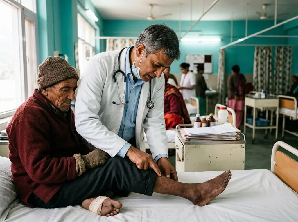
A recent cross-sectional study reveals that prolonged residence at high altitude significantly escalates foot frostbite risk among lowland migrants, with chronic mountain sickness acting as a significant physiological mediator through hyperviscosity and microcirculatory stasis.
Today
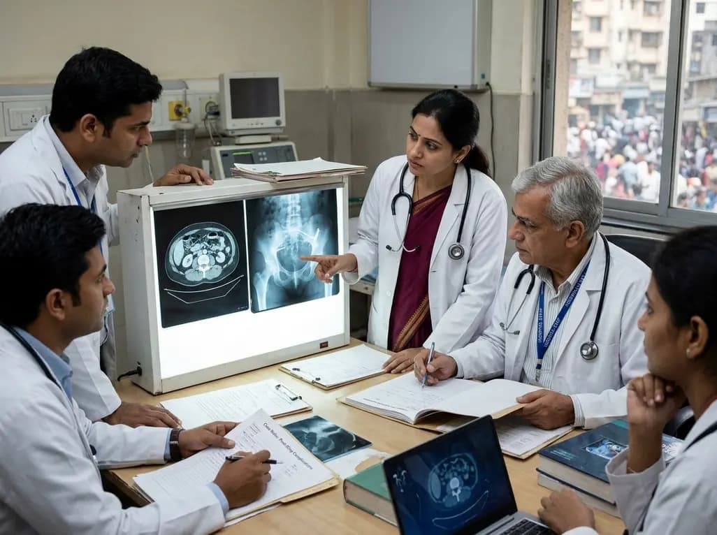
A litigation-derived series of 252 women highlights the risks of repeat revisions after midurethral sling surgery. Recurrent incontinence occurred in up to 60% of patients, and repeat revision rates reached 70%, emphasizing cautious patient selection and skilled multidisciplinary management.
Today
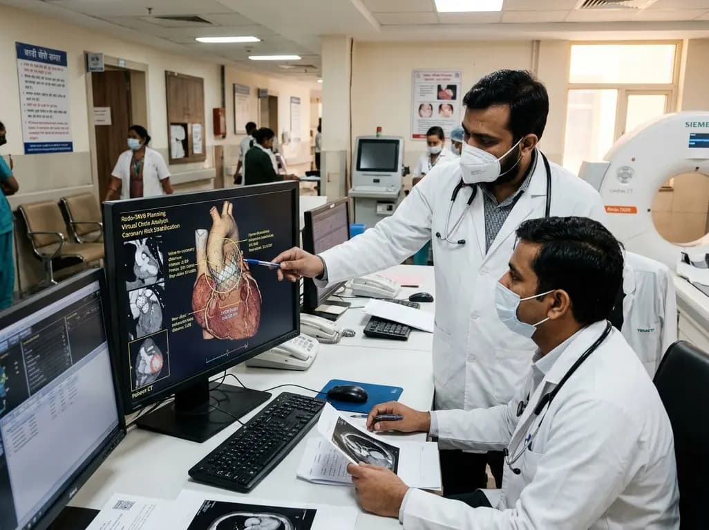
A multicenter study evaluating paired cardiac CT scans reveals that the virtual circle method reliably predicts index transcatheter valve frame expansion and changes in valve-to-aorta distance during redo-TAVR, offering accurate or conservative coronary risk stratification in 85% of cases.
Today
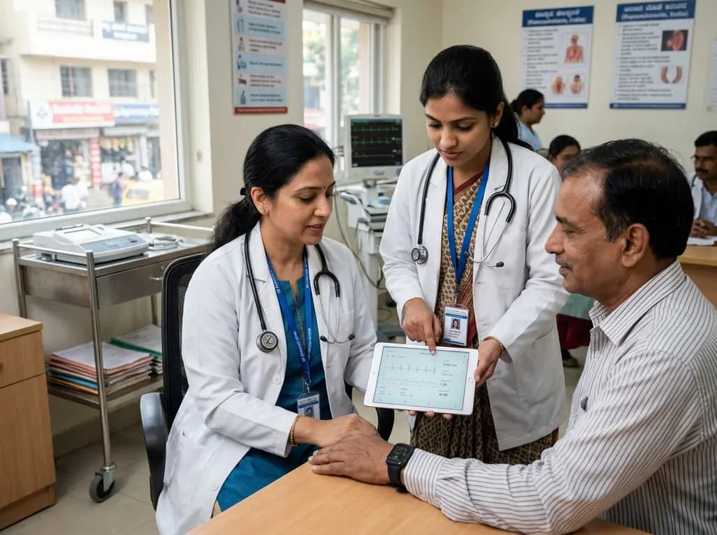
India is poised to transform chronic disease management through wearable health technology. Discover how continuous remote monitoring and predictive algorithms empower clinicians to detect early cardiovascular signals, personalize metabolic interventions, and deliver equitable preventive care across populations.
Today
