

Loading, please wait...
Loading, please wait...

Aortic aneurysmal disease represents a systemic vasculopathy rather than an isolated structural failure. Clinicians frequently encounter complex remodeling across multiple vascular territories. However, comparative evidence regarding intracranial aneurysm risk between thoracic and abdominal aneurysms has historically remained sparse. A landmark nationwide study evaluated 114,805 surgical hospitalizations to address these critical clinical questions. Consequently, the findings provide vital guidance regarding screening strategies and perioperative risk stratification.
Historically, surgeons viewed thoracic and abdominal aneurysms as completely independent vascular pathologies. Nevertheless, emerging epidemiological data suggest shared vulnerabilities throughout the arterial circulation. In this nationwide cohort of 114,805 hospitalizations, researchers compared concurrent cerebral aneurysms across both patient groups. Overall, administratively coded intracranial aneurysms occurred in a modest subset of patients. However, the unadjusted prevalence was tenfold higher in thoracic aneurysms compared to abdominal lesions. Specifically, thoracic patients exhibited a coded frequency of 1.0 percent versus 0.1 percent in abdominal cases. Furthermore, after propensity score matching balanced clinical differences, thoracic disease maintained a significant association with cerebral aneurysms. Therefore, clinicians must recognize that aneurysmal degeneration often extends directly into the intracranial vasculature. Vascular specialists should maintain a high index of suspicion whenever they diagnose proximal aortic enlargement. This perspective shifts patient assessment toward a comprehensive whole-body vascular evaluation. Consequently, identifying concurrent lesions early can fundamentally improve clinical management plans. In addition, early detection allows teams to coordinate multidisciplinary treatment before vascular complications develop.
The anatomical divide between thoracic and abdominal segments mirrors notable developmental and structural variations. Smooth muscle cells within the thoracic aorta originate from the neural crest and second heart field. Conversely, abdominal aortic smooth muscle cells derive primarily from the embryonic paraxial mesoderm. Because of these distinct lineages, thoracic aneurysms frequently reflect intrinsic genetic abnormalities and matrix dysregulation. In contrast, abdominal aortic aneurysms correlate strongly with tobacco exposure, advanced age, and severe atherosclerosis. This developmental dichotomy explains why intracranial aneurysm risk demonstrates a stronger predilection for thoracic cohorts. Nationwide hospital data revealed that thoracic patients presented at younger ages than abdominal aneurysm patients. Furthermore, multivariable regression and machine learning analyses identified female sex and younger age as key predictors. Thus, systemic vascular vulnerability does not distribute equally across all demographic subgroups. Clinicians must integrate these phenotypic nuances when evaluating aneurysmal disease across different arterial beds. Recognizing these risk patterns helps physicians identify candidates who require targeted imaging.
Shared molecular mechanisms directly link thoracic aortic enlargement with intracranial aneurysmal degeneration. Both vascular beds depend on preserved transforming growth factor-beta signaling and intact extracellular matrix scaffolding. Mutations affecting fibrillin, collagen, and contractile vascular smooth muscle elements impair arterial wall homeostasis. Consequently, structural elastolysis reduces vascular compliance and predisposes vessels to focal outward remodeling. In addition, both proximal aortic and cerebral vessels face continuous high pulsatile mechanical stress from ventricular ejection. This relentless hemodynamic shear stress accelerates localized vessel wall fatigue in genetically susceptible individuals. Meanwhile, abdominal aortic aneurysms display extensive transmural inflammation, macrophage infiltration, and severe lipid deposition. Therefore, thoracic aortic walls share far greater histological and biological kinship with the circle of Willis. Syndromic conditions such as Marfan and Loeys-Dietz syndromes highlight this interconnected pathological mechanism. Understanding these shared genetic pathways provides biological plausibility for cross-system vascular surveillance. Furthermore, ongoing genetic discoveries continue to illuminate common molecular drivers across divergent arterial sites.
Beyond anatomical associations, cerebrovascular complications represent a devastating hazard during major aortic repair. Surgical interventions on the thoracic aorta naturally carry substantial neurological perils. Indeed, national registry data demonstrated elevated rates of ischemic stroke and subarachnoid hemorrhage following thoracic repair. Thoracic aneurysm hospitalizations also exhibited significantly higher in-hospital mortality compared to abdominal procedures. Moreover, patients with coexisting intracranial lesions experienced longer hospitalizations and markedly higher healthcare charges. These adverse trends arise because thoracic procedures often involve aortic cross-clamping, hypothermic arrest, or arch reconstruction. Such complex intraoperative maneuvers create severe risks of cerebral hypoperfusion and solid microembolism. Additionally, pre-existing intracranial aneurysms may rupture during sudden hypertensive swings in the perioperative phase. Therefore, anesthesiologists and surgical teams must execute meticulous hemodynamic control throughout the perioperative window. Implementing neuromonitoring and neuroprotective strategies remains essential to reduce perioperative neurological morbidity. Consequently, multidisciplinary vigilance safeguards brain perfusion during high-risk cardiovascular reconstructions. These efforts substantially improve patient survival and optimize resource utilization across intensive care units.
The dramatic variance in intracranial aneurysm frequency carries immediate clinical screening implications. Current cardiovascular guidelines do not recommend universal cerebral imaging for all patients with abdominal aortic aneurysms. The extremely low baseline rate of 0.1 percent in abdominal cohorts confirms that broad screening yields minimal benefit. However, the ten-fold higher prevalence in thoracic cohorts justifies a more vigilant screening strategy. Specifically, vascular teams should consider selective magnetic resonance angiography for high-risk thoracic patients. Key indications include younger age, female sex, known connective tissue disorders, and a family history of aneurysms. Furthermore, identifying unruptured intracranial aneurysms prior to thoracic repair enables precise perioperative planning. Clinicians can tailor blood pressure targets and avoid rapid blood pressure spikes during systemic anticoagulation. In addition, neurointerventional specialists can secure fragile cerebral aneurysms before elective aortic reconstruction begins. Thus, selective neurovascular screening balances preventative clinical benefits against unnecessary healthcare expenditures. This targeted strategy ensures that diagnostic imaging focuses directly on patients facing the greatest risk.
Managing multicentric aneurysmal disease demands seamless collaboration across traditional medical and surgical boundaries. Cardiologists, vascular surgeons, neurologists, and interventional radiologists must coordinate care closely for these complex patients. Because vascular remodeling progresses over time, patients require sustained long-term surveillance rather than isolated procedural care. Physicians should emphasize intensive medical management, including strict blood pressure control and aggressive smoking cessation. Furthermore, modern genetic testing panels can uncover familial mutations that affect multiple family members. Machine learning algorithms and computational modeling also show tremendous promise in forecasting aneurysm growth and rupture. Consequently, innovative technologies will help clinicians anticipate vascular failure before irreversible complications occur. Prospective longitudinal registries are currently needed to refine surveillance intervals and validate risk-scoring tools. Meanwhile, institutional multidisciplinary aortic boards can provide personalized therapeutic recommendations for challenging presentations. By integrating neurovascular and cardiovascular expertise, healthcare systems can optimize outcomes across all vascular territories. Ultimately, proactive systemic vigilance remains the cornerstone of modern vascular disease management.
Thoracic aortic aneurysms and intracranial aneurysms share common embryonic origins and biological mechanisms, including transforming growth factor-beta signaling defects and extracellular matrix fragility. Both vascular territories also experience intense cyclic pulsatile stress. In contrast, abdominal aortic aneurysms arise primarily from acquired atherosclerotic inflammation, resulting in lower rates of concurrent cerebral arterial remodeling.
Routine cerebral screening is not recommended for all abdominal aneurysm patients due to a very low co-occurrence rate of 0.1 percent. However, clinicians should consider targeted magnetic resonance angiography screening for thoracic aortic aneurysm patients, especially younger individuals, women, and those with a family history of aneurysms or connective tissue disorders.
Thoracic aortic repairs carry significantly higher ischemic and hemorrhagic stroke risks than abdominal repairs due to arch vessel manipulation, aortic clamping, and circulatory arrest. Furthermore, preexisting intracranial aneurysms elevate rupture hazards during acute perioperative blood pressure fluctuations, requiring strict neurocritical monitoring and tight hemodynamic control throughout the perioperative period.
Disclaimer: This content is for informational and educational purposes only and should not be considered medical advice. Always consult a qualified healthcare professional before making clinical decisions. Refer to the latest local and national guidelines for clinical practice.
References

Read summarized clinical updates, watch expert medical content, and earn CME certifications right from your smartphone.
Open your smartphone camera to scan and install.





A national analysis of 114,805 hospitalizations reveals that thoracic aortic aneurysms carry a significantly higher intracranial aneurysm risk and greater cerebrovascular morbidity compared to abdominal aortic aneurysms, underscoring the need for selective neurovascular screening.
Today

A validated natural language processing pipeline leverages clinical notes and intermediate annotations to identify risky alcohol use across surgical patients, offering automated preoperative risk stratification.
Today

The GESS-AEEM consensus establishes a clear clinical implementation framework for testosterone therapy in postmenopausal women with hypoactive sexual desire disorder, focusing on transdermal dosing, precise laboratory monitoring, and safety communication.
Yesterday
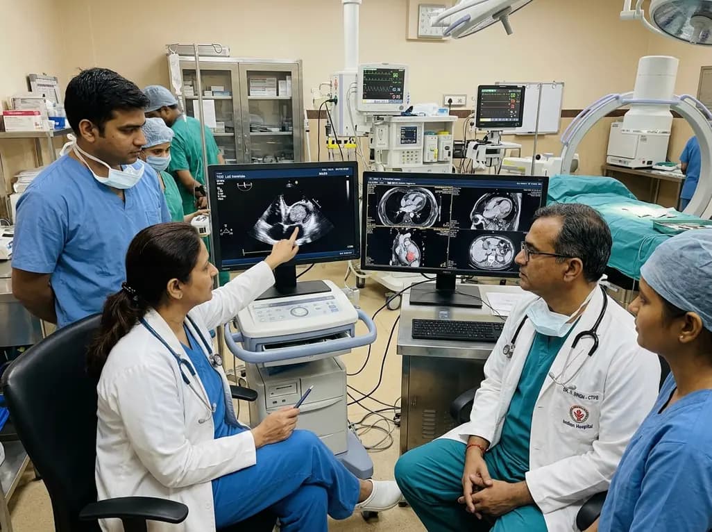
A rare case of an asymptomatic left ventricular hemangioma highlights the vital role of multimodal imaging in differentiating vascular tumors from thrombus. Complete surgical resection under transesophageal echocardiography guidance prevents thromboembolism and delivers optimal outcomes.
Today

A landmark study evaluates long-term quality of life in adolescents and adults with brachial plexus birth injury. Severe functional limitation and limited access to care significantly worsen outcomes, underscoring the urgent need for persistent, multidisciplinary management across adult healthcare transitions.
Today

An analysis of 1,000 posts by macro- and mega-influencers on Instagram and TikTok revealed that 78.5% contained nutrition misinformation, while only 24% of creators held valid credentials. This article examines the clinical risks of viral dietary fads and provides actionable guidance for healthcare professionals.
Today
