

Loading, please wait...
Loading, please wait...

Glioblastoma multiforme remains one of the most aggressive and lethal primary brain malignancies encountered in clinical neuro-oncology. Despite aggressive maximal surgical resection, temozolomide chemotherapy, and radiotherapy, tumor recurrence remains nearly universal. Clinicians continually seek precise biomarkers to forecast therapeutic responses and patient outcomes. Recent breakthroughs highlight the prognostic utility of glioblastoma metabolic signatures derived from distinct intratumoral regions. Specifically, spatial metabolic variations across the tumor architecture provide critical clues regarding disease biology. Understanding these localized metabolic phenotypes allows clinicians to appreciate how cellular adaptation directly governs overall patient survival.
Traditionally, clinicians have classified glioblastoma based on histopathology and distinct genomic alterations, including IDH mutation status and MGMT promoter methylation. However, genomic classifications often fail to explain why patients with identical genetic profiles exhibit divergent clinical survival times. In response to this limitation, spatial metabolomics offers a dynamic view of functional cellular activity. Glioblastoma tumors display profound intratumoral heterogeneity, creating diverse physiological microenvironments within a single lesion. For example, the contrast-enhancing tumor core experiences profound vascular disruption and nutrient fluctuations. In contrast, the surrounding peritumoral brain zone features infiltrating glioma cells embedded among functional neurons and reactive astrocytes. Furthermore, these distinct microenvironments force malignant cells to alter their metabolic programs to survive diverse local stresses. Consequently, evaluating biochemical changes across these distinct anatomical habitats provides vital prognostic information that traditional genomics cannot capture alone. Researchers have demonstrated that spatial metabolic mapping exposes critical metabolic dependencies across different tumor niches. Therefore, examining these metabolic signatures equips neuro-oncologists with a more nuanced physiological roadmap of tumor aggressiveness and clinical course.
A landmark translational investigation by Goodin and colleagues examined whether region-specific metabolic variations directly correlate with patient survival duration. The investigators collected paired surgical biopsies from thirty-seven glioblastoma patients. Specifically, neurosurgeons extracted tissue from both the contrast-enhancing tumor core and the T2/FLAIR hyperintense peritumoral edge. The team then analyzed these samples using high-resolution two-dimensional liquid chromatography-mass spectrometry. Notably, the investigators compared metabolite abundance between patients with short survival, defined as less than or equal to the median, and long survival. The findings revealed striking spatial divergence between these two compartments. Core metabolite levels generally increased in long-term survivors, whereas edge metabolite levels decreased significantly. Even more remarkably, metabolic signatures derived from the peritumoral edge predicted patient survival duration far more accurately than core tissue signatures. Because the peritumoral margin harbors the invasive front responsible for local recurrence, its metabolic status strongly influences clinical prognosis. Consequently, these findings challenge the conventional practice of focusing primarily on the resected tumor core during prognostic biomarker evaluation.
The metabolites that distinguished short-term survivors from long-term survivors mapped directly to crucial biochemical pathways within the Kyoto Encyclopedia of Genes and Genomes database. In particular, the analysis uncovered severe dysregulation in fatty acid synthesis, amino acid catabolism, glycolysis, gluconeogenesis, and mitochondrial ATP synthesis. Highly aggressive tumors in short-term survivors displayed elevated metabolic activity at the peritumoral margin. This hyperactive metabolic state enables invasive glioma cells to rapidly generate biomolecules and cellular energy. For instance, enhanced glycolysis allows infiltrating glioma cells to thrive in hypoxic edge tissue while producing lactate that suppresses local immune defenses. Additionally, altered fatty acid metabolism supports rapid membrane synthesis and promotes therapy resistance in migrating cells. In contrast, long-term survivors exhibited lower metabolite levels in peritumoral edge tissue, indicating a less aggressive invasive periphery. These patients maintained higher core metabolite levels, which may reflect contained metabolic stress within non-disseminating tumor segments. Thus, survival duration depends heavily on whether metabolic hyperactivity remains confined to the core or spreads into the surrounding brain tissue.
To ensure the clinical reliability of these metabolic signatures, the investigators implemented a rigorous machine learning framework. They utilized repeated nested cross-validation alongside independent holdout test set evaluations. As a result, the predictive models avoided common overfitting biases that often hinder translational biomarker discovery. The computational models demonstrated robust accuracy when categorizing patients into short versus long overall survival groups. Furthermore, the machine learning algorithms consistently weighted edge-specific metabolites as the most influential prognostic variables. This automated analytical pipeline proves that localized metabolic changes represent reproducible biological signals rather than random technical noise. Moreover, the integration of advanced bioinformatics with mass spectrometry highlights the growing role of computational oncology in patient care. Clinicians can potentially incorporate these validated predictive models into existing diagnostic workflows to refine risk stratification. In addition, automated algorithms could soon analyze tissue metabolomes within hours after resection. Ultimately, pairing machine learning with spatial metabolomics brings neuro-oncologists closer to genuine precision oncology for glioblastoma management.
These spatial metabolomic insights carry immediate clinical relevance for neurosurgeons, medical oncologists, and radiation oncologists. Currently, standard surgical resection targets the contrast-enhancing tumor core while attempting to spare adjacent functional brain tissue. However, malignant glioma cells routinely infiltrate beyond the surgical margin into the peritumoral edge. Therefore, targeting metabolic vulnerabilities specific to this invasive zone could suppress disease recurrence. For example, pharmacological inhibitors targeting dysregulated fatty acid synthesis or aberrant glutamine pathways could eliminate dormant infiltrative cells. Additionally, radiation oncologists might use advanced magnetic resonance spectroscopic imaging to map high-risk metabolic zones. Consequently, clinicians could adapt radiotherapy margins to deliver targeted boosts to biologically aggressive peritumoral regions. In everyday neuro-oncology practice, recognizing that the peritumoral edge dictates survival will guide the development of regional adjuvant treatments. Furthermore, these regional differences suggest that combination therapies must address distinct metabolic phenotypes simultaneously. In summary, characterizing localized metabolic profiles provides an actionable framework to prolong patient survival and overcome therapeutic resistance.
As multi-omics profiling advances, integrating spatial metabolomics with transcriptomics and proteomics will deepen our understanding of glioma biology. Currently, neuro-oncologists rely heavily on tissue biopsies collected during the initial craniotomy. However, non-invasive liquid biopsies and advanced metabolic neuroimaging could soon track these regional metabolic signatures longitudinally. For instance, tracking altered circulating metabolites in cerebrospinal fluid or blood may signal early tumor progression before conventional magnetic resonance imaging detects structural recurrence. Furthermore, researchers are exploring how metabolic reprogramming influences the glioma microenvironment, particularly antitumor immune surveillance. Notably, immunosuppressive metabolites like lactate and kynurenine impair cytotoxic T-lymphocyte function within the peritumoral boundary. Therefore, combining metabolic inhibitors with immunotherapies could restore immune efficacy and improve therapeutic responses. Ultimately, multicenter clinical trials must validate these regional metabolic signatures across diverse patient populations. Such translational research will ensure that metabolic stratification successfully transitions from academic discovery to bedside clinical practice.
Glioblastoma metabolic signatures represent distinct biochemical profiles of metabolites within distinct tumor compartments. Specifically, these molecular fingerprints reveal how tumor cells process nutrients, alter energy production, and adapt to microenvironmental stressors. Consequently, oncologists can utilize these regional metabolic patterns to stratify survival outcomes and identify actionable biological pathways for individualized therapy.
The peritumoral edge contains actively invading glioma cells that directly interact with adjacent neural parenchyma. Consequently, this dynamic border zone reflects early treatment resistance and tumor recurrence potential. In contrast to necrotic core tissue, metabolic alterations at the invasive edge offer a more accurate physiological indicator of disease progression and overall patient survival duration.
Metabolic profiling can uncover specific enzymatic vulnerabilities in pathways such as amino acid synthesis, glycolysis, and lipid metabolism. Therefore, clinicians can design targeted metabolic inhibitors tailored to residual invasive rim cells. Furthermore, pairing these regional signatures with modern imaging techniques may enhance surgical planning, radiotherapy margins, and longitudinal therapeutic response tracking.
Disclaimer: This content is for informational and educational purposes only. It is not intended to be a substitute for professional medical advice, diagnosis, or treatment. Always seek the advice of your physician or another qualified health provider with any questions you may have regarding a medical condition. Refer to the latest local and national guidelines for clinical practice.
References

Read summarized clinical updates, watch expert medical content, and earn CME certifications right from your smartphone.
Open your smartphone camera to scan and install.





Recent research reveals that glioblastoma metabolic signatures within peritumoral edge tissue accurately reflect patient survival duration. Spatial metabolomics shows distinct dysregulation in glycolysis, fatty acids, and amino acids, opening new avenues for personalized prognostic stratification and targeted therapy.
Today
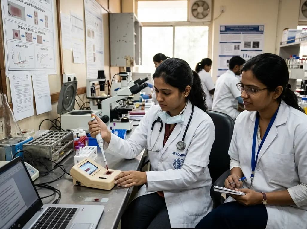
Researchers at IIT Bombay have engineered an automated, cost-effective point-of-care device that diagnoses anaemia and sickle cell disease from a single drop of blood. By measuring blood smear spreading length, this portable tool assesses blood viscosity to offer rapid screening in resource-limited healthcare centers.
Today

A multicenter study evaluates whether standardized simulation training reduces cup detachment during vacuum-assisted vaginal birth. Evidence-based simulation equips obstetricians to improve cup placement, reduce maternal-fetal trauma, and optimize clinical decision-making during operative deliveries.
Today
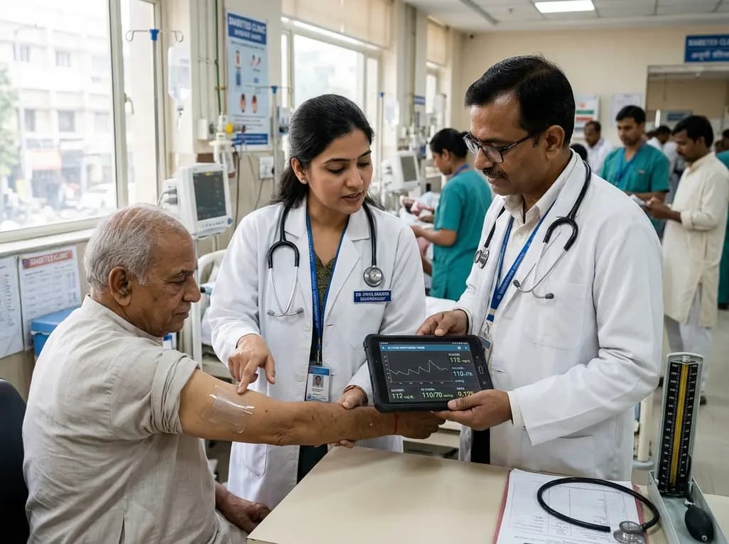
A breakthrough ultrathin multimodal sensor decouples physical hemodynamic monitoring from disposable electrochemical calibration, achieving long-term wearable glucose prediction with 81.36% accuracy and a 9.12% MARD. This offers a robust, drift-free solution for outpatient metabolic surveillance.
Today

A comparative review of tension band wiring and intramedullary screw fixation in thumb metacarpophalangeal arthrodesis, evaluating union rates, hardware complications, and clinical outcomes.
Today

Following regulatory scrutiny from FSSAI regarding promotional representations and lean muscle preservation claims, Dr. Reddy's Laboratories and Nestlé Health Science are currently reviewing the branding and marketing strategy for their joint nutritional supplement Celevida GLP+.
Today
