.webp&w=384&q=75)

Loading, please wait...
Loading, please wait...
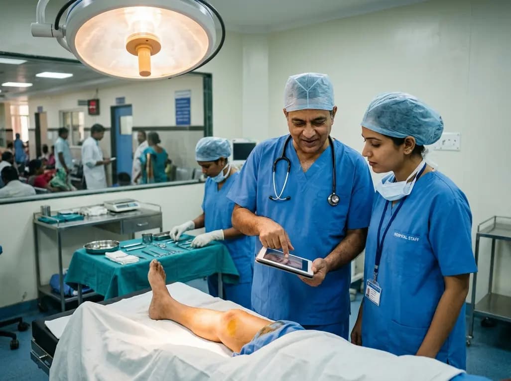
Reconstructive microsurgeons continually seek donor sites that minimize patient morbidity while maximizing long-term outcomes. In patients presenting with chronic lower extremity paresis or plegia, free flap harvesting from affected extremities represents an innovative surgical strategy. Reconstructive teams historically avoided paretic limbs due to theoretical concerns regarding vessel caliber, chronic hypoperfusion, and compromised wound healing. However, emerging clinical evidence demonstrates that neurogenic muscle atrophy does not impair microvascular integrity. By utilizing tissues from an already non-functional extremity, surgeons can spare healthy donor sites. Consequently, this technique preserves the functional capacity of neurologically intact extremities, which remains critical for patient independence.
Selecting an appropriate donor site requires balancing reconstructive tissue requirements against functional donor site morbidity. When patients present with baseline neuromuscular deficits, harvesting tissue from a healthy extremity can severely impair their remaining mobility. For instance, taking an anterolateral thigh flap or a fibular graft from an unaffected leg may compromise independent ambulation or wheelchair transfers. Therefore, performing free flap harvesting from the paralyzed limb shields the functional extremity from unnecessary surgical trauma. Clinicians have successfully applied this approach in patients with poliomyelitis sequelae, spinal cord pathology, and hereditary myopathies. Furthermore, these individuals often depend entirely on their functional limbs to maintain independence in daily activities. Sparing healthy muscle groups and innervation prevents secondary functional decline. Reconstructive teams thus achieve robust soft tissue coverage while respecting the overall functional biomechanics of the patient.
A frequent clinical concern in denervated extremities involves potential microvascular hypoplasia and hemodynamic compromise. Longstanding neuromuscular paralysis causes extensive skeletal muscle atrophy and fibrotic tissue remodeling. However, anatomical investigations confirm that major axial blood vessels and cutaneous perforators remain remarkably patent and reliable. Systematic evaluations reveal that dominant vascular pedicles, including the lateral circumflex femoral system and tibial vessels, maintain adequate luminal diameter. Furthermore, fasciocutaneous and musculocutaneous perforators show caliber and flow velocity comparable to healthy controls. Histological examinations demonstrate intact endothelial structures and normal vascular architecture despite decades of motor denervation. Consequently, microsurgeons can perform microvascular anastomoses using standard microsurgical instruments and techniques. High flap survival rates across published cohorts confirm that denervation does not degrade peripheral tissue perfusion or compromise graft viability.
Surgeons utilize diverse flap configurations from paralyzed donor extremities depending on recipient wound characteristics. The anterolateral thigh flap remains the most frequently selected reconstructive modality due to its versatile design and predictable vascular anatomy. Because skeletal muscle mass diminishes markedly in paretic extremities, harvesting thin fasciocutaneous flaps becomes substantially easier. Additionally, surgeons harvest posterior tibial artery perforator flaps and vascularized fibular osteoseptocutaneous flaps when bony structural support is required. The thin subcutaneous tissue layer in atrophic donor beds provides pliable coverage for complex head, neck, or extremity defects. Reconstructive teams can tailor flap dimensions precisely without encountering excessive muscular bulk. Therefore, paralyzed donor beds offer distinct anatomical advantages for contour matching during complex oncologic and traumatic reconstructions.
Clinical studies indicate that donor site morbidity remains remarkably low when harvesting tissue from paralyzed extremities. Systematic reviews demonstrate an overall flap survival rate approaching one hundred percent across published cohorts. Furthermore, minor postoperative complications remain infrequent and respond well to standard wound care protocols. Reported minor issues primarily include delayed wound healing, localized seroma formation, and superficial infection. Importantly, objective clinical assessments demonstrate zero postoperative decline in baseline donor limb motor function or joint stability. Because the donor extremity already exhibits established neuromuscular deficits, sacrificing non-contractile muscular elements causes no functional loss. Moreover, primary closure of donor sites is frequently easier because diminished muscle volume reduces compartment tension. As a result, healing rates at the donor site remain comparable to standard donor territories.
Rigorous surgical planning remains essential when selecting a paralyzed limb as a reconstructive donor territory. Clinicians must conduct comprehensive preoperative vascular imaging to delineate vascular anatomy and exclude subclinical peripheral vascular disease. Specifically, computed tomography angiography and color duplex ultrasonography provide precise identification of cutaneous perforator trajectories and pedicle lengths. Surgeons should also evaluate skin quality, joint contractures, and chronic trophic changes before operating. In addition, reconstructive teams must account for previous orthopaedic surgeries or chronic pressure areas on the paretic extremity. Intraoperatively, delicate dissection techniques are critical because altered anatomical landmarks require meticulous tissue handling. Consequently, combining rigorous preoperative imaging with refined microvascular technique ensures safe flap harvest and optimal reconstruction.
Chronic motor denervation induces significant skeletal muscle atrophy, but major axial blood vessels and cutaneous perforators typically retain normal anatomical integrity. High-resolution diagnostic imaging confirms adequate vessel caliber and patent vascular lumens comparable to neurologically intact limbs. Consequently, microvascular anastomosis proceeds smoothly without an increased risk of arterial thrombosis or anastomotic failure, provided that systemic peripheral vascular disease is ruled out beforehand.
The anterolateral thigh flap offers exceptional anatomical versatility, consistent pedicle length, and predictable perforator patterns. In paralyzed lower limbs, marked muscle atrophy naturally creates a thinner, more pliable soft tissue paddle. Consequently, reconstructive surgeons can easily contour the flap to reconstruct complex defects without requiring secondary debulking procedures, while preserving functional tissue in healthy extremities.
Published clinical evidence demonstrates exceptionally low donor site morbidity following tissue harvest from paralyzed extremities. The most frequently observed complications are minor and manageable, including delayed wound edge healing, localized seroma formation, and superficial surgical site infections. Importantly, patients experience no postoperative functional decline or loss of baseline mobility, because non-functional tissue is utilized rather than active motor units.
Disclaimer: This content is for informational and educational purposes only. It is not intended to substitute for professional medical advice, diagnosis, or treatment. Always seek the advice of your physician or other qualified health provider with any questions you may have regarding a medical condition or surgical plan. Refer to the latest local and national guidelines for clinical practice.
References

Read summarized clinical updates, watch expert medical content, and earn CME certifications right from your smartphone.
Open your smartphone camera to scan and install.





A systematic review confirms that free flap harvesting from paralyzed limbs is safe, reliable, and preserves vital functional tissue in healthy extremities without increasing donor site morbidity.
last month
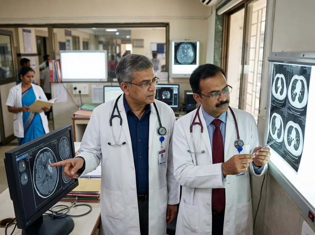
A recent clinical report identifies a novel GDF2 frameshift mutation in a patient with hereditary hemorrhagic telangiectasia presenting with acute cerebral infarction secondary to microscopic pulmonary arteriovenous malformations, emphasizing the necessity of comprehensive screening and genetic evaluation.
Today
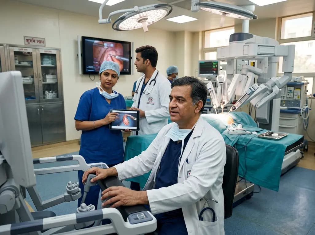
India experiences an exponential rise in robotic-assisted procedures, with Delhi NCR leading system installations and clinical volume. Discover how multidisciplinary surgical adoption, remote telesurgery breakthroughs, and standardized international consensus guidelines are shaping surgical care nationwide.
Today

Radiofrequency catheter ablation of the cavotricuspid isthmus successfully terminates typical atrial flutter. This article reviews the mechanisms driving coronary sinus electrogram changes, wavefront activation pathways, and clinical criteria to verify bidirectional block during electrophysiology studies.
Today

A study comparing four knee flexion strength tests after ACL reconstruction using hamstring tendon autografts reveals marked variability in limb symmetry indices. Clinicians must recognize that different strength testing modalities yield divergent return-to-sport clearance rates.
Today

A breakthrough study leverages natural language processing to accurately identify incident medication-related osteonecrosis of the jaw and track antiresorptive drug holidays from clinical narratives in electronic health records, advancing osteoporosis pharmacovigilance and dental safety.
Today
