.webp&w=384&q=75)

Loading, please wait...
Loading, please wait...

Hereditary hemorrhagic telangiectasia represents a multifaceted genetic vascular disorder characterized by direct arteriovenous shunts. Most patients inherit mutations in classical genes such as ENG or ACVRL1. However, modern genetic sequencing now identifies uncommon pathogenic variations in the GDF2 gene. The GDF2 gene encodes bone morphogenetic protein 9, a circulating ligand that binds endothelial receptors. Consequently, disruption of this critical signaling cascade impairs vascular remodeling and cellular integrity. A recent medical report highlights a 64-year-old female presenting with acute ischemic stroke secondary to microscopic pulmonary malformations. Notably, genetic analysis uncovered a novel heterozygous frameshift mutation in GDF2 that expanded her underlying diagnostic picture. Because her mother experienced similar bleeding episodes, the pedigree confirmed an autosomal dominant inheritance pattern. Furthermore, this discovery underscores that uncommon genetic variations can produce severe vascular phenotypes. Clinicians must therefore recognize that vascular fragility in these patients extends beyond classic presentation lines. In addition, comprehensive molecular panels help identify rare disease alleles that explain complex neurovascular manifestations. As a result, recognizing uncommon variants allows physicians to individualize surveillance strategies across diverse patient populations.
Patients who suffer from hereditary hemorrhagic telangiectasia frequently develop abnormal communications between arteries and veins. In normal pulmonary physiology, the pulmonary capillary bed acts as an efficient physical sieve. Therefore, healthy lung capillaries successfully filter out small venous thromboemboli, bacteria, and particulate debris. In contrast, pulmonary arteriovenous malformations create low-resistance right-to-left conduits that bypass this essential capillary filtration. As a consequence, venous microthrombi can travel directly into systemic arterial circulation and occlude intracranial cerebral arteries. Furthermore, even microscopic vascular shunts that evade standard radiologic detection can still cause catastrophic embolic infarctions. The reported case illustrates this subtle pathological mechanism directly. The patient experienced an acute cerebral infarction despite having subtle microscopic pulmonary vascular alterations. Additionally, chronic hypoxemia and secondary polycythemia often increase baseline blood viscosity in these individuals. This hyperviscosity further compounds the ongoing risk of spontaneous clot formation within sluggish venous circuits. Consequently, clinicians evaluating cryptogenic strokes must investigate hidden pulmonary shunting actively. Identifying these anomalous channels early prevents recurrent neurovascular morbidity and preserves vital neurological function.
Accurate clinical identification begins with the internationally recognized Curaçao criteria. Under these established guidelines, a definitive diagnosis requires at least three distinct clinical manifestations. First, physicians look for spontaneous and recurrent epistaxis occurring without mechanical trauma. Second, clinicians examine mucosal surfaces for multiple mucocutaneous telangiectasias across the oral cavity, lips, and fingertips. Third, specialists evaluate visceral organ involvement involving pulmonary, cerebral, or hepatic vascular architecture. Finally, examiners confirm a positive family history featuring an affected first-degree relative. In the present case report, the patient exhibited classic recurrent epistaxis and distinct cutaneous vascular lesions. Moreover, her mother had a verified personal history of identical hemorrhagic symptoms. Therefore, the patient fulfilled three major criteria, confirming definite hereditary hemorrhagic telangiectasia before genetic validation. However, clinicians often overlook subtle mucosal findings during routine primary care consultations. Consequently, diagnostic delays remain widespread in healthcare settings globally. Physicians must maintain vigilance when evaluating patients with persistent unexplained nosebleeds. Early recognition allows timely intervention, reducing the risk of catastrophic vascular events.
Pulmonary vascular lesions represent a major source of preventable neurological morbidity. Therefore, international consensus guidelines mandate proactive screening for all confirmed or suspected patients. Contrast transthoracic echocardiography serves as the primary noninvasive screening modality of choice. Specifically, clinicians administer agitated saline intravenously while tracking bubble transit through the cardiac chambers. If microbubbles appear in the left atrium after three cardiac cycles, the test confirms intrapulmonary right-to-left shunting. Furthermore, chest computed tomography provides high-resolution anatomical visualization of discrete arteriovenous nidi and feeding vessels. However, microscopic pulmonary malformations may produce significant physiological shunts without forming distinct macroscopic nodules. In such instances, clinicians must combine physiological gas exchange measurements with contrast studies to confirm shunting. In addition, serial pulse oximetry in supine and standing positions helps identify orthodeoxia. Detecting these occult pulmonary shunts enables targeted therapeutic planning before neurological deficits emerge. Consequently, structured screening protocols prevent life-threatening complications, including brain abscesses and ischemic strokes. Periodic surveillance ensures that physicians detect evolving vascular lesions before irreversible ischemic injury occurs.
Effective management requires coordinated intervention across multiple specialty disciplines. Interventional radiologists typically treat macroscopic pulmonary arteriovenous malformations using transcatheter embolotherapy. By deploying mechanical coils or vascular plugs into feeding arteries, clinicians successfully close high-flow shunts. Consequently, embolization substantially lowers lifetime risks of paradoxical cerebral embolism and intracranial abscess. However, microscopic vascular channels that lack discrete arterial feeders require medical and preventive strategies instead. Specifically, healthcare providers advise strict prophylactic antibiotic regimens before dental or invasive procedures. This practice prevents transient bacteremia from causing cerebral infections across pulmonary shunts. Additionally, clinicians must manage epistaxis through topical humidification, hemostatic therapies, or local cautery. When severe mucocutaneous bleeding causes profound iron deficiency, intravenous iron supplementation becomes essential. Moreover, antiangiogenic medications, such as systemic bevacizumab, offer substantial therapeutic promise for refractory bleeding. Ultimately, personalized medical care and regular clinical follow-up minimize vascular complications significantly. Regular communication between pulmonologists, neurologists, and geneticists ensures optimal long-term outcomes for affected patients.
The GDF2 gene encodes bone morphogenetic protein 9, an essential circulating ligand for the ALK1 endothelial receptor complex. When pathogenic variants disrupt this signaling cascade, normal vascular remodeling fails. Consequently, fragile capillaries and abnormal arteriovenous malformations develop across multiple organ systems. Although GDF2 mutations account for only a small percentage of cases, identifying them confirms the genetic diagnosis, guides family screening, and clarifies clinical vascular risks.
Microscopic pulmonary arteriovenous malformations establish direct, low-resistance shunts between pulmonary arteries and veins that bypass normal capillary filters. Consequently, systemic venous microthrombi evade capillary entrapment and enter arterial circulation directly. These paradoxical emboli can travel swiftly into cerebral arteries, causing acute ischemic stroke. In addition, chronic intrapulmonary shunting often induces hypoxemia and secondary erythrocytosis, further elevating thrombosis risks within sluggish venous vascular channels.
Clinicians should screen every patient with confirmed or suspected disease using transthoracic contrast echocardiography with agitated saline. The delayed appearance of microbubbles in the left atrium confirms an intrapulmonary right-to-left shunt. If testing demonstrates significant shunting, physicians order high-resolution thoracic computed tomography to evaluate vascular architecture. Additionally, specialists measure arterial oxygenation in supine and upright postures to detect orthodeoxia and quantify physiological shunting accurately.
Disclaimer: This content is for informational and educational purposes only... Refer to the latest local and national guidelines for clinical practice.
References

Read summarized clinical updates, watch expert medical content, and earn CME certifications right from your smartphone.
Open your smartphone camera to scan and install.





A recent clinical report identifies a novel GDF2 frameshift mutation in a patient with hereditary hemorrhagic telangiectasia presenting with acute cerebral infarction secondary to microscopic pulmonary arteriovenous malformations, emphasizing the necessity of comprehensive screening and genetic evaluation.
Today
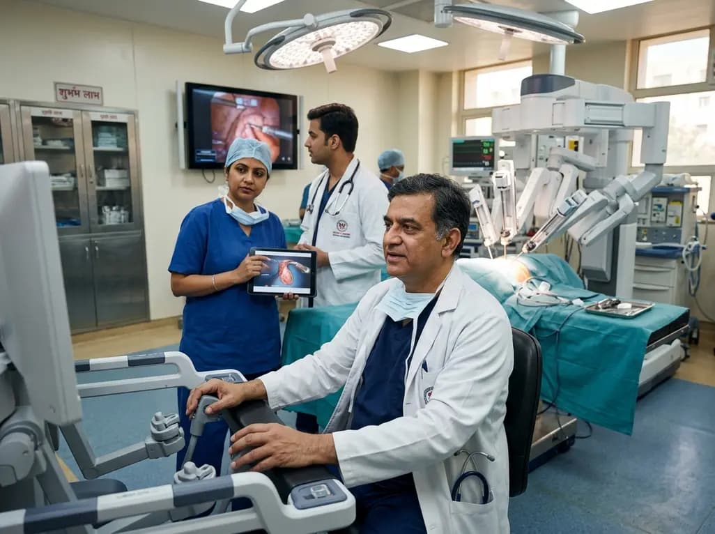
India experiences an exponential rise in robotic-assisted procedures, with Delhi NCR leading system installations and clinical volume. Discover how multidisciplinary surgical adoption, remote telesurgery breakthroughs, and standardized international consensus guidelines are shaping surgical care nationwide.
Today

Radiofrequency catheter ablation of the cavotricuspid isthmus successfully terminates typical atrial flutter. This article reviews the mechanisms driving coronary sinus electrogram changes, wavefront activation pathways, and clinical criteria to verify bidirectional block during electrophysiology studies.
Today
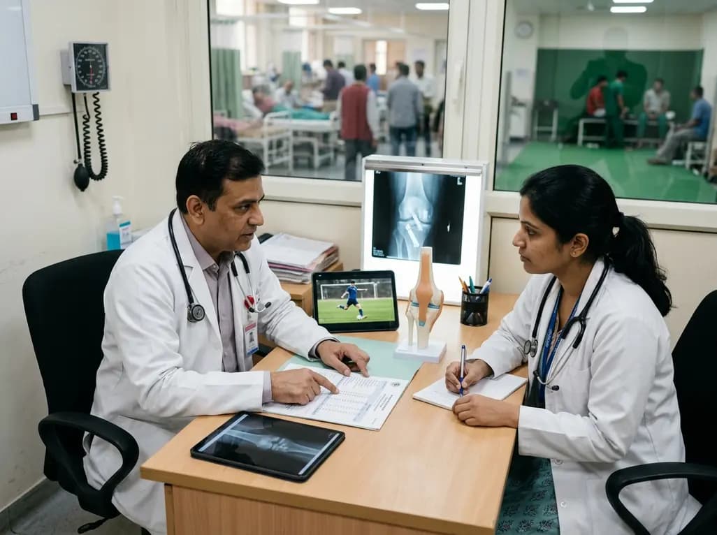
A media-based study of 278 European professional football players finds no clear association between time to return-to-play after ACL reconstruction and the hazard of a first additional injury or matches missed, reinforcing the need for criteria-based clearance over calendar timelines.
Today

A breakthrough study leverages natural language processing to accurately identify incident medication-related osteonecrosis of the jaw and track antiresorptive drug holidays from clinical narratives in electronic health records, advancing osteoporosis pharmacovigilance and dental safety.
Today
