.webp&w=384&q=75)

Loading, please wait...
Loading, please wait...

Gliomas remain among the most formidable therapeutic challenges in modern neuro-oncology. Diffuse infiltrative growth, profound intratumoral heterogeneity, and robust immunosuppression complicate patient care. Moreover, the blood-brain barrier severely restricts drug delivery and obstructs the shed of diagnostic molecules into peripheral blood. Consequently, establishing noninvasive diagnostics has stalled. Innovative integration of focused ultrasound liquid biopsy provides a breakthrough approach to overcome this physical impediment. Clinicians now have unprecedented opportunities to sample intracranial biology safely and systematically.
Malignant gliomas, particularly glioblastomas, display marked cellular heterogeneity and aggressive local infiltration. Neurosurgeons frequently encounter infiltrative neoplastic margins that blend indistinguishably into eloquent brain regions. Therefore, complete microscopic surgical resection remains virtually impossible in routine practice. In addition, systemic chemotherapy frequently fails to reach cytotoxic concentrations within infiltrating margins. This failure stems directly from the tight junctions of the neurovascular unit, commonly called the blood-brain barrier.
Furthermore, standard clinical surveillance relies almost entirely on serial neuroimaging. Magnetic resonance imaging identifies structural changes but frequently fails to separate true tumor recurrence from treatment-induced inflammation. Radiation necrosis and pseudoprogression mimic aggressive disease progression on routine scans. Stereotactic tissue re-biopsy carries distinct procedural risks, including intracranial hemorrhage and persistent neurological deficits. Hence, clinicians need reliable molecular surrogates to detect residual clonal disease early. Peripheral blood sampling provides an attractive alternative, yet intact cerebral vasculature prevents substantial tumor DNA from entering peripheral circulation.
Low-intensity focused ultrasound combined with intravascular microbubbles presents a noninvasive solution to this biological impasse. When ultrasound acoustic waves hit circulating microbubbles, they induce stable volumetric oscillations known as stable cavitation. Consequently, this mechanical vibration exerts gentle shear stress on brain endothelial cells. This physical force temporarily disassembles tight junction proteins, including claudin-5, occludin, and zonula occludens-1. As a result, the vessel wall becomes permeable for several hours.
Importantly, this bioacoustic effect creates a bidirectional conduit across the previously impenetrable blood-brain barrier. Systemic chemotherapeutic agents and targeted antibodies can enter the tumor microenvironment with greater bioavailability. Simultaneously, tumor-derived molecules escape from the interstitial parenchymal space directly into systemic circulation. Therefore, focused ultrasound liquid biopsy transforms an inaccessible intracranial compartment into an accessible peripheral biomarker source. Patients avoid repeating invasive cranial surgeries while neuro-oncologists acquire real-time access to evolving neoplastic genomics.
Preclinical and early human studies confirm substantial increases in peripheral tumor biomarkers following focused acoustic disruption. For instance, post-sonication plasma samples exhibit pronounced surges in cell-free tumor DNA, microRNAs, and intact extracellular vesicles. Circulating tumor cells also enter the vascular stream in measurable quantities. Consequently, laboratory platforms can isolate glioblastoma-specific mutations, including telomerase reverse transcriptase promoter variants, isocitrate dehydrogenase alterations, and epidermal growth factor receptor amplifications.
Moreover, extracellular vesicles harvested after acoustic opening carry preserved transmembrane surface proteins and noncoding transcriptomes. Novel microfluidic isolation chips readily capture these nanoscale vesicles from standard phlebotomy specimens. Researchers can therefore monitor downstream metabolic adaptations and transcriptional remodeling over time. In contrast to static tissue specimens taken during index resection, sonopermeation-enabled blood sampling captures ongoing temporal clonal shifts. As a result, clinicians can identify therapeutic resistance weeks or months before gross macroscopic changes appear on standard neuroimaging scans.
Sonication protocols generate complex multidimensional datasets encompassing acoustic parameters, longitudinal sequencing reads, and functional magnetic resonance sequences. Manual interpretation of these vast registries presents significant cognitive and logistical hurdles. Therefore, artificial intelligence algorithms have become indispensable computational tools. Machine learning models systematically synthesize acoustic cavitation signatures alongside temporal biomarker surges. This integration ensures personalized acoustic energy calibration while maintaining high safety margins.
Additionally, deep learning architectures correlate liquid biomarker dynamics with advanced radiomic features. Algorithms evaluate post-treatment fluid changes against perfusion-weighted imaging metrics to eliminate ambiguities surrounding pseudoprogression. Neural networks also analyze fragmented plasma DNA methylation signatures to project overall survival outcomes. Ultimately, artificial intelligence pipelines translate dense biophysical and genomic profiles into actionable therapeutic recommendations. Clinicians can rapidly determine whether an ongoing systemic regimen successfully controls active subclonal expansion.
Despite promising exploratory trials, widespread clinical adoption requires clearing substantial technological and logistical hurdles. First, optimal acoustic parameter standardization remains variable across clinical devices. Implantable transducer arrays provide dependable repeated access, whereas transcranial stereotactic systems offer noninvasive flexibility. Each platform alters vascular permeability through differing acoustic spectra and pressure distributions. Therefore, multi-center trials must validate standardized dosing schemes to ensure consistent, reproducible biomarker release across varied brain regions.
Second, laboratory assay thresholds require higher analytical sensitivity. Peripheral clearance mechanisms rapidly eliminate cell-free tumor fragments, creating a narrow temporal window for blood collection. Clinicians must standardize phlebotomy timing, usually within thirty to ninety minutes following sonication. Furthermore, resource constraints in developing health ecosystems present meaningful access boundaries. Advanced focused ultrasound installations require major capital investment and multidisciplinary infrastructure. Establishing multi-institutional registries and clear clinical trial frameworks will ultimately allow global neuro-oncology teams to standardize this innovative paradigm.
Low-intensity focused ultrasound interacts with circulating intravenous microbubbles to cause stable acoustic cavitation. This mechanical motion transiently opens tight junctions within cerebral capillary endothelial cells. As the blood-brain barrier permeability increases, tumor-derived molecules like cell-free DNA, messenger RNA, and extracellular vesicles leak into systemic blood vessels. Phlebotomy performed shortly after sonication allows clinicians to harvest high-quality genetic biomarkers without invasive neurosurgical procedures.
Yes, acoustic liquid biopsy offers substantial diagnostic value in distinguishing true tumor growth from inflammatory pseudoprogression. Routine neuroimaging frequently yields equivocal contrast enhancement patterns following concurrent radiation and temozolomide chemotherapy. By measuring rapid changes in circulating glioma-derived mutations or tumor-specific extracellular vesicles after acoustic opening, oncologists can objectively verify biological tumor proliferation and alter systemic interventions appropriately.
Key translation hurdles include determining optimal sonication parameters, harmonizing specialized blood sampling intervals, and establishing assay analytical sensitivities. Furthermore, specialized hardware requires significant financial capital and rigorous operator training. Large-scale randomized clinical trials must continue validating how biomarker-guided treatment adjustments directly improve overall progression-free survival before regulatory agencies approve the technology for standard clinical practice.
Disclaimer: This content is for informational and educational purposes only... Refer to the latest local and national guidelines for clinical practice.
References
Liu CJ et al. Converging Focused Ultrasound and Liquid Biopsy for Glioma Management: Next Steps. Hematol Oncol Clin North Am. 2026 Oct 09. doi: undefined. PMID: 42855393.
Sonabend AM, Youngblood M, Kumari A, et al. Focused ultrasound-enabled liquid biopsy for monitoring glioblastoma response to therapy. Nat Commun. 2026;17(1):112-124.
Meng Y, Pople CB, Lea-Banks H, et al. MRI-guided focused ultrasound liquid biopsy enriches circulating biomarkers in patients with brain tumors. Neuro Oncol. 2021;23(10):1789-1797.

Read summarized clinical updates, watch expert medical content, and earn CME certifications right from your smartphone.
Open your smartphone camera to scan and install.





Integrating focused ultrasound with liquid biopsy offers a noninvasive paradigm for glioma management. By transiently disrupting the blood-brain barrier, clinicians can harvest circulating tumor biomarkers, assess treatment response early, and guide personalized neuro-oncological interventions.
Today
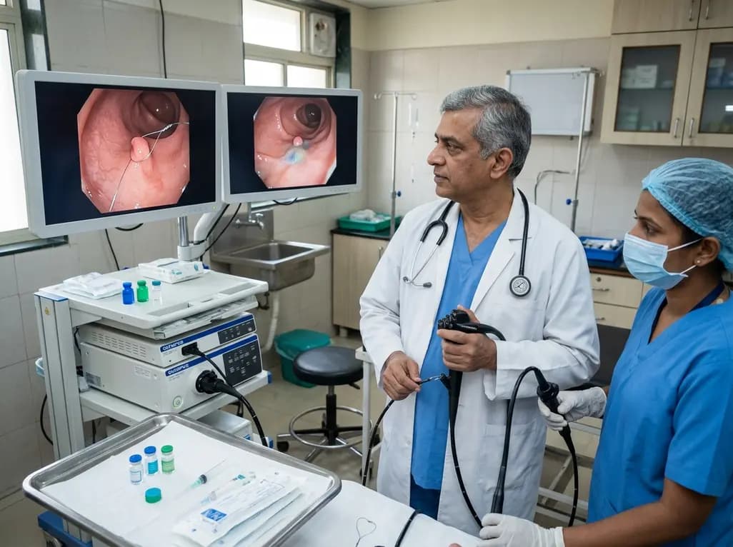
A randomized controlled trial compared conventional cold snare polypectomy with submucosal injection for 3-9 mm polyps. Both techniques demonstrated exceptionally low incomplete resection rates, though injection lowered the need to convert to endoscopic mucosal resection when forced pull was avoided.
Today

Maternal anaemia contributes to nearly half of pregnancy-related fatalities across India. To prevent complications, Uttar Pradesh has added single-dose parenteral iron to its essential medicines list. This clinical transition aims to restore haemoglobin rapidly and protect high-risk mothers across rural facilities.
Today
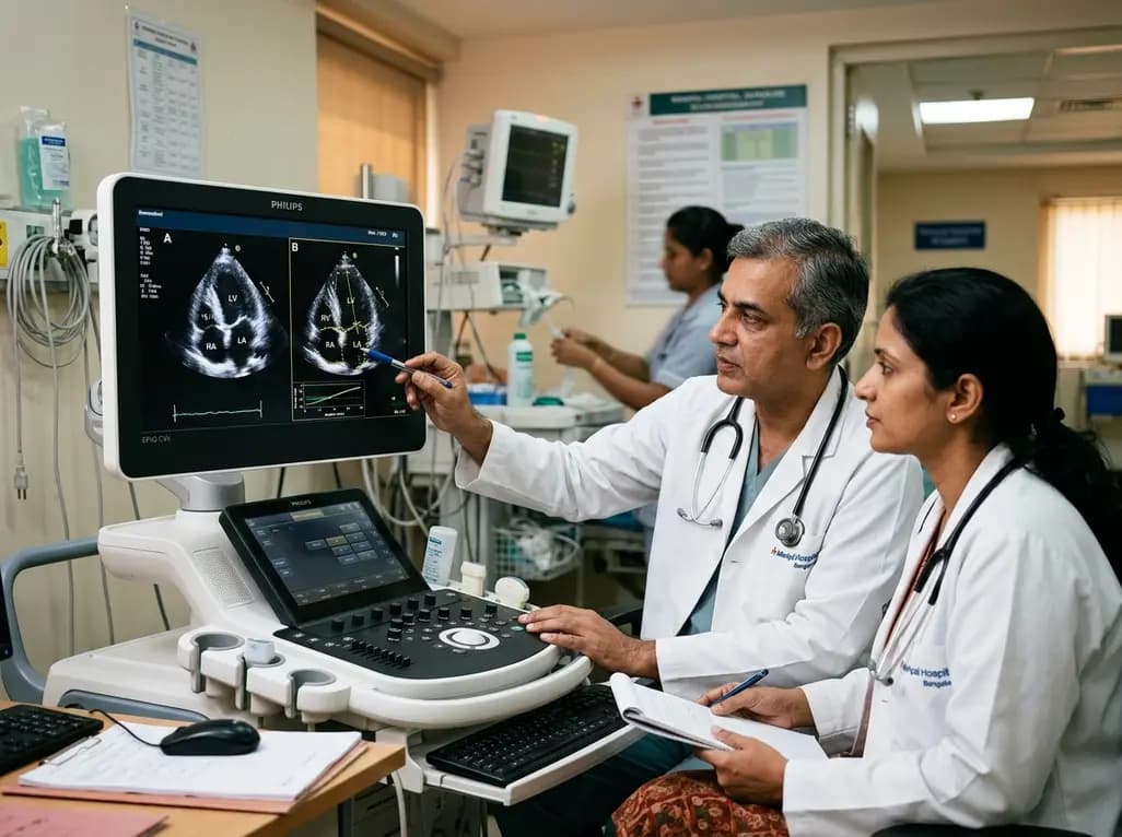
A multi-expert study shows pairwise deep learning accurately assesses echocardiographic rotational alignment in apical four-chamber views, outperforming pointwise regression. This technique improves automated image quality control and standardizes functional cardiac assessment.
Today

A longitudinal study of 1,055 chronic low back pain patients reveals that higher patient activation fosters constructive pain medication attitudes, reduces disability, and decreases short- and long-term opioid use through collaborative, multimodal care.
Today

A landmark cross-sectional study in Asian adults with type 2 diabetes identifies novel urinary metabolites linked to mild cognitive impairment. Elevated urinary sucrose and altered metabolites highlight non-invasive pathways for early cognitive risk stratification and targeted metabolic management.
Today
