

Loading, please wait...
Loading, please wait...

Brain-computer interface technology establishes a direct, bidirectional communication conduit between cerebral neural circuits and external computational hardware. In clinical neurotechnology, flexible BCI electrodes have emerged as transformative tools designed to overcome the historical limitations of rigid silicon or metal microelectrodes. Neurologists and neurosurgeons increasingly recognize that chronic interface stability depends fundamentally on mechanical compliance with delicate cerebral parenchyma. Consequently, material scientists and biomedical engineers are developing compliant interfaces that capture high-fidelity signals without inducing destructive chronic tissue reactions.
Traditional neural implants rely on rigid metals and silicon shanks. However, these stiff substrates possess a Young's modulus several orders of magnitude higher than soft cerebral tissue. Brain parenchyma exhibits an elastic modulus typically between one and ten kilopascals. In contrast, standard silicon probes possess moduli exceeding one hundred gigapascals. This stark biomechanical mismatch induces chronic shear strain during physiological brain micromotion, arterial pulsation, and patient movement. Consequently, mechanical shearing damages surrounding neural microvasculature and triggers robust neuroinflammatory cascades. Activated microglia and reactive astrocytes encapsulate the rigid shanks within a dense, non-conductive glial scar. Over time, this biological encapsulation increases electrical impedance and dramatically degrades the signal-to-noise ratio. As a result, chronic recordings often fail within several months. Flexible BCI electrodes eliminate this mismatch by matching tissue compliance. By utilizing biocompatible polymers, these soft interfaces conform smoothly to curvilinear cortical surfaces. Thus, flexible architectures significantly attenuate microglial activation, minimize neuronal loss around the implant, and preserve clear electrophysiological recording channels over sustained implantation periods.
Engineers utilize several sophisticated fabrication methodologies to construct compliant neural arrays. Microelectromechanical systems lithography represents the traditional gold standard, enabling precise microscale patterning of thin-film polymeric substrates. For example, microfabrication facilities routinely utilize polyimide, parylene-C, and polydimethylsiloxane to construct multi-layered electrode ribbons. Furthermore, high-resolution printing and transfer techniques have gained substantial clinical interest. Inkjet printing and transfer methods allow rapid prototyping of complex conductive pathways directly onto ultrathin elastomeric membranes. In addition, researchers increasingly deploy laser micromachining and thermal drawing processes to create high-density, multi-material fiber probes. Thermal drawing combines conductive cores and insulating cladding into flexible microscale fibers capable of simultaneous recording and optical stimulation. Meanwhile, electrospinning methods produce fibrous, biomimetic scaffolds that mimic the extracellular matrix. These porous electrospun meshes promote seamless cell adhesion and direct neurite outgrowth. Consequently, these microfabrication paradigms provide clinicians with tailored tools that accommodate varied surgical approaches across diverse neural monitoring paradigms.
Substrate flexibility alone cannot guarantee exceptional recording fidelity; low electrochemical impedance is equally essential. Because microscale recording sites exhibit very small geometric surface areas, their baseline electrical impedance remains inherently high. Therefore, surface modification and nanomaterial deposition strategies are indispensable for lowering interface resistance. Investigators routinely deposit conductive polymers, such as poly(3,4-ethylenedioxythiophene) doped with polystyrene sulfonate, onto microelectrode sites. These conductive polymer coatings expand the effective electrochemical surface area and facilitate efficient capacitive charge transfer. Similarly, clinicians benefit from arrays modified with platinum nanoparticles, carbon nanotubes, or graphene flakes. These nanostructured interfaces substantially reduce thermal noise and boost charge-injection capacity. Moreover, biochemical surface functionalization provides critical anti-inflammatory protection. Researchers incorporate anti-inflammatory agents, such as dexamethasone, or tether bioactive neural adhesion peptides onto electrode surfaces. These bioactive modifications actively suppress local foreign body reactions, prevent biofouling, and preserve local neuronal viability. Consequently, optimizing surface chemistry and nanoscale topography ensures stable, long-term electrophysiological signal acquisition with high signal-to-noise ratios.
Flexible neural interfaces exhibit remarkable clinical versatility across invasive, semi-invasive, and non-invasive medical domains. In invasive intracortical applications, ultra-flexible microwires and mesh electronics enter the cortex via transient stiffening shuttles or microfluidic delivery systems. Once deployed, these microscopic ribbons interweave with neuropil, enabling single-unit action potential recordings without disrupting microvascular integrity. Semi-invasive configurations, particularly flexible micro-electrocorticography grids, rest directly upon the subdural or epidural cortex. These high-density arrays record broad local field potentials with outstanding spatial resolution. Consequently, they prove invaluable during epilepsy resective surgery and awake craniotomy speech mapping. Furthermore, non-invasive flexible interfaces are revolutionizing scalp electroencephalography. Traditional scalp monitoring mandates abrasive skin preparation and messy electrolytic pastes that dry out and degrade signal quality rapidly. In contrast, modern epidermal electronic tattoos and breathable dry elastomeric electrodes conform intimately to skin micro-contours. These soft epidermal sensors minimize motion artifacts and maintain low contact impedance over prolonged sessions. Thus, flexible architectures expand translational monitoring capabilities across acute neurosurgical procedures, long-term neurointensive care monitoring, and outpatient motor rehabilitation programs.
Despite rapid preclinical breakthroughs, several technical obstacles impede widespread clinical adoption. Large-scale manufacturing consistency remains a primary engineering bottleneck. Fabricating defect-free, high-channel-count flexible ribbons requires stringent cleanroom quality control to prevent moisture delamination and pinhole insulation breakdown. Furthermore, delivering ultra-floppy microelectrodes precisely into deep brain targets poses ongoing neurosurgical challenges. To address this, surgical teams utilize biodegradable polymer shuttles, such as silk fibroin or gelatin needles. These temporary guides provide necessary stiffness during cortical insertion and rapidly dissolve in vivo. Looking forward, the next generation of neural technology will integrate fully autonomous, self-powered, and wireless architectures. Onboard application-specific integrated circuits will process neural signals directly at the interface, thereby eliminating cumbersome transcutaneous trans-dural percutaneous wiring. Additionally, machine learning algorithms embedded in neuromorphic edge hardware will enable real-time, closed-loop neuromodulation. These systems hold vast therapeutic promise for treatment-resistant epilepsy, neuropsychiatric illness, and spinal cord injuries. Consequently, sustained collaboration between neurosurgeons, biomedical engineers, and regulatory agencies will soon accelerate these soft bioelectronic tools toward mainstream clinical application.
Traditional rigid shanks possess high mechanical stiffness, which clashes with natural pulsatile brain micromotion. This continuous mechanical friction damages delicate cerebral microvessels and triggers persistent microglial activation, leading to thick glial scarring. Conversely, flexible electrodes match the soft Young's modulus of cerebral parenchyma. By eliminating chronic shear strain and tissue tethering, flexible substrates dramatically minimize foreign body reactions, suppress neuroinflammatory cascades, and maintain healthy neuronal density adjacent to the recording sites.
Because flexible microelectrodes lack structural rigidity, they easily buckle upon contacting the brain surface. Clinicians overcome this physical limitation using temporary stiffening strategies. Engineers embed flexible microfilaments inside dissolvable, biocompatible matrices like silk fibroin, gelatin, or polyethylene glycol. These polymer shuttles provide adequate insertion force to penetrate pia mater cleanly. Once implanted, body fluids dissolve the temporary coating within minutes, leaving only the ultra-compliant, flexible electrode array safely integrated within target neural structures.
Flexible micro-electrocorticography arrays conform seamlessly across complex gyral and sulcal cortical topologies without applying focal pressure on underlying brain tissue. Furthermore, their high electrode packing density records local field potentials with exceptional spatial and temporal precision. Intraoperatively, this fidelity enables surgical teams to map epileptogenic foci and functional motor or language boundaries accurately. Consequently, surgeons can maximize pathological resection margins while preserving critical eloquent brain areas during awake craniotomy or epilepsy surgical procedures.
Disclaimer: This content is for informational and educational purposes only. It is not intended to be a substitute for professional medical advice, diagnosis, or treatment. Always seek the advice of your physician or other qualified health provider with any questions you may have regarding a medical condition. Refer to the latest local and national guidelines for clinical practice.
References

Read summarized clinical updates, watch expert medical content, and earn CME certifications right from your smartphone.
Open your smartphone camera to scan and install.





Flexible BCI electrodes offer enhanced mechanical compatibility, lower immune reactivity, and chronic signal stability over traditional rigid arrays. This review outlines modern microfabrication, interface optimization, and translational prospects in clinical neurology and neurosurgery.
Today
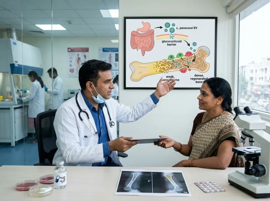
A novel oral nanoplatform using Lacticaseibacillus paracasei-derived extracellular vesicles delivers alendronate, reversing glucocorticoid-induced bone loss and restoring gut-immune-bone homeostasis.
Today

A study in JAMIA Open shows lightweight open-source reasoning LLMs achieve up to 95% sensitivity and accelerate evidence synthesis screening by nearly twentyfold, presenting a cost-effective, scalable AI solution for biomedical research and systematic reviews.
Yesterday

A landmark study details how an enterprise pediatric remote patient management model expanded from a single cardiology pilot to 16 diverse specialty programs by combining centralized digital operations with specialty clinical leadership.
Yesterday

A comparative review of tension band wiring and intramedullary screw fixation in thumb metacarpophalangeal arthrodesis, evaluating union rates, hardware complications, and clinical outcomes.
Yesterday

A real-world trial evaluating a near-infrared spectroscopy noninvasive glucose monitoring device in adults with type 2 diabetes demonstrated a combined MAD/MARD of 16.3 across 2,616 paired readings, highlighting its clinical feasibility.
Today
