

Loading, please wait...
Loading, please wait...

Fishbone gastrointestinal perforation represents an uncommon yet life-threatening emergency that frequently challenges acute care surgeons and gastroenterologists. Because accidental ingestion happens unnoticed during meals, most patients cannot recall swallowing a bone. Consequently, individuals present days or weeks later with non-specific abdominal discomfort rather than classic signs of peritonitis. Recent observational data reveal that diagnostic delays substantially elevate surgical morbidity. Therefore, clinicians practicing in areas with high fish intake must maintain acute clinical suspicion when investigating unexplained abdominal pain.
Dietary foreign body ingestion occurs frequently across adult populations, but transmural perforation develops in fewer than one percent of presentations. Among sharp objects that pierce the digestive canal, fishbones constitute the leading causative foreign material. A recent retrospective investigation from southern Vietnam examined twenty-one consecutive patients treated for perforative fishbone injuries. Within this cohort, thirteen patients were female, representing nearly sixty-two percent of cases. Furthermore, the mean patient age was approximately fifty-eight years, ranging from thirty-nine to eighty-five years. Elderly individuals and patients using removable dentures face an increased risk because reduced oral tactile sensation impairs bone detection. In addition, culinary preparations featuring whole, unfilleted fish substantially compound accidental swallowing. Because early clinical signs mimic ordinary diverticulitis or acute appendicitis, primary clinicians often delay surgical referral. Consequently, patients frequently arrive at emergency departments after localized peritonitis has already progressed. Clinicians must therefore elicit thorough dietary histories from elderly patients who present with subacute peritonitis.
Preoperative detection remains notoriously challenging on conventional radiographs because fishbones possess minimal radiopacity. Moreover, surrounding soft tissue inflammation and overlying bowel gas easily mask thin bones on plain films. Contrast-enhanced computed tomography currently serves as the definitive diagnostic standard for acute foreign body perforations. In the Vietnamese series, radiologists performed computed tomography in twelve patients, successfully identifying the offending fishbone in over eighty-three percent of cases. Specifically, thin-slice helical scans allow clinicians to detect linear, hyperdense foreign structures traversing inflamed intestinal walls. Furthermore, computed tomography clearly demonstrates secondary inflammatory hallmarks, such as localized extraluminal air, fat stranding, focal wall thickening, and phlegmon. Bedside ultrasonography occasionally aids rapid triage, but luminal gas artifacts limit complete anatomical visualization. Therefore, emergency physicians should immediately secure high-resolution computed tomography whenever peritonitis lacks an evident cause. Prompt imaging avoids catastrophic delays and facilitates targeted surgical interventions.
Sharp foreign bodies can penetrate any digestive segment, but physiological narrowings, sharp angulations, and ileocecal junctions present the highest perforation risk. In the Vietnamese series, the large intestine represented the most common perforation location, involving thirteen patients or sixty-two percent. Specifically, the sigmoid colon proved especially vulnerable due to its tortuous caliber and firm fecal consistency. In contrast, the small intestine represented the second most frequent site, accounting for six cases or twenty-nine percent. The terminal ileum routinely traps slender bones as progressive peristalsis drives sharp points through the mucosa. Furthermore, gastric perforations occurred in two patients, representing under ten percent of cases. Although gastric impaction remains less frequent, penetrating bones occasionally provoke localized omental masses or pyogenic liver abscesses. Consequently, operating surgeons must carefully inspect both colonic segments and small bowel loops during exploration. Precise anatomical localization ensures targeted repair and prevents residual intra-abdominal infection.
Operative intervention represents the definitive cornerstone of management once foreign bodies cause perforative peritonitis. Minimally invasive diagnostic laparoscopy provides significant benefits, including superior visualization, lower wound infection rates, and accelerated bowel recovery. In the analyzed clinical series, surgeons attempted diagnostic laparoscopy in seventeen patients, representing eighty-one percent of admissions. Laparoscopic exploration successfully identified the exact perforation site in all attempted cases. However, surgical teams completed the procedure laparoscopically in eight patients, while nine individuals required conversion to open surgery. In total, extensive fibrinous adhesions, severe tissue friability, and purulent abscesses necessitated conversion in over fifty percent of attempted cases. Furthermore, four patients underwent primary open laparotomy due to severe sepsis. Surgeons performed localized foreign body removal followed by primary suture repair, bowel resection, or diverting stoma construction. Consequently, while laparoscopy serves as an invaluable diagnostic modality, surgeons must promptly convert to open surgery whenever severe inflammation impairs operative safety.
Fishbone perforations cause notable postoperative morbidity, especially when delayed presentations permit diffuse intra-abdominal sepsis to evolve. In the Vietnamese cohort, postoperative complications occurred in five patients, accounting for nearly twenty-four percent of cases. Observed complications included surgical site infections, delayed wound healing, prolonged paralytic ileus, and systemic sepsis. Unfortunately, one patient died, reflecting an overall mortality rate of roughly five percent. Furthermore, the mean postoperative hospital stay reached nearly nine days, with durations ranging from three to twenty-six days. Prolonged hospitalizations emphasize the complexity of eradicating secondary peritonitis through broad-spectrum antimicrobial therapy and nutritional support. In addition, prompt surgical source control decisively interrupts systemic inflammatory cascades. Medical teams must initiate prompt intravenous antibiotics targeting enteric gram-negative bacilli and anaerobic pathogens in all suspected cases. Therefore, early multidisciplinary care involving surgeons, intensivists, and gastroenterologists remains vital for achieving optimal recovery and preventing avoidable mortality.
Most patients do not remember swallowing a fishbone because the ingestion occurs accidentally during regular meals. Small or slender bones easily blend with solid food boluses, bypassing conscious oral detection. Furthermore, elderly individuals with dental prostheses or missing dentition exhibit reduced palatal tactile sensitivity. Consequently, patients swallow sharp bones without choking sensations. Symptoms only arise days or weeks later when the foreign body pierces the intestinal wall, completely detaching the incident from clinical awareness.
Contrast-enhanced computed tomography provides the highest diagnostic sensitivity and specificity for fishbone perforations. Standard abdominal radiographs frequently miss fishbones due to inadequate radiopacity and overlapping intestinal gas. Conversely, thin-slice computed tomography accurately identifies slender, linear hyperdensities penetrating the intestinal wall. In addition, computed tomography scans evaluate secondary complications, including localized extraluminal air, fat stranding, phlegmon, or intra-abdominal abscess formation. Therefore, computed tomography serves as the primary diagnostic imaging modality of choice whenever foreign body perforation is suspected.
Surgeons should convert from laparoscopy to laparotomy when severe inflammation, dense fibrinous adhesions, or obscured visibility compromise patient safety. Extensive purulent contamination and marked bowel wall friability make safe laparoscopic manipulation difficult. Furthermore, hemodynamic instability, retroperitoneal abscess extension, or the inability to locate the perforating bone necessitate rapid conversion. Prioritizing patient outcomes over laparoscopic completion ensures thorough peritoneal lavage, safe foreign body extraction, and reliable bowel repair without causing secondary iatrogenic enterotomies.
Disclaimer: This content is for informational and educational purposes only... Refer to the latest local and national guidelines for clinical practice.
References

Read summarized clinical updates, watch expert medical content, and earn CME certifications right from your smartphone.
Open your smartphone camera to scan and install.





Fishbone-induced gastrointestinal perforation is a rare, life-threatening emergency often presenting with non-specific abdominal symptoms. This review examines diagnosis via thin-slice CT, anatomical perforation patterns, laparoscopic exploration, conversion triggers, and surgical outcomes.
Today
The KASID clinical guidelines provide an evidence-based roadmap for the diagnosis and monitoring of inflammatory bowel disease, highlighting objective treat-to-target strategies, non-invasive biomarker utilization, cross-sectional imaging advancements, nutritional optimization, and colorectal cancer surveillance.
Today
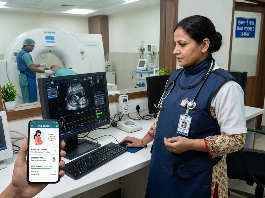
PregiDose is an innovative, user-centred mobile app designed to support pregnant radiographers by enhancing fetal dosimetry, occupational safety education, and psychosocial well-being. It bridges gaps in clinical radiation monitoring and aligns with global healthcare standards.
Today

A comparative non-contact study reveals that high-power radiofrequency delivery with the QDOT MICRO catheter induces an artifactual contact force rise. Electrophysiologists must understand this power-dependent thermal and electrical phenomenon to avoid inadvertent catheter withdrawal and ineffective ablation.
Today
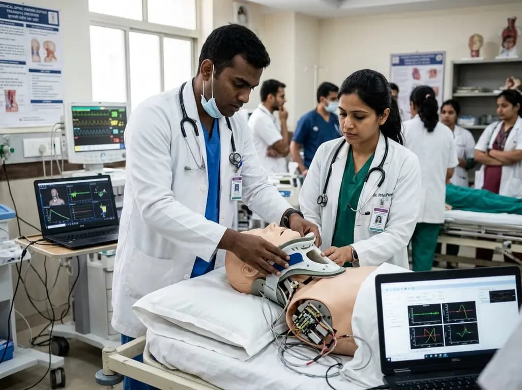
A novel proof-of-concept study presents a six-degree-of-freedom articulated manikin neck equipped with linear potentiometers. The system provides real-time kinematic motion feedback during cervical immobilisation, delivering sub-degree tracking accuracy to improve pre-hospital trauma training and patient safety.
Today
Caffeoylquinic acids, such as chlorogenic acid, offer broad antioxidant, antidiabetic, and anti-inflammatory properties. Advanced plant cell and organ culture methods now enable scalable, sustainable, and pure production of these high-value therapeutic metabolites for pharmaceutical and clinical applications.
Today
