

Loading, please wait...
Loading, please wait...
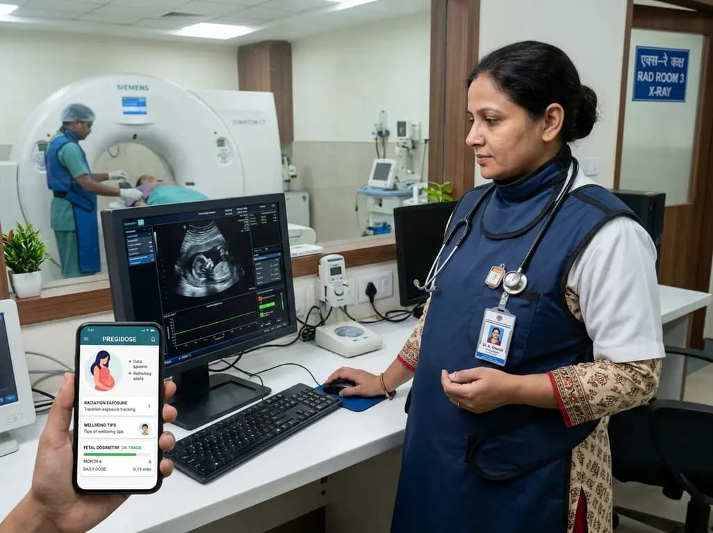
Occupational radiation protection remains a critical priority for diagnostic imaging facilities worldwide, particularly when safeguarding expectant healthcare workers. Female radiographers face unique clinical challenges during gestation because rapidly dividing embryonic and fetal tissues exhibit pronounced sensitivity to ionizing radiation. Consequently, implementing accurate fetal dosimetry and targeted health communication is vital to maintain regulatory compliance while supporting maternal confidence. Recent technological advancements demonstrate how digital innovations can transform routine radiation safety management into proactive, user-centered clinical support systems.
Ionizing radiation poses established biological risks to developing human tissues, primarily due to DNA damage that can cause deterministic or stochastic effects. During early organogenesis, excessive exposure may cause structural malformations, growth restriction, or severe neurodevelopmental deficits. Later gestational stages still present heightened risks for radiation-induced childhood malignancies. Therefore, international radiological bodies classify the conceptus as a member of the general public rather than an occupational worker. This vital distinction requires immediate application of much stricter dose thresholds. Conventional staff monitoring involves single dosimeters worn at the neck collar or chest level. However, fetal dosimetry demands specialized assessment through dedicated monitors placed on the lower abdomen beneath lead aprons. Furthermore, clinicians must evaluate beam energy, workload distribution, and room geometry to calculate accurate intrauterine attenuation. In addition, healthcare facilities must implement continuous surveillance to ensure that exposure rates remain uniform throughout pregnancy. By tracking abdominal absorption meticulously, radiology departments eliminate guesswork and substantiate protective shielding protocols. Consequently, systematic fetal dose calculation upholds maternal safety while preserving departmental workflow efficiency.
Global radiological organizations provide explicit guidelines to shield pregnant workers and their developing fetuses from occupational hazards. The International Commission on Radiological Protection recommends that once a worker declares pregnancy, the equivalent dose to the fetus should not exceed 1 millisievert for the remainder of gestation. Similarly, national authorities, including the Atomic Energy Regulatory Board in India, endorse this stringent benchmark to prevent excess public exposure. Moreover, regulatory standards advise keeping abdominal surface exposure below 2 millisieverts over the entire gestational period. Facility radiation safety officers must review personal exposure histories every month. Therefore, safety teams should quickly reassign expectant staff if cumulative trends approach administrative intervention levels. In addition, hospitals must offer formal counseling regarding voluntary declaration procedures, confidential health documentation, and work modifications. Employers cannot penalize pregnant radiographers financially or professionally for declaring their status. Instead, management teams must collaborate closely with clinical supervisors to optimize shift allocations and reduce high-scatter exposure. Consequently, clear institutional adherence to statutory radiation standards safeguards legal compliance, enhances clinical governance, and fosters a robust occupational safety culture.
Despite clear regulatory directives, practical implementation of occupational radiation safety often encounters substantial barriers in busy hospital environments. Many healthcare institutions struggle with delayed dosimetry reports, which third-party monitoring laboratories often issue weeks after exposure occurs. Furthermore, pregnant radiographers frequently experience significant psychological distress, fearing invisible radiation risks while lacking immediate feedback on their fetal shielding effectiveness. Ineffective monitoring practices in resource-limited clinical settings compound these workplace anxieties and lead to unnecessary absenteeism. In addition, traditional educational materials often lack accessible digital formats, leaving clinical staff with unresolved questions regarding fluoroscopy safety, lead apron integrity, and fetal biology. Therefore, modern healthcare systems require real-time, interactive tools that connect daily clinical tasks with validated dosimetry data. By leveraging portable digital technology, departments can replace cumbersome paper logs with dynamic tracking applications. Moreover, mobile interfaces empower frontline healthcare workers by providing direct access to safety statistics, risk stratification algorithms, and supportive professional communities. Consequently, technological innovation bridges the historical gap between abstract radiation physics and everyday clinical practice, improving adherence across dynamic healthcare environments.
To resolve chronic operational shortcomings in maternal monitoring, researchers developed PregiDose, a tailored mobile application engineered specifically for pregnant radiographers. The development team executed a user-centered design approach, initiating the project through participatory design workshops with practicing clinical radiographers. Furthermore, software engineers and medical radiation physicists collaborated throughout the application lifecycle to translate practical user feedback into functional mobile software. This comprehensive methodology established three foundational operational modules: dose tracking, educational dissemination, and psychosocial support. The dose-tracking module allows clinicians to log clinical procedures, monitor cumulative abdominal badge readings, and receive automated alerts when approaching institutional safety thresholds. Additionally, the educational repository supplies evidence-based literature explaining radiobiology, proper personal protective equipment utilization, and statutory regulations. The well-being module incorporates peer discussion forums and mental health screening tools to alleviate occupational stress. Furthermore, iterative user testing verified intuitive interface navigation and high data security compliance. Thus, PregiDose exemplifies how participatory research methodologies produce clinically relevant digital artefacts that directly resolve the complex operational realities of modern radiation medicine.
Occupational radiation protection extends beyond physical lead shielding and numerical threshold compliance; it also encompasses maternal psychological welfare. Fear of accidental fetal irradiation frequently triggers severe emotional distress among expectant healthcare workers, sometimes prompting unjustified career pauses or premature resignations. However, when clinical facilities implement transparent dosimetry platforms like PregiDose, healthcare providers gain continuous visibility into actual exposure parameters. This direct access replaces ambiguity with tangible reassurance, confirming that ambient doses remain negligible under standard operating procedures. Moreover, providing structured psychosocial resources empowers radiographers to manage workplace anxieties proactively. In addition, structured mobile education demystifies radiation exposure myths and clarifies the biological safety mechanisms of diagnostic shielding. Healthcare administrators benefit simultaneously, as streamlined digital tracking reduces administrative overhead and minimizes the risk of regulatory noncompliance. Furthermore, supporting maternal healthcare professionals aligns directly with broader institutional commitments to gender equity and worker retention in specialized medical fields. Therefore, adopting comprehensive digital dosimetry tools transforms occupational safety from a bureaucratic obligation into an empowering, holistic employee health strategy.
Regulatory guidelines from the International Commission on Radiological Protection and national bodies establish that the equivalent dose to the fetus must not exceed 1 millisievert for the remainder of the pregnancy once declared. Furthermore, employers must maintain abdominal surface exposure below 2 millisieverts throughout gestation. Consequently, radiation safety officers schedule duties to avoid high-dose environments, ensuring that exposure remains evenly distributed and well below statutory occupational safety thresholds without causing professional interruption.
Standard occupational monitoring calculates whole-body equivalent dose using a chest or collar badge worn outside protective apparel. In contrast, fetal dosimetry tracks radiation exposure directly at the maternal abdominal surface, underneath lead aprons. This dedicated abdominal dosimeter estimates real fetal exposure rather than occupational ambient dose. Therefore, regular monitoring verifies that protective shielding remains effective and provides objective reassurance that ionizing exposure does not exceed the strict limits established for fetal health.
Pregnant radiographers can continue performing diagnostic and interventional duties if safety precautions remain optimized. However, department heads often reassign pregnant personnel from high-dose fluoroscopy or radionuclide therapies to general radiography or ultrasound suites. Furthermore, if workers continue assisting in fluoroscopy rooms, they must wear double lead aprons with specialized abdominal shielding and an under-apron fetal monitor. Consequently, consistent compliance ensures that cumulative exposures remain well within safe, acceptable limits.
Disclaimer: This content is for informational and educational purposes only and should not be considered medical advice. Always consult a qualified healthcare professional regarding any medical condition or treatment. Refer to the latest local and national guidelines for clinical practice.
References

Read summarized clinical updates, watch expert medical content, and earn CME certifications right from your smartphone.
Open your smartphone camera to scan and install.





PregiDose is an innovative, user-centred mobile app designed to support pregnant radiographers by enhancing fetal dosimetry, occupational safety education, and psychosocial well-being. It bridges gaps in clinical radiation monitoring and aligns with global healthcare standards.
Today

The 2026 KASID clinical practice guidelines present evidence-based recommendations for ulcerative colitis medical management. This update details induction and maintenance strategies, biologic and small molecule sequencing, the urgent handling of acute severe colitis, and indications for timely surgical intervention.
Today

A comparative non-contact study reveals that high-power radiofrequency delivery with the QDOT MICRO catheter induces an artifactual contact force rise. Electrophysiologists must understand this power-dependent thermal and electrical phenomenon to avoid inadvertent catheter withdrawal and ineffective ablation.
Today
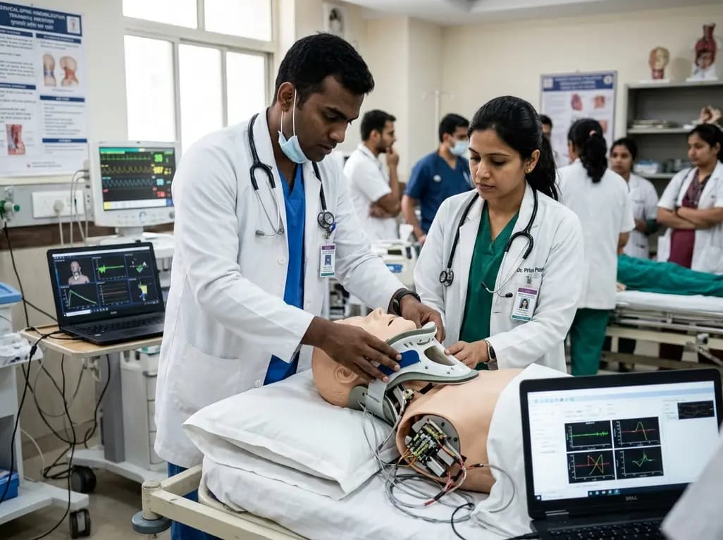
A novel proof-of-concept study presents a six-degree-of-freedom articulated manikin neck equipped with linear potentiometers. The system provides real-time kinematic motion feedback during cervical immobilisation, delivering sub-degree tracking accuracy to improve pre-hospital trauma training and patient safety.
Today

Novel glucose-responsive mPEG-PSA nanocarriers deliver insulin aspart on demand, achieving dual postprandial and basal glycemic control without hypoglycemia in diabetic models. This biomaterial breakthrough offers promising clinical implications for physiologic diabetes management.
Today
