

Loading, please wait...
Loading, please wait...

Inflammatory bowel disease encompasses chronic, relapsing conditions that present substantial diagnostic hurdles worldwide. Recently, the Korean Association for the Study of Intestinal Diseases introduced updated recommendations to streamline clinical workflows and optimize patient outcomes. Systematic inflammatory bowel disease monitoring and accurate early diagnosis help clinicians avoid misdiagnosis, prevent irreversible intestinal damage, and limit inappropriate therapies. Moreover, clinical presentations remain highly heterogeneous across diverse patient populations. Therefore, clinicians must adopt structured strategies that combine clinical assessment, laboratory testing, and advanced imaging modalities.
Diagnosing ulcerative colitis and Crohn's disease requires a meticulous, multifaceted approach. First, physicians must obtain a detailed clinical history along with routine laboratory workups. These initial tests should evaluate complete blood counts, renal function, liver enzymes, and systemic inflammatory markers. In addition, practitioners must routinely perform microbiological stool assays to exclude common infectious causes of enterocolitis. For example, testing should rule out Clostridioides difficile, parasitic infestations, and bacterial pathogens before establishing a definitive diagnosis.
Furthermore, differential diagnosis holds paramount importance in regions with endemic gastrointestinal infections. Clinicians frequently encounter diagnostic dilemmas when distinguishing Crohn's disease from intestinal tuberculosis. Consequently, initial workups must incorporate chest radiography, interferon-gamma release assays, and comprehensive endoscopic biopsies. Endoscopy allows direct visualization of mucosal architecture and facilitates multiple tissue samplings from inflamed and uninflamed bowel segments. As a result, histopathological review can confirm chronic architectural distortion and identify classic features such as non-caseating granulomas. Therefore, a comprehensive baseline assessment secures diagnostic accuracy and sets a reliable benchmark for therapeutic efficacy.
Accurate phenotypic classification guides subsequent therapeutic decision-making in intestinal disorders. Once practitioners confirm an inflammatory bowel disease diagnosis, they must promptly assess the anatomical extent and disease severity. For ulcerative colitis, practitioners categorize disease distribution into proctitis, left-sided colitis, or extensive colitis according to the Montreal classification. Similarly, physicians stratify Crohn's disease according to age at diagnosis, anatomical location, and specific inflammatory behavior.
Additionally, identifying structural complications early prevents emergency surgical interventions. Crohn's disease frequently progresses from an uncomplicated inflammatory phenotype into stricturing or penetrating patterns. Thus, clinicians must identify deep transmural ulcerations, fistulas, or fibrostenotic strictures at baseline. Validated clinical scoring tools and standardized endoscopic indices offer objective measurements of baseline inflammatory burden. Moreover, stratifying patients based on initial risk profiles helps physicians select between stepped-care paradigms and early top-down biologic interventions. Consequently, thorough phenotypic stratification ensures appropriate therapy while reducing the risk of progressive bowel damage.
Modern disease management has shifted from purely symptomatic control toward objective treat-to-target paradigms. Historically, clinicians relied on subjective symptom reports to judge therapeutic response. However, clinical symptoms correlate poorly with ongoing mucosal inflammation, potentially permitting silent bowel destruction. Consequently, updated frameworks prioritize regular inflammatory bowel disease monitoring to secure sustained endoscopic and transmural healing.
Furthermore, dynamic monitoring protocols demand regular reassessments following treatment initiation or medication adjustments. Clinicians should evaluate symptomatic improvement within initial weeks, followed by objective biomarker reassessments at two to three months. In addition, gastroenterologists recommend scheduling repeat endoscopic examinations between six and twelve months to verify mucosal healing. If an individual fails to achieve predefined targets, clinicians must promptly optimize medical dosing or switch therapeutic classes. Therefore, systematic tracking eliminates therapeutic inertia, decreases hospital admissions, and preserves intestinal function over the long term.
Non-invasive diagnostic modalities have transformed longitudinal patient assessment. Specifically, fecal calprotectin provides high sensitivity and specificity for detecting active gastrointestinal inflammation. Serial calprotectin testing enables early detection of subclinical flares well before overt symptomatic relapses occur. Moreover, serum C-reactive protein serves as an accessible surrogate marker of systemic inflammation, although its sensitivity varies among specific phenotypes.
Additionally, cross-sectional imaging modalities play indispensable roles in managing transmural bowel disorders. Computed tomography enterography and magnetic resonance enterography reliably evaluate small bowel lesions beyond the reach of conventional colonoscopes. Magnetic resonance enterography offers exceptional soft-tissue resolution without exposing patients to ionizing radiation. Meanwhile, intestinal ultrasound is emerging as an agile, non-invasive bedside tool for point-of-care assessment. Consequently, serial ultrasound evaluations allow clinicians to track bowel wall thickness, hypervascularity, and mesenteric inflammation in real time. Therefore, combining non-invasive biomarkers with cross-sectional imaging drastically limits the need for invasive, repeated colonoscopies.
Comprehensive care in chronic intestinal inflammation extends beyond mucosal healing. In particular, malnutrition and micronutrient deficiencies remain highly prevalent due to chronic diarrhea, anorexia, and malabsorption. Clinicians must routinely screen patients for protein-energy malnutrition, iron deficiency anemia, and bone mineral density loss. Furthermore, physicians should provide targeted nutritional support, dietary counseling, and aggressive mineral repletion to prevent skeletal fragility.
Similarly, long-standing colonic inflammation substantially escalates the risk of dysplasia and colorectal cancer. Therefore, clinical guidelines recommend initiating colonoscopic surveillance eight to ten years after symptom onset for extensive colitis. Physicians should utilize high-definition chromoendoscopy with targeted biopsies rather than random mucosal sampling. Moreover, shorter surveillance intervals become necessary if concurrent primary sclerosing cholangitis, severe active inflammation, or extensive pseudopolyps exist. In conclusion, integrating routine nutritional screening and rigorous surveillance provides a holistic management framework that protects patient longevity.
Modern management focuses on achieving objective treat-to-target goals. Initially, clinicians aim for rapid symptomatic relief and normalization of systemic inflammatory markers like C-reactive protein. Subsequently, intermediate targets require normalization of fecal calprotectin levels to demonstrate resolving intestinal inflammation. Ultimately, long-term monitoring targets complete mucosal healing on endoscopic evaluation and transmural remission on cross-sectional imaging. Achieving these objective benchmarks prevents cumulative bowel damage, minimizes hospitalization, and reduces surgical intervention rates significantly.
Clinicians should initiate colonoscopic surveillance eight to ten years following symptom onset in patients with extensive ulcerative colitis or left-sided colitis. Thereafter, follow-up colonoscopy intervals range from one to five years depending on risk stratification. High-risk features, such as concomitant primary sclerosing cholangitis, severe active inflammation, or extensive pseudopolyps, necessitate annual surveillance. Conversely, patients with mild inflammation and no adverse risk factors can safely undergo surveillance colonoscopies every three to five years using chromoendoscopy.
Cross-sectional imaging provides vital anatomical insights that conventional colonoscopy cannot achieve. Endoscopic evaluations remain limited to the mucosal surface and cannot reach most segments of the jejunum or ileum. In contrast, modalities like magnetic resonance enterography and intestinal ultrasound assess transmural bowel wall thickness, penetrating fistulas, intra-abdominal abscesses, and fibrostenotic strictures. Furthermore, regular cross-sectional imaging detects active extramural inflammation without requiring repeated invasive endoscopic procedures, guiding timely medical escalation or prompt surgical planning when necessary.
Disclaimer: This content is for informational and educational purposes only... Refer to the latest local and national guidelines for clinical practice.
References

Read summarized clinical updates, watch expert medical content, and earn CME certifications right from your smartphone.
Open your smartphone camera to scan and install.





The KASID clinical guidelines provide an evidence-based roadmap for the diagnosis and monitoring of inflammatory bowel disease, highlighting objective treat-to-target strategies, non-invasive biomarker utilization, cross-sectional imaging advancements, nutritional optimization, and colorectal cancer surveillance.
Today
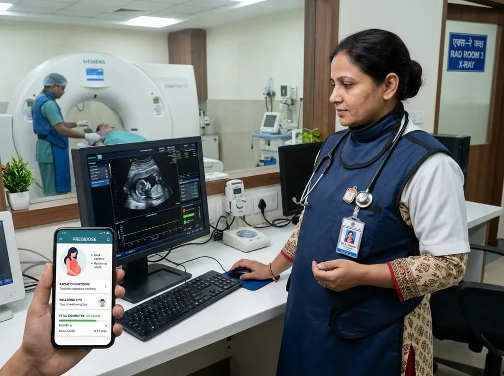
PregiDose is an innovative, user-centred mobile app designed to support pregnant radiographers by enhancing fetal dosimetry, occupational safety education, and psychosocial well-being. It bridges gaps in clinical radiation monitoring and aligns with global healthcare standards.
Today

A comparative non-contact study reveals that high-power radiofrequency delivery with the QDOT MICRO catheter induces an artifactual contact force rise. Electrophysiologists must understand this power-dependent thermal and electrical phenomenon to avoid inadvertent catheter withdrawal and ineffective ablation.
Today
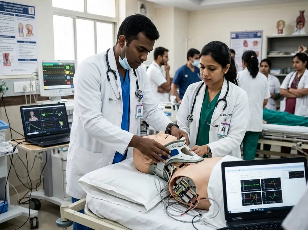
A novel proof-of-concept study presents a six-degree-of-freedom articulated manikin neck equipped with linear potentiometers. The system provides real-time kinematic motion feedback during cervical immobilisation, delivering sub-degree tracking accuracy to improve pre-hospital trauma training and patient safety.
Today
Caffeoylquinic acids, such as chlorogenic acid, offer broad antioxidant, antidiabetic, and anti-inflammatory properties. Advanced plant cell and organ culture methods now enable scalable, sustainable, and pure production of these high-value therapeutic metabolites for pharmaceutical and clinical applications.
Today
