

Loading, please wait...
Loading, please wait...
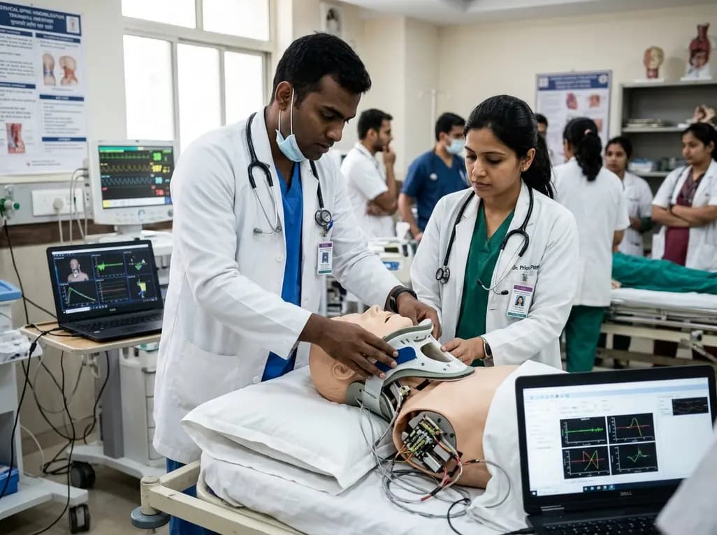
Trauma management relies heavily on effective cervical spine immobilisation training to minimize secondary neurological injury during pre-hospital handling. Consequently, emergency medical providers require rigorous cervical spine immobilisation training to safeguard vulnerable patients during extrication and transfer. Conventional training manikins fail to replicate human neck flexibility and lack objective feedback mechanisms. Therefore, learners often practice vital manual in-line stabilisation techniques without knowing if they exceeded safe motion thresholds. A novel proof-of-concept investigation introduces an articulated, six-degree-of-freedom manikin neck equipped with linear potentiometers. This innovative medical training tool measures real-time displacement, providing clear feedback that can transform emergency education worldwide.
Standard patient simulator manikins offer stiff cervical segments that do not resemble actual human anatomy. As a result, trainees rarely encounter the true flaccidity or resistance seen in unconscious trauma victims. Furthermore, existing simulators lack electronic tracking to record subtle linear and rotational displacements during spinal motion restriction. Instructors must rely on visual assessment alone, which frequently misses dangerous rotational micro-movements. In addition, manual evaluation introduces significant subjective bias during clinical competency exams. Medical educators understand that excessive neck flexion, extension, or rotation during log-rolling procedures can trigger irreversible quadriplegia. For this reason, simulation centers need bio-fidelic models that replicate multi-planar kinematics. Recent bioengineering efforts bridge this gap by replacing rigid monolithic necks with dynamic robotic assemblies. By mimicking realistic joint properties, new simulators allow trainees to experience authentic tactile feedback. Consequently, clinicians develop refined muscle memory and learn to adapt their manual stabilisation force during high-stress scenarios. These upgrades significantly improve educational quality across global emergency departments.
To overcome existing mechanical shortcomings, bioengineers manufactured a custom articulated neck utilizing advanced 3D printing techniques. Specifically, the neck architecture features interlocking ball-and-socket joints that replicate multi-vertebral cervical mobility. Integrated extension springs deliver passive stiffness, ensuring that the manikin head remains compliant throughout early movement arcs. Moreover, internal mechanical stoppers simulate natural physiological constraints to prevent unphysical anatomical displacement. The engineers also fabricated custom aluminium adaptor plates and locking mechanisms to interface smoothly with existing simulator chassis. Consequently, training facilities can retrofit their current adult manikins without purchasing entirely new full-body systems. This modular design dramatically lowers institutional costs while expanding access to high-fidelity simulation. In addition, the design accommodates complex multi-axis motion including flexion, extension, lateral bending, and axial rotation. Trainees who practice on this articulated prototype experience authentic tactile resistance during cervical collar placement and scoop stretcher transfers. Therefore, the physical interface closely mirrors the biomechanical dynamics encountered during field extrication.
Objective kinematic assessment requires robust sensor instrumentation capable of detecting minimal head displacement in real time. For this purpose, researchers implemented a passive Stewart-Gough platform arrangement using high-precision linear slide potentiometers. As the trainee handles the head, the potentiometers extend and retract along geometric vectors. Subsequently, a forward kinematics algorithm calculates the exact three-dimensional spatial coordinates and angular orientation of the head relative to the torso. Furthermore, investigators validated the prototype against laboratory-grade 3D optoelectronic motion capture cameras. The experimental results demonstrated outstanding tracking accuracy, achieving a root-mean-squared error of less than 0.89 degrees across all movement axes. Because potentiometers provide immediate electrical signals, the system processes kinematic deviations instantly without lag. Therefore, simulation facilitators can observe live telemetry during complex maneuvers such as rapid extrication, helmet removal, and cervical collar application. Notably, this objective sensor integration eliminates observer subjectivity from high-stakes paramedic certification exams.
Effective cervical spine immobilisation training demands immediate feedback so practitioners can correct improper manual handling techniques instantly. During prototype testing, investigators recorded the neck's operational range of motion across various anatomical planes. The potentiometer assembly permitted 25.0 degrees of flexion, 29.3 degrees of extension, and lateral bending of over 21 degrees. Meanwhile, the mechanical stoppers safely constrained left and right axial rotation to 52.5 degrees and 48.4 degrees, respectively. In addition, the team analyzed the prototype's passive stiffness profile during movement. The neck exhibited remarkably low stiffness during initial motion, ranging from 5.8 to 28.3 Newton-millimeters per degree. However, as movement approached the end of the range, stiffness spiked by up to ten times, reaching up to 404.5 Newton-millimeters per degree. This progressive mechanical resistance mirrors real human ligamentous tension. As a result, trainees quickly learn to recognize when their maneuvers apply unsafe strain to an injured cervical column.
Spinal cord trauma remains a major cause of mortality and permanent disability across India and developing healthcare systems. In particular, road traffic accidents frequently cause severe cervical injuries that require meticulous pre-hospital stabilization. Unfortunately, many first responders and hospital triage staff receive limited hands-on practice with instrumented simulation tools. Implementing sensorized, six-degree-of-freedom necks can drastically upgrade regional trauma life support training programs. Furthermore, the ability to retrofit existing training manikins makes this technology viable for resource-conscious medical colleges. Clinical educators can deploy these articulated heads during interdisciplinary drills involving emergency physicians, orthopedic trainees, neurosurgeons, and nursing personnel. Moreover, the quantifiable kinematic metrics encourage friendly competition and objective skill mastery among emergency medical technicians. Ultimately, mastering gentle, aligned stabilization on responsive manikins reduces secondary spinal cord transection during real-world ambulance transfers. Improving educational fidelity therefore translates directly into superior neurological outcomes for vulnerable trauma victims.
Although this proof-of-concept manikin neck demonstrates exceptional accuracy, developers must implement several refinements prior to routine clinical deployment. For instance, future prototypes should expand the available range of motion to match human anatomical extremes more completely. Furthermore, engineers plan to refine the spring stiffness gradient to reflect the hypermobility of ligamentous disruption or elderly degenerative spines. In addition, incorporating soft tissue silicone coverings will improve physical realism during airway management and cervical collar sizing. Simulation researchers must also evaluate the prototype across diverse paramedic cohorts in high-stress outdoor extrication scenarios. By gathering qualitative user feedback, developers can optimize the ergonomics and durability of internal potentiometer rods. Concurrently, developing mobile tablet applications will allow instructors to visualize real-time angular displacement graphs at the bedside. Through these continuous technological innovations, simulation centers will prepare healthcare professionals to deliver safer, more compassionate emergency trauma care.
Real-time motion feedback allows trainees to recognize subtle spinal displacements that instructors cannot detect with the naked eye. Consequently, learners receive immediate objective warnings when their manual stabilisation exceeds safe thresholds. Furthermore, this dynamic feedback helps emergency responders correct their hand placement and grip strength during high-risk transfers. Ultimately, immediate telemetry builds reliable muscle memory, which prevents secondary neurological damage and spinal cord transection during real trauma patient extrications.
Conventional training manikins feature rigid neck joints that severely restrict natural multi-planar movement. In contrast, a six-degree-of-freedom articulated neck allows authentic flexion, extension, lateral bending, and axial rotation. In addition, internal springs and stoppers mimic human ligamentous resistance and flaccidity. Therefore, healthcare providers encounter realistic physical resistance while applying cervical collars or performing log-rolls, which bridges the gap between simulated laboratory exercises and authentic patient clinical emergencies.
Yes, researchers designed this articulated neck with custom aluminium adaptor plates and modular locking mechanisms. Consequently, institutions can attach the six-degree-of-freedom neck directly to their existing simulator attachment points without modifying the original manikin chassis. This retrofitting capability significantly lowers procurement costs for simulation centers and teaching hospitals. Furthermore, it allows medical colleges in resource-constrained environments to modernize their current trauma training equipment efficiently without purchasing costly new simulators.
Disclaimer: This content is for informational and educational purposes only, and does not substitute professional medical advice, diagnosis, or treatment. Refer to the latest local and national guidelines for clinical practice.
References

Read summarized clinical updates, watch expert medical content, and earn CME certifications right from your smartphone.
Open your smartphone camera to scan and install.





A novel proof-of-concept study presents a six-degree-of-freedom articulated manikin neck equipped with linear potentiometers. The system provides real-time kinematic motion feedback during cervical immobilisation, delivering sub-degree tracking accuracy to improve pre-hospital trauma training and patient safety.
Today
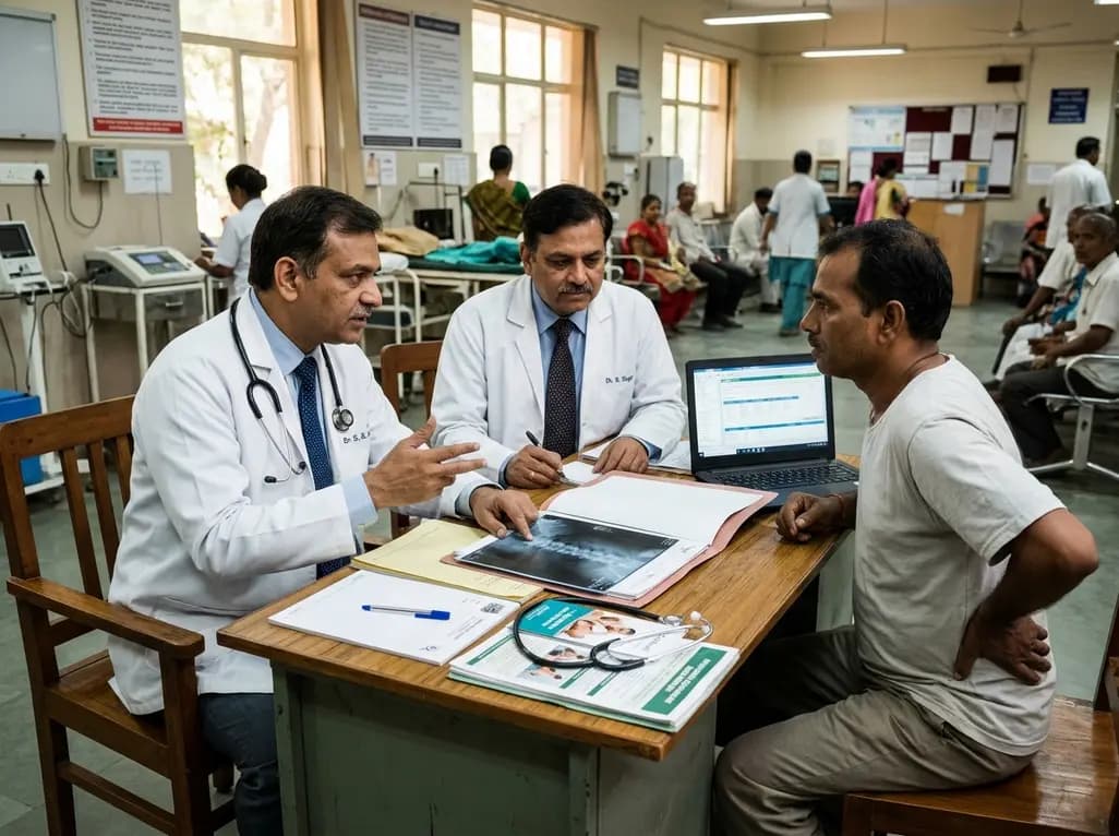
A landmark 12-year longitudinal study highlights significant reductions in opioid prescribing for nonspecific low back pain following clinical guidelines and prescription monitoring systems, emphasizing non-opioid multimodal pain care.
Today
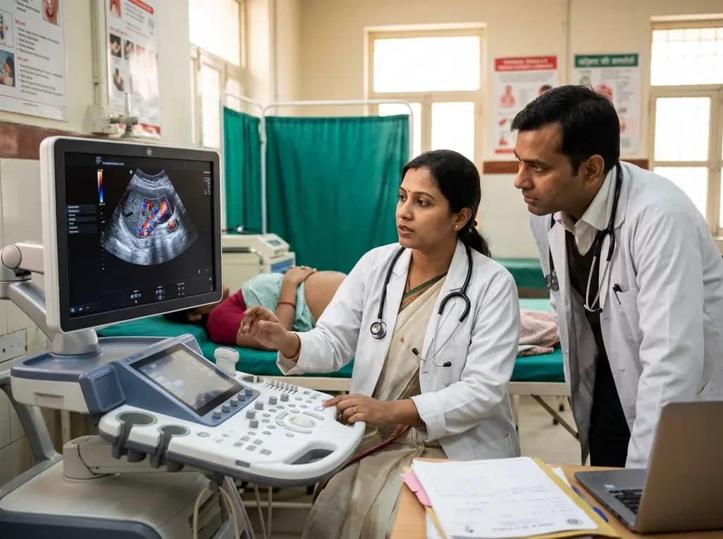
A study in Ultrasound in Obstetrics & Gynecology shows that quantitative vascularization of the anterior fornix/cervix with transvaginal ultrasound reliably identifies placenta accreta spectrum and predicts severe intraoperative hemorrhage, offering vital objective risk stratification.
Today

A landmark study details how an enterprise pediatric remote patient management model expanded from a single cardiology pilot to 16 diverse specialty programs by combining centralized digital operations with specialty clinical leadership.
Yesterday
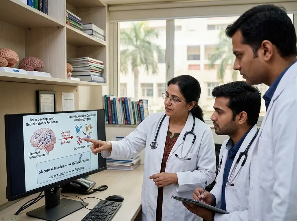
Protein O-GlcNAcylation couples cellular nutrient status to vital neuronal functions. This review explores how disrupted O-GlcNAc cycling drives both neurodevelopmental delays and neurodegenerative cascades, positioning metabolic post-translational modifications as therapeutic targets in clinical neurology.
Today
