.webp&w=384&q=75)

Loading, please wait...
Loading, please wait...

Spindle cell lipoma represents an established benign mesenchymal neoplasm that classically demonstrates mature adipocytes, bland spindle cells, and dense collagen bundles. However, atypical histological presentations frequently complicate the diagnostic evaluation. Specifically, pathologists occasionally encounter a fat-free spindle cell lipoma, wherein mature adipocytic components are virtually or entirely undetectable. This dramatic histological variation removes the defining feature clinicians expect to see in lipomatous tumors. Consequently, when this lesion arises in an unexpected demographic, diagnostic uncertainty escalates rapidly. Healthcare providers must recognize this deceptive entity promptly during daily clinical practice. Clinicians can thereby prevent aggressive, unnecessary surgical interventions and ease patient anxiety effectively.
Pathologists first characterized spindle cell lipoma as a distinct entity several decades ago. Classically, the tumor consists of three cardinal histological elements in variable proportions. These components include mature adipose cells, uniform spindle cells, and thick collagen bundles resembling frayed ropes. In typical specimens, mature adipocytes dominate the architectural pattern throughout the lesion. Nevertheless, the proportion of adipose tissue can fluctuate substantially across individual clinical cases. Some tumors display a prominent myxoid stroma, whereas others feature extensive collagenous bands. In extreme presentations, adipocytes decrease to less than five percent or vanish completely. When fat cells disappear entirely, pathologists classify the lesion as a fat-free variant. Consequently, the absence of adipocytes strips away the primary histological clue of a lipomatous neoplasm. This morphological transformation shifts the microscopic appearance toward purely fibroblastic or neural proliferations. Therefore, clinicians who examine such lesions often fail to consider lipoma initially. Recognizing this broad morphological spectrum remains essential for accurate pathological interpretation. Familiarity with this rare variant prevents severe misinterpretations of benign soft tissue architecture. Furthermore, understanding this structural variation assists multidisciplinary teams in separating innocent mimickers from malignant lesions before initiating treatment.
Classic spindle cell lipomas arise predominantly in male patients between fifty and seventy years of age. Furthermore, these lesions typically localize to the subcutaneous tissues of the posterior neck, upper back, and shoulders. These anatomical areas represent well-documented predilection zones in routine surgical practice. However, uncommon variants frequently defy these historical demographic parameters. For example, recent clinical reports describe fat-free lesions occurring in young adult females. Additionally, these atypical presentations can develop in unusual anatomical sites such as the extremities, face, or anterior trunk. When a firm, slowly expanding subcutaneous nodule develops in a young woman, physicians rarely suspect a lipomatous tumor. Instead, clinicians often entertain diagnoses like dermatofibroma, schwannoma, or cutaneous sarcoma. Moreover, physical examination alone cannot distinguish between these distinct pathological processes. Diagnostic imaging frequently reveals a well-circumscribed, non-infiltrative mass without characteristic fat attenuation. Because radiologic findings mirror other solid soft tissue neoplasms, histopathological assessment becomes indispensable. Clinicians must maintain a heightened index of suspicion whenever atypical demographics intersect with unusual subcutaneous masses. Accordingly, comprehensive tissue sampling provides the single reliable pathway toward accurate identification.
The microscopic analysis of these unusual lesions demands meticulous morphological scrutiny. Even when mature fat disappears, certain structural clues persist to guide the discerning pathologist. For instance, the tumor typically exhibits short, uniform spindle cells with parallel bipolar cytoplasmic processes. These cells reside within a variable background of loose myxoid matrix and thick, hypereosinophilic collagen bands. Furthermore, these collagen bundles frequently display a distinct rope-like or shredded-carrot appearance. Nuclear atypia, hyperchromasia, and atypical mitotic figures remain conspicuously absent throughout the entire specimen. Nevertheless, diagnostic pitfalls abound during routine microscopic evaluation. Without recognizable adipocytes, the lesion closely mimics solitary fibrous tumors, dermatofibrosarcoma protuberans, and low-grade fibromyxoid sarcomas. In particular, dermatofibrosarcoma protuberans demonstrates prominent spindle cell fascicles that can confuse unwary evaluators. Similarly, schwannomas and neurofibromas share overlapping cellular characteristics with this benign entity. Mistaking a benign spindle cell proliferation for an aggressive sarcoma exposes patients to radical resections. Therefore, pathologists must correlate cytological blandness with specialized supplementary testing before finalizing a formal diagnosis. Consequently, this detailed morphological appraisal avoids costly overtreatment. Additionally, careful histological examination protects patients from unnecessary physical trauma.
Immunohistochemistry plays a pivotal role in confirming this challenging diagnostic entity. Specifically, the spindle cells demonstrate strong and diffuse cytoplasmic reactivity for CD34. Conversely, the tumor cells consistently lack expression of S100 protein, SOX10, desmin, and smooth muscle actin. While CD34 positivity confirms the mesenchymal nature, it also raises concern for other spindle cell lesions. For example, solitary fibrous tumors also express CD34 abundantly. However, nuclear STAT6 staining remains completely negative in spindle cell lipoma, effectively ruling out solitary fibrous neoplasms. Additionally, evaluating nuclear retinoblastoma protein provides decisive diagnostic clarity. Most spindle cell lipomas harbor monosomy or partial deletions involving chromosome 13q14. Consequently, immunohistochemical staining reveals a distinctive loss of nuclear RB1 expression in the lesional spindle cells. In contrast, benign mimics like neurofibromas retain intact nuclear RB1 staining. Furthermore, testing for MDM2 amplification distinguishes this tumor from well-differentiated liposarcoma. Because liposarcomas retain RB1 and express MDM2, this focused panel readily resolves complex diagnostic dilemmas. Integrating these molecular and immunohistochemical findings guarantees precise classification and prevents misdiagnosis. Therefore, molecular confirmation serves as an indispensable tool during borderline cases.
Clinical management of this uncommon benign neoplasm remains reassuringly conservative. Once the pathologist establishes the correct diagnosis, complete surgical excision provides definitive therapy. Surgeons perform simple marginal enucleation or local excision with narrow margins under local anesthesia. Because the tumor maintains well-circumscribed borders, complete removal rarely presents significant technical difficulties. Furthermore, extensive wide resections or regional lymph node dissections are entirely unnecessary. Postoperative recovery proceeds smoothly for the vast majority of patients. Moreover, local recurrence rates remain exceptionally low, even when histological margins demonstrate close approximation. Long-term studies confirm that spindle cell lipomas possess zero potential for malignant transformation or metastatic dissemination. Therefore, routine oncological surveillance imaging adds unnecessary financial burden and emotional distress. Clinicians should reassure patients regarding the entirely benign biological behavior of their condition. Periodic clinical inspection during routine primary care visits offers sufficient postoperative follow-up. Through accurate initial diagnosis and conservative intervention, clinicians ensure optimal patient outcomes while avoiding overtreatment. Consequently, patients quickly resume normal activities with complete peace of mind. Overall, conservative surgical intervention ensures excellent physical and cosmetic recovery.
Genetic alterations drive this distinctive phenotype, primarily involving deletions of chromosome 13q14. This chromosomal loss inactivates the RB1 gene and alters downstream mesenchymal differentiation pathways. Consequently, neoplastic progenitor cells fail to complete mature adipogenesis during tumor growth. Instead of producing recognizable fat vacuoles, the lesional cells proliferate predominantly as bland spindle cells surrounded by collagen. These biological changes produce an entirely non-lipomatous morphological appearance without altering the tumor's benign natural clinical course.
Pathologists rely on a concise immunohistochemical panel to establish diagnostic certainty. Lesional spindle cells display diffuse and strong cytoplasmic positivity for CD34 alongside a characteristic loss of nuclear RB1 expression. Meanwhile, negative staining for S100 protein and SOX10 reliably excludes neural neoplasms. Furthermore, the absence of STAT6 expression rules out solitary fibrous tumors, while negative MDM2 testing excludes liposarcoma. This coordinated immunoprofile allows clinicians to confidently confirm the benign diagnosis.
Surgeons recommend simple, conservative marginal excision as the standard curative management strategy. Because this neoplasm behaves in an entirely benign manner, wide oncological safety margins are unnecessary. Surgeons typically perform the procedure under local anesthesia with minimal morbidity. Even if microscopic margins show focal tumor approximation, local recurrence remains exceedingly rare. Consequently, patients achieve complete resolution following uncomplicated excision and do not require postoperative radiation, adjuvant chemotherapy, or repetitive oncological surveillance scans.
Disclaimer: This content is for informational and educational purposes only... Refer to the latest local and national guidelines for clinical practice.
References

Read summarized clinical updates, watch expert medical content, and earn CME certifications right from your smartphone.
Open your smartphone camera to scan and install.





Fat-free spindle cell lipoma presents an intriguing diagnostic challenge when classical adipocytes are entirely absent. Clinicians and pathologists must evaluate immunohistochemical markers like CD34 and RB1 loss to distinguish this benign entity from soft tissue sarcomas and guide conservative surgical management.
Today
The phase II DAPA-AIC trial demonstrates that dapagliflozin 10 mg daily significantly preserves left ventricular ejection fraction compared to placebo over four months in adults undergoing anthracycline chemotherapy, highlighting a potential preventive strategy in cardio-oncology.
Today

India's national digital health service achieved a historic milestone of fifty crore virtual consultations. Notably, female patients account for over fifty-seven percent of utilisation, demonstrating improved healthcare accessibility for remote communities, chronic disease management, and primary triage.
Today
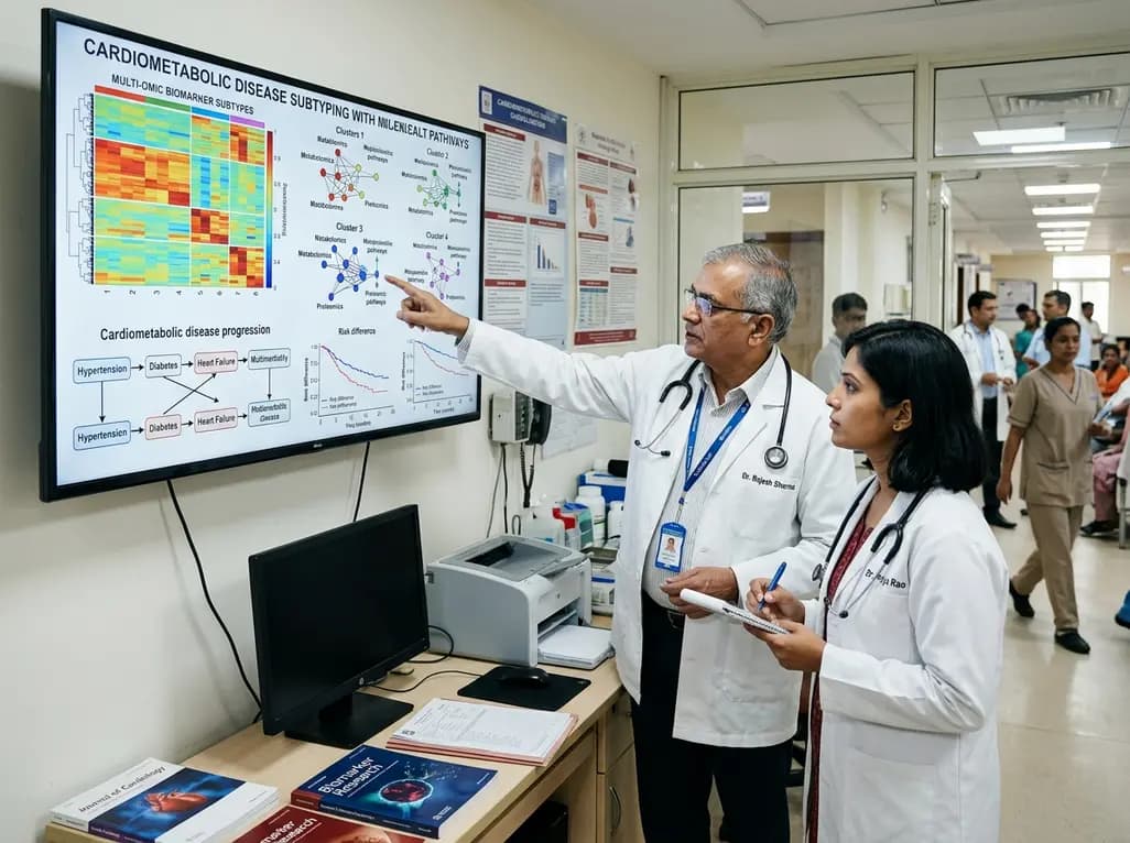
A large UK Biobank study identified four routine biomarker subtypes that predict cardiometabolic disease progression to multimorbidity. Integrating multi-state modelling, metabolomics, and proteomics revealed distinct biological pathways, offering new avenues for tailored clinical risk stratification.
Today

Dual-nanofiber interpenetrating network scaffolds combining silica and PLLA/gelatin nanofibers with sodium alginate achieve complementary mechanical stability, immunomodulatory M2 macrophage polarization, and enhanced osteogenesis for advanced bone repair.
Today

Researchers at IISER Berhampur have engineered a covalent organic framework immunosensor that detects urinary NGAL. This novel nanotechnology platform distinguishes diabetic nephropathy from other renal conditions, enabling rapid, minimally invasive stratification and earlier clinical intervention for patients.
Today
