.webp&w=384&q=75)

Loading, please wait...
Loading, please wait...
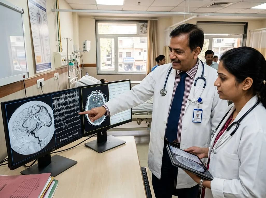
Restoring motor autonomy remains a major challenge in neurorehabilitation for individuals living with progressive neurodegenerative diseases. An endovascular brain-computer interface now provides an innovative pathway to capture high-resolution neural commands without traditional open craniotomies. By recording cortical dynamics directly from cerebral venous sinuses, this emerging neurotechnology bridges the gap between invasive cortical arrays and non-invasive surface sensors. Consequently, clinicians are closely following trial data to evaluate its long-term diagnostic fidelity and decoding accuracy.
For decades, clinical neuroscientists relied on either non-invasive scalp electroencephalography or invasive open-skull electrode grids to decode motor volition. While scalp electrodes provide exceptional safety, the thick cranial vault severely attenuates high-frequency neuroelectric activity. Conversely, subdural electrocorticography and penetrating microelectrode arrays require invasive craniotomies that carry significant risks of hemorrhage, infection, and tissue scarring.
To circumvent these surgical barriers, bioengineers designed stent-electrode arrays that neurointerventionalists can navigate endovascularly through the jugular vein. Specifically, operators guide the flexible mesh catheter directly into the superior sagittal sinus adjacent to the primary motor cortex. Once deployed, the self-expanding metallic stent apposes the inner endothelial wall, placing recording contacts mere millimeters from cortical pyramidal cell columns. Furthermore, the vessel wall shields the brain parenchyma from direct mechanical trauma. Recent clinical protocols, such as the early feasibility COMMAND trial, confirm the practical viability of this transvenous approach. Consequently, this endovascular platform delivers a stable, durable bridge for chronic neural decoding. Clinicians now have an unprecedented tool to evaluate cortical motor intent while minimizing open-skull neurosurgical morbidity. Therefore, understanding how this vascular signal compares directly against standard scalp electrophysiology represents a pivotal milestone in neural engineering.
In a recent investigative trial, researchers conducted a direct, simultaneous recording comparison between vascular electrocorticography and 64-channel gel-based scalp electroencephalography. The study evaluated an adult participant presenting with severe upper limb tetraplegia secondary to amyotrophic lateral sclerosis. Although the participant suffered from profound functional motor loss, residual subcortical and cortical pathways preserved intermittent voluntary motor activation.
During two structured experimental recording sessions, computer screens presented visual cues prompting the participant to attempt specific motor movements. In particular, the participant executed repetitive ankle dorsiflexion and plantarflexion across multiple controlled trials. In addition, the protocol examined cognitive motor imagery, wherein the participant vividly imagined the movement without physical exertion. Simultaneously, recording software captured raw signal streams across both modalities under identical temporal conditions. Investigators then applied rigorous spectral analyses to extract task-related frequency bands, quantifying power changes during active effort versus baseline rest. Furthermore, researchers evaluated spatial lateralization to determine if vascular sensors could distinguish between left and right limb commands. This meticulous paradigm provided an objective benchmark to analyze real-world signal characteristics in paralyzed users. Thus, the direct comparison eliminated physiological variance, ensuring valid performance metrics across both diagnostic modalities.
The comparative analysis revealed substantial differences in spectral dynamics and raw signal strength between the two systems. Specifically, both modalities captured significant neural modulation during attempted ankle movement when contrasted against baseline rest intervals. However, vascular electrocorticography channels exhibited markedly greater modulation depth per channel across high-frequency bands. Because the stent-electrode array resides beneath the cranium, it completely bypasses the bone-induced low-pass filtering that severely degrades scalp recordings.
Interestingly, attempted physical movement generated significantly stronger spectral shifts than covert motor imagery across both recording systems. When the participant merely imagined ankle movements, signal amplitudes dropped substantially in both intravascular contacts and surface electrodes. Moreover, spatial source localization failed to demonstrate statistically significant lateralization between left and right ankle attempts in either modality. The midline venous anatomy of the superior sagittal sinus records bilaterally from both paracentral lobules, inherently limiting hemispherically separated spatial resolution. Nevertheless, the elevated signal-to-noise ratio in vascular channels permitted robust classification of movement onset. Consequently, the stent-electrode array demonstrated a superior capacity to detect raw motor intent compared to traditional surface electrodes. Therefore, vascular recordings provide an exceptionally stable biological substrate for high-fidelity computer algorithms.
Every neurorecording modality contends with distinct environmental and biological noise profiles that challenge machine learning decoders. In this comparative trial, investigators analyzed the disruptive impact of cranial muscle contraction, ocular movements, 60-Hertz electrical lines, and cardiac rhythms. Consequently, the researchers discovered divergent vulnerability patterns across the intravascular and surface sensors.
Scalp electroencephalography exhibited substantial vulnerability to superficial myogenic artifacts and involuntary cranial movements. Specifically, simple actions such as jaw clenching, vocalization, and ocular blinks introduced massive electrical deflections that masked underlying cortical rhythms. In contrast, the stent-electrode array remained completely impervious to external cranial muscle activity and eye movement artifacts. Because the skull and intracranial fluid mechanically insulate the venous sinus, superficial muscle potentials cannot penetrate the vascular recording field. However, vascular electrocorticography presented its own unique biological contaminant in the form of cardiac pulse waveforms. Pulsatile blood flow and cardiac electrical conduction periodically contaminated the intravascular channels. Fortunately, automated digital filtering and adaptive template subtraction algorithms eliminated these cardiac waveforms without sacrificing critical motor frequencies. Thus, intravascular sensors provide a far cleaner spectrum across the target frequency domains. In addition, eliminating line noise proved significantly easier within the shielded intracranial vascular compartment.
These comparative findings deliver vital practical insights for clinicians managing progressive neuromuscular diseases such as amyotrophic lateral sclerosis. Currently, patients suffering from severe motor impairment face profound communication barriers as speech and limb functions deteriorate. While non-invasive brain-computer interfaces offer helpful assistive options, daily operational hurdles frequently limit their real-world adoption. For instance, scalp caps require conductive gels, tedious skin preparation, and constant technical recalibration by skilled caregivers. Furthermore, superficial sweating and electrode displacement degrade signal quality over time.
Conversely, endovascular brain-computer interfaces present a permanent, fully implantable solution that operates seamlessly in domestic environments. Once implanted via routine neurointerventional catheterization, the vascular array functions continuously without requiring daily montage setup. Moreover, because the sensor maintains high signal stability across months, decoding algorithms retain their calibration without frequent retraining. Patients can independently operate digital communication software, send text messages, browse assistive websites, and control smart environmental units. In addition, neurosurgeons and interventional neurologists can perform the implantation within standard catheterization laboratories under local anesthesia. Therefore, transvenous neuroprosthetics represent a viable, highly scalable clinical pathway toward restoring autonomy in severely paralyzed individuals. Consequently, clinicians anticipate broader therapeutic applications as endovascular neuroprosthetic systems complete late-stage pivotal trials.
An endovascular brain-computer interface utilizes a flexible stent-electrode array placed inside the superior sagittal sinus rather than external skin electrodes. Consequently, the vascular sensor sits directly beneath the skull, eliminating the severe signal attenuation caused by bone and scalp tissue. While conventional scalp electroencephalography is non-invasive, it requires daily gel maintenance and suffers from cranial muscle artifacts. In contrast, transvenous implants deliver permanent, stable recording channels with substantially superior modulation depth.
Intravascular electrocorticography recordings remain completely shielded from superficial myogenic activity, meaning eye blinks, jaw clenching, and speech do not distort neural signals. However, because the array resides within a major venous sinus, it encounters prominent cardiac pulse contamination from adjacent arterial pulsations and venous flow. Fortunately, clinical software algorithms readily identify and filter these repetitive cardiac waveforms, ensuring clean spectral data across the high-frequency gamma bands essential for accurate motor intent classification.
Spatial lateralization failed to reach statistical significance primarily due to the midline anatomical position of the superior sagittal sinus. The sinus runs directly along the longitudinal fissure, positioning the recording electrodes equidistant between the bilateral motor homunculi in the paracentral lobules. Consequently, the sensor captures overlapping neural activity from both cerebral hemispheres during unilateral ankle movements. Nevertheless, robust modulation timing and amplitude still permit reliable binary intent classification for assistive communication devices.
Disclaimer: This content is for informational and educational purposes only... Refer to the latest local and national guidelines for clinical practice.
References:

Read summarized clinical updates, watch expert medical content, and earn CME certifications right from your smartphone.
Open your smartphone camera to scan and install.





A landmark study compares endovascular brain-computer interface recordings via stent-electrode arrays with scalp EEG in an ALS patient. Endovascular vECoG demonstrated superior signal modulation without skull attenuation, offering a reliable, minimally invasive neural pathway for restoring communication and mobility.
Today
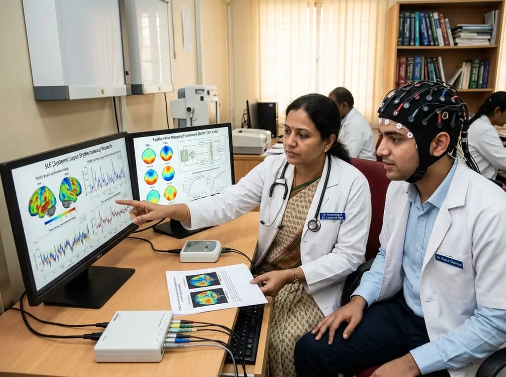
A dual-site study demonstrates that an EEG spatial prior mapping framework (MPW-CSP-SVM) standardizes fNIRS biomarkers in SLE across different optode configurations. The model achieved high classification accuracy (AUC up to 0.839) and correlated with disease activity and emotional distress.
Today

A landmark randomized clinical trial published in NEJM reveals that high maternal intake of eggs and peanuts during pregnancy and lactation does not lower the risk of infant egg or peanut allergy at one year of age, reinforcing existing dietary recommendations for balanced maternal nutrition without restrictions.
Today
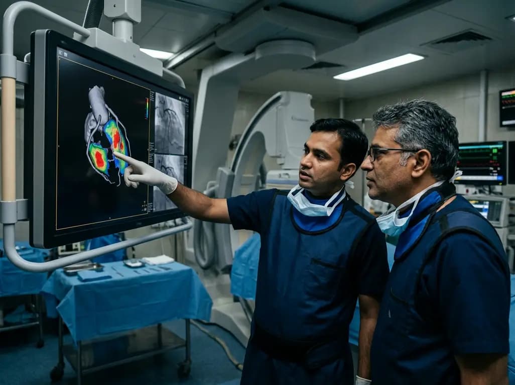
A groundbreaking study demonstrates that coronary angiography-derived radial wall strain reflects plaque morphology, predicting future rupture and erosion.
Today

The Delhi Cabinet has cleared an advanced seven-storey critical care expansion at Guru Tegh Bahadur Hospital under PMSSY Phase-IV. Costing Rs 244.87 crore, the new wing features 250 beds, specialized intensive care units, modern operation theatres, and round-the-clock emergency diagnostics for East Delhi.
Today
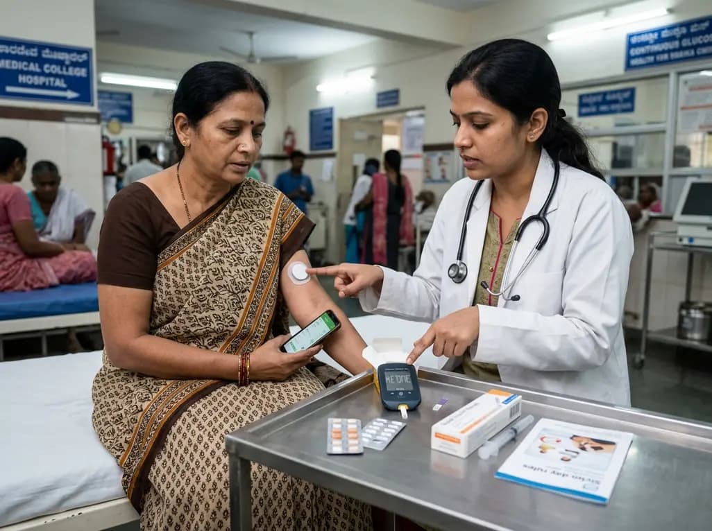
A landmark case demonstrates that favorable continuous glucose monitoring metrics cannot rule out SGLT2 inhibitor-associated euglycemic diabetic ketoacidosis. Clinicians must educate patients on symptom-based sick-day rules, uninterrupted basal insulin therapy, and proactive blood ketone testing.
Today
