.webp&w=384&q=75)

Loading, please wait...
Loading, please wait...

Systemic lupus erythematosus often affects the central nervous system, producing subtle cognitive dysfunction and affective disturbances. Unfortunately, conventional neuropsychiatric assessments remain largely subjective and time-consuming. Clinicians frequently struggle to distinguish lupus fog from primary mood disorders. Consequently, medical researchers are actively evaluating non-invasive optical neuroimaging to identify objective functional signatures. Recent investigations into fNIRS biomarkers in SLE demonstrate immense potential for bedside hemodynamic monitoring during cognitive activation tasks. Functional near-infrared spectroscopy measures localized cortical oxygenation variations with remarkable safety and portability. However, physical variations among optode montages across hospital networks have historically prevented cross-center biomarker validation.
Moreover, patients in tertiary lupus clinics frequently present with fluctuating complaints of executive dysfunction and memory impairment. Standard structural neuroimaging often reveals unremarkable findings in early central nervous system involvement. Therefore, physiological monitoring during real-time cognitive stress provides vital diagnostic clarity. By quantifying hemodynamic changes during a standardized verbal fluency task, clinicians can capture frontotemporal network disruptions. Furthermore, functional spectroscopy offers lower sensitivity to motion artifacts than magnetic resonance imaging. Thus, establishing robust analytical pipelines represents a pivotal milestone for lupus clinical neurophysiology.
Multi-site optical neuroimaging studies inevitably encounter substantial heterogeneity in hardware architecture and probe geometries. For instance, different commercial fNIRS devices utilize distinct channel counts, detector placements, and inter-optode distances. In a dual-site cohort, researchers examined 77 patients with lupus alongside 54 healthy controls using a 37-channel device at Site 1. In parallel, Site 2 evaluated 75 lupus patients and 38 control subjects using a 50-channel system. Direct comparison of raw optical signals across these disparate configurations produces significant analytical bias. Therefore, investigators constructed a standardized spatial mapping framework to align divergent optical arrays into a shared coordinate space.
Specifically, the investigators established spatial prior operators based on standard international 10-20 electroencephalography coordinates. Native optical channels were mapped to standardized cranial locations using fixed mathematical transformations. The computational pipeline utilized equal-weight regional averaging for Site 1 data. Meanwhile, the pipeline applied inverse-distance-squared weighting for Site 2 recordings. Furthermore, this spatial normalization bridged hardware differences without sacrificing local signal resolution. Consequently, the strategy transformed heterogeneous optical datasets into uniformly structured spatial representations. Thus, this mathematical standardization provides a reproducible roadmap for collaborative multicentric neuroimaging trials.
Following spatial alignment, the researchers implemented a machine learning architecture combining weighted common spatial patterns with support vector machines. Furthermore, this MPW-CSP-SVM framework extracted targeted spatial features reflecting differential frontotemporal oxygenation dynamics during the verbal fluency task. The resulting model achieved outstanding diagnostic accuracy across both independent institutions. At Site 1, the classifier achieved an area under the receiver operating characteristic curve of 0.786. Meanwhile, Site 2 demonstrated an impressive area under the curve of 0.839. Importantly, repeated stratified cross-validation confirmed that these classification metrics remained remarkably stable and robust.
Additionally, the MPW-CSP-SVM algorithm significantly outperformed alternative analytical strategies. Baseline support vector machines trained solely on raw optical features yielded inferior predictive accuracy. Similarly, advanced few-shot Transformer neural architectures exhibited poorer generalization across the datasets. Therefore, spatial prior constraints proved essential for filtering physiological noise and focusing on authentic disease dynamics. Permutation testing and bootstrap analyses further pinpointed specific prefrontal cortical clusters driving discriminative performance. Furthermore, these findings confirm that standardized optical signatures generalize across diverse clinical environments. Thus, rigorous validation confirms that fNIRS biomarkers in SLE offer reliable discriminative performance across independent clinical settings.
Beyond binary diagnostic discrimination, a clinically useful neuroimaging biomarker must reflect underlying disease severity and patient symptomatology. Therefore, the investigative team correlated the extracted spatial neural features with validated clinical scoring systems. These metrics included the Systemic Lupus Erythematosus Disease Activity Index, the Hamilton Depression Rating Scale, and the Hamilton Anxiety Rating Scale. The analyses revealed meaningful statistical associations between abnormal prefrontal hemodynamic responses and elevated clinical indices. Consequently, the imaging findings directly mirrored systemic inflammatory activity as well as subjective affective distress.
Importantly, prefrontal hemodynamic blunting exhibited pronounced links with anxiety and depressive scores among affected individuals. In clinical practice, distinguishing primary psychological morbidity from organic neuropsychiatric involvement remains exceptionally difficult. These correlation findings suggest that microvascular frontotemporal alterations contribute to mood dysregulation in systemic autoimmunity. Furthermore, tracking hemodynamic oscillations could provide an objective metric for treatment responsiveness. Clinicians managing refractory symptoms might utilize serial spectroscopy to monitor cerebral neurovascular recovery. Additionally, quantitative cerebral mapping helps clinicians evaluate whether targeted immunosuppression improves cognitive performance. Thus, optical functional biomarkers bridge the divide between peripheral immune activity, cerebral microvascular alterations, and psychiatric manifestations.
The implementation of standardized optical neuroimaging brings substantial practical advantages to rheumatology and neurology outpatient practice. Magnetic resonance imaging provides exquisite anatomic detail but remains expensive, immobile, and vulnerable to motion artifact. In contrast, functional near-infrared spectroscopy offers rapid, non-invasive, and cost-effective functional brain evaluations directly at the clinic bedside. Moreover, the integration of EEG coordinate mapping eliminates hardware dependency. This mathematical solution permits tertiary hospitals with different equipment to exchange and pool clinical research data seamlessly. Therefore, multi-center international registries can finally leverage functional spectroscopy for large-scale observational studies.
Furthermore, early identification of cognitive vulnerability empowers clinicians to initiate individualized neuroprotective or immunomodulatory regimens promptly. Physicians can also adjust glucocorticoid therapy or immunosuppressive agents based on measurable neurovascular physiological recovery. Additionally, longitudinal monitoring can assess cognitive rehabilitation therapies and pharmacological interventions with objective precision. As computational workflows mature into automated diagnostic software, bedside optical scanning could become routine in comprehensive lupus evaluations. Ultimately, such objective platforms will elevate the precision of interdisciplinary lupus care. Consequently, this study represents a decisive conceptual advance toward standardized, objective neurofunctional diagnostics in everyday rheumatology management.
Functional near-infrared spectroscopy non-invasively evaluates cortical hemodynamics directly at the bedside with high temporal resolution and low susceptibility to movement. Unlike conventional neuroimaging techniques, the apparatus functions safely without claustrophobia, radioactive tracers, or high operational costs. Therefore, clinicians can readily deploy optical spectroscopy during cognitive tasks to identify early prefrontal microvascular dysregulation. Consequently, this versatile technology offers an accessible, practical method for assessing subtle lupus-related cognitive dysfunction in routine clinical practice.
Different functional spectroscopy devices possess variable optode channel layouts and detector spacings, which impairs cross-center reproducibility. By mathematically projecting raw optical channels onto standardized international electroencephalography coordinates, the framework standardizes disparate recording geometries into a common spatial reference space. Furthermore, applying site-specific weighting matrices preserves regional physiological fidelity while eliminating hardware-specific variance. Consequently, algorithms reliably process and compare neural signals across independent institutions without requiring identical physical headsets or proprietary acquisition hardware.
Blunted prefrontal oxygenation during verbal fluency tasks correlates significantly with elevated Systemic Lupus Erythematosus Disease Activity Index scores and heightened depression and anxiety ratings. These optical biomarkers demonstrate that cerebral microvascular and metabolic alterations mirror systemic autoimmune inflammation and affective distress. Therefore, serial functional near-infrared spectroscopy enables clinicians to track subclinical central nervous system involvement objectively. Consequently, these metrics provide valuable quantitative guidance when evaluating neuropsychiatric flares and monitoring therapeutic responses over time.
Disclaimer: This content is for informational and educational purposes only... Refer to the latest local and national guidelines for clinical practice.
References

Read summarized clinical updates, watch expert medical content, and earn CME certifications right from your smartphone.
Open your smartphone camera to scan and install.





A dual-site study demonstrates that an EEG spatial prior mapping framework (MPW-CSP-SVM) standardizes fNIRS biomarkers in SLE across different optode configurations. The model achieved high classification accuracy (AUC up to 0.839) and correlated with disease activity and emotional distress.
Today

A landmark randomized clinical trial published in NEJM reveals that high maternal intake of eggs and peanuts during pregnancy and lactation does not lower the risk of infant egg or peanut allergy at one year of age, reinforcing existing dietary recommendations for balanced maternal nutrition without restrictions.
Today
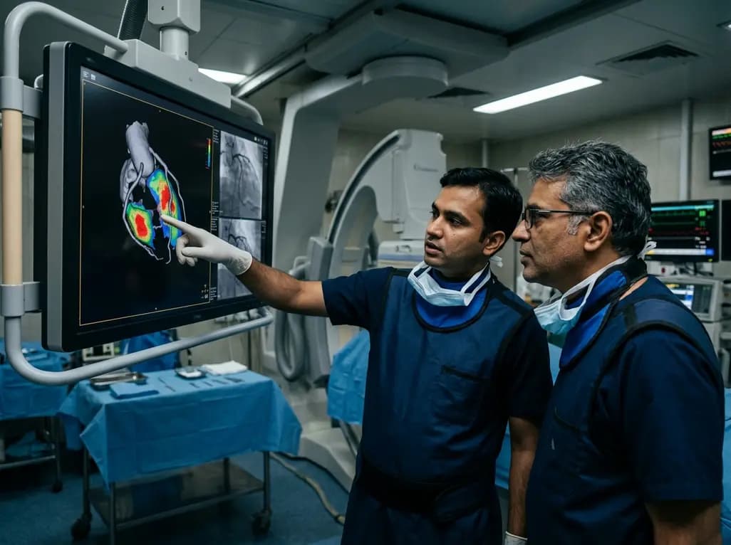
A groundbreaking study demonstrates that coronary angiography-derived radial wall strain reflects plaque morphology, predicting future rupture and erosion.
Today

The Delhi Cabinet has cleared an advanced seven-storey critical care expansion at Guru Tegh Bahadur Hospital under PMSSY Phase-IV. Costing Rs 244.87 crore, the new wing features 250 beds, specialized intensive care units, modern operation theatres, and round-the-clock emergency diagnostics for East Delhi.
Today

A landmark case demonstrates that favorable continuous glucose monitoring metrics cannot rule out SGLT2 inhibitor-associated euglycemic diabetic ketoacidosis. Clinicians must educate patients on symptom-based sick-day rules, uninterrupted basal insulin therapy, and proactive blood ketone testing.
Today
