.webp&w=384&q=75)

Loading, please wait...
Loading, please wait...

Atherosclerotic cardiovascular disease remains a leading cause of premature mortality worldwide. Although standard invasive catheterization reliably delineates anatomical luminal narrowing, it frequently fails to assess biomechanical lesion instability. Recently, clinical researchers identified radial wall strain as an innovative physiological marker that measures coronary arterial deformability during cardiac cycles. By quantifying dynamic lumen variations from routine angiograms, interventional cardiologists can gauge focal mechanical stress. Consequently, this computational approach bridges the gap between baseline angiography and future acute coronary events.
Coronary plaque disruption triggers catastrophic myocardial infarctions across diverse patient cohorts. Historically, interventional operators evaluated coronary disease severity primarily through percentage diameter stenosis. However, large clinical registries confirm that acute coronary syndromes often arise from mild-to-intermediate, non-obstructive plaques. Therefore, understanding intrinsic plaque composition and physical stress distribution is essential for modern cardiovascular risk assessment.
Biomechanical stress within the coronary arterial wall determines whether a fibrous cap remains intact or ruptures. In this dynamic vascular environment, radial wall strain serves as a dependable surrogate of arterial compliance. When cyclic blood pressure distends the vessel, diseased segments exhibit heterogeneous structural deformation. Specifically, lipid-rich necrotic cores covered by thin fibrous caps undergo pronounced stretching compared to dense fibrotic tissue. Consequently, elevated localized strain exposes vulnerable caps to critical fatigue over repetitive contractions. Because standard fluoroscopy omits intramural architecture, strain calculation adds vital functional depth to two-dimensional angiograms. Thus, measuring cyclic wall displacement enables early detection of biomechanically fragile lesions long before acute thrombotic occlusion occurs.
A recent study by Masuda and colleagues evaluated angiography-derived radial wall strain in acute coronary presentations. The researchers analyzed ninety-eight patients who underwent index coronary angiography up to five years before their acute event. During index catheterization, operators classified intermediate lesions into future culprit and non-culprit sites.
Subsequently, optical coherence tomography identified whether culprit lesions suffered plaque rupture or erosion. Interestingly, conventional angiographic parameters showed no differences between groups. However, maximum radial wall strain was significantly higher in culprit lesions than non-culprit lesions. Specifically, culprit rupture lesions demonstrated an average strain of 15.8 percent versus 12.6 percent in non-culprit sites. Furthermore, plaque erosions displayed an elevated strain of 14.1 percent. Multivariable Cox models confirmed that higher strain independently predicted subsequent plaque ruptures and erosions. Therefore, baseline coronary angiograms harbor critical information regarding lesion-specific mechanical vulnerability.
Acute coronary syndromes stem from distinct pathological mechanisms, primarily plaque rupture and plaque erosion. Plaque rupture typically features a voluminous necrotic core, intense macrophage infiltration, and a fractured fibrous cap. In contrast, plaque erosion preserves fibrous cap continuity, presenting with superficial endothelial loss and platelet thrombi. In the Masuda study, multinomial logistic regression demonstrated distinct strain distributions across these histological phenotypes.
Higher maximum radial wall strain strongly favored future plaque rupture over non-culprit lesions. Conversely, lower strain values reliably characterized stable non-culprit plaques. Meanwhile, the estimated probability of plaque erosion remained modest across the entire strain spectrum. These results highlight key mechanistic variations in coronary atherothrombosis. Because plaque rupture involves marked cap thinning, elevated cyclic stress directly causes mechanical tearing. On the other hand, plaque erosion arises primarily from local shear stress disruptions rather than structural wall strain. Consequently, biomechanical profiling assists clinicians in identifying lesions prone to physical structural failure. This discrimination provides vital guidance for targeted patient management.
Interventional cardiologists frequently encounter mild-to-intermediate coronary narrowings during routine diagnostic procedures. Deciding which moderate lesions require aggressive intervention remains a major clinical challenge. Fractional flow reserve reliably detects ischemia-causing stenoses, but it cannot predict acute plaque rupture in non-flow-limiting lesions. Herein lies the profound utility of evaluating radial wall strain directly from standard cineangiography.
By measuring luminal variation during cardiac contraction without requiring pressure wires, this technique provides immediate structural risk assessment. Furthermore, it uncovers vulnerable plaques that appear visually benign on two-dimensional silhouettes. As a result, physicians can intensify medical therapy before adverse clinical events occur. High-risk patients presenting with elevated strain values benefit substantially from high-intensity statins, PCSK9 inhibitors, or antiplatelet escalation. In addition, recognizing biomechanically vulnerable phenotypes helps interventionalists avoid stenting stable lesions while closely monitoring precarious plaques. Therefore, integrating dynamic strain assessment into catheterization laboratory protocols substantially refines clinical decision-making. Ultimately, this paradigm improves outcomes by linking preventative strategies to plaque vulnerability.
Coronary artery disease presents an escalating public health crisis across South Asia, particularly among Indian patients with early-onset disease. Clinicians frequently evaluate younger individuals who exhibit diffuse coronary atherosclerosis with high lipid burdens. In this demanding environment, cost-effective diagnostic tools deliver exceptional value for timely risk stratification. Because radial wall strain relies on standard angiographic projections, catheterization laboratories require no expensive supplementary equipment or specialized catheters.
Consequently, high-volume centers can implement this computational method without increasing procedural expenses or extending patient radiation exposure. Furthermore, interventional teams can seamlessly combine physiological evaluation with diagnostic catheterization. When physicians identify intermediate lesions displaying elevated strain, they can initiate aggressive plaque-stabilizing pharmacotherapy promptly. Clinicians can also recommend closer non-invasive monitoring to track anatomical progression over time. Additionally, patient compliance improves when cardiologists share objective biomechanical data demonstrating plaque vulnerability. Thus, incorporating vessel wall strain analytics into routine Indian interventional cardiology workflows bridges a crucial preventative gap. Ultimately, this innovative approach empowers physicians to optimize long-term clinical care.
Radial wall strain measures the maximum relative percentage change in coronary luminal diameter throughout a single cardiac cycle. Clinicians derive this metric directly from standard coronary angiographic cine projections using automated edge-detection algorithms. By tracking systolic and diastolic vascular dimensions across cardiac contraction, the software calculates localized structural deformation. Higher values signify substantial vessel compliance mismatch and localized wall stress, highlighting plaques that carry elevated mechanical vulnerability for downstream clinical rupture.
Distinguishing between plaque rupture and plaque erosion is crucial because their underlying pathophysiology and therapeutic requirements differ substantially. Plaque rupture involves structural fibrous cap disruption overlying a necrotic lipid core, frequently demanding mechanical stenting. Conversely, plaque erosion features intact fibrous caps with overlying thrombi, which often heal effectively with potent antithrombotic pharmacotherapy alone. Identifying biomechanical strain variations helps cardiologists predict specific lesion behavior, avoid unnecessary stent implantations, and tailor long-term medical management strategies.
Radial wall strain complements rather than completely replaces high-resolution intravascular imaging modalities like optical coherence tomography or intravascular ultrasound. While intravascular imaging delivers microscopic visualization of fibrous cap thickness, lipid content, and macrophage accumulation, it requires invasive intracoronary instrumentation and added procedural cost. Conversely, strain analysis provides physiological biomechanical deformation data directly from routine angiograms. Combining angiographic strain screening with targeted optical coherence tomography yields an optimal, highly cost-effective strategy for comprehensive cardiovascular risk assessment.
Disclaimer: This content is for informational and educational purposes only... Refer to the latest local and national guidelines for clinical practice.
References

Read summarized clinical updates, watch expert medical content, and earn CME certifications right from your smartphone.
Open your smartphone camera to scan and install.





A groundbreaking study demonstrates that coronary angiography-derived radial wall strain reflects plaque morphology, predicting future rupture and erosion.
Today

A landmark randomized clinical trial published in NEJM reveals that high maternal intake of eggs and peanuts during pregnancy and lactation does not lower the risk of infant egg or peanut allergy at one year of age, reinforcing existing dietary recommendations for balanced maternal nutrition without restrictions.
Today
A nationwide Japan AMI Registry study reveals right ventricular involvement in RCA-related STEMI treated with PCI increases 12-month mortality (HR 2.05) and major bleeding (HR 2.28). In patients with RVI, major bleeding was tied to a nearly fivefold risk of death, highlighting crucial access and antithrombotic management.
Today

The SOLARIO trial shows that a short course (≤7 days) of systemic antibiotic therapy is noninferior to long courses (≥4 weeks) in orthopedic infections treated with local antibiotic carriers, with significantly fewer adverse events.
Today
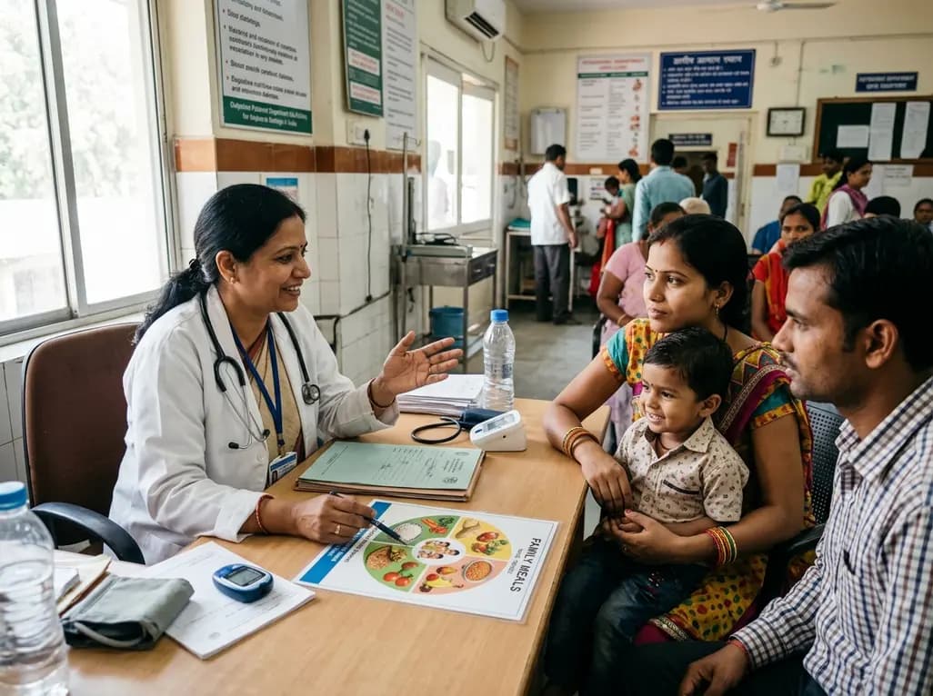
Gestational diabetes doubles the future risk of type 2 diabetes for mothers and offspring. Adopting a life-course approach centered on family meals and brief, supportive nutrition messaging empowers households to reduce metabolic risks and establish sustainable dietary habits across generations.
Today
