.webp&w=384&q=75)

Loading, please wait...
Loading, please wait...

Neurodegenerative disorders such as Alzheimer's disease and frontotemporal dementia present major diagnostic hurdles during their earliest stages. Novel computational techniques utilizing EEG centrality metrics provide an objective window into functional network deterioration. Clinicians recognize that Alzheimer's disease predominantly impairs episodic memory networks, whereas frontotemporal dementia dismantles fronto-insular socioemotional circuits. Consequently, both diseases disrupt large-scale functional communication across diverse cerebral regions long before structural atrophy manifests on standard neuroimaging. Resting-state electroencephalography offers high temporal resolution, wide accessibility, and cost-effectiveness. Therefore, neuroscientists increasingly utilize non-invasive scalp recordings to investigate functional connectivity. Rather than viewing dementia solely as localized cellular loss, modern neurology conceptualizes these conditions as complex disconnection syndromes. However, pathological protein aggregates progressively degrade delicate synaptic pathways across cortical hubs. As communication channels fail, the topological organization of the human connectome undergoes profound reshuffling. Early detection remains paramount because disease-modifying therapies yield optimal benefits before extensive neuronal destruction occurs. Developing sensitive electrophysiological biomarkers represents an urgent clinical priority.
Graph theory treats individual electrode positions as discrete nodes and their mutual phase synchronization as connecting edges. Within this mathematical framework, EEG centrality metrics quantify the relative importance and communication influence of specific nodes across the entire connectome. A recent pilot study comprehensively examined four distinct graph metrics: closeness centrality, betweenness centrality, leverage centrality, and weighted leverage measure. Specifically, the investigators evaluated resting-state electroencephalography recordings from forty-five participants, including fifteen patients with Alzheimer's disease, fifteen with frontotemporal dementia, and fifteen healthy controls. The research protocol utilized a standardized nineteen-channel electrode montage. Subsequently, the researchers constructed functional connectivity networks for every individual to determine whether stacked centrality metrics could reliably distinguish between neurodegenerative cohorts. Betweenness centrality evaluates how frequently a node falls along the shortest path between nodal pairs. Conversely, closeness centrality reflects how rapidly a node interacts with all other nodes across the network. Furthermore, leverage-based metrics appraise node influence relative to immediate anatomical neighbors.
The empirical findings revealed distinct topological reorganization patterns across both patient groups compared with healthy individuals. Notably, closeness centrality provided the strongest false discovery rate-corrected evidence of disease-related network alteration. Regional analyses confirmed that these significant alterations localized predominantly to the frontal, occipital, and temporal lobes. Furthermore, hemispheric evaluations identified prominent bilateral disruptions in closeness centrality, especially among patients suffering from Alzheimer's disease. In contrast, betweenness centrality demonstrated a corrected left-hemisphere alteration when comparing Alzheimer's disease subjects against healthy controls. Temporal region findings across multiple metrics aligned with known neurodegenerative progression pathways, although these regional trends remained partly exploratory after statistical correction. Moreover, the study highlighted clear pathophysiological divergence between the two conditions. Patients with frontotemporal dementia displayed localized frontotemporal disturbances, reflecting classic executive deficits. Meanwhile, patients with Alzheimer's disease exhibited widespread bilateral disconnectivity across extended cortical networks. Thus, stacked topological evaluations successfully identified distinct regional signatures reflecting specific neurodegenerative pathology.
Beyond identifying diagnostic group differences, the investigators correlated network abnormalities directly with clinical indicators. Specifically, leverage-based metrics displayed a strong negative correlation with patient age in frontotemporal dementia. This striking association suggests that younger individuals presenting with frontotemporal dementia may suffer accelerated functional network collapse. Additionally, both betweenness centrality and closeness centrality exhibited moderate positive associations with Mini-Mental State Examination scores. Consequently, these metrics directly mirror the severity of general cognitive decline observed at the bedside. As synaptic failure spreads, vital network hubs lose their efficient shortest-path communication channels. Therefore, information processing slows substantially, impairing attention, memory consolidation, and executive decision-making. These quantitative correlations emphasize that mathematical graph metrics capture genuine neurobiological degeneration rather than non-specific recording artifacts. Furthermore, tracking functional degradation through scalable electrophysiological assessments may help clinicians monitor therapeutic responses during future clinical trials. When pharmaceutical interventions stabilize synaptic communication, corresponding topological improvements should theoretically manifest across these centrality measures.
The clinical diagnosis of dementia syndromes remains exceedingly challenging in routine outpatient practice. For instance, early-stage behavioral frontotemporal dementia often mimics primary psychiatric illnesses, while atypical Alzheimer's disease variants can confuse experienced clinicians. Advanced neuroimaging modalities like positron emission tomography and volumetric magnetic resonance imaging provide valuable anatomical and molecular information. However, these tools carry high financial costs and limited availability across secondary healthcare facilities. In contrast, resting-state electroencephalography represents an inexpensive, non-invasive, and widely accessible neurodiagnostic platform. Clinicians can readily perform standard nineteen-channel recordings in diverse outpatient environments without subjecting elderly patients to radiation or claustrophobic scanners. If automated computational algorithms can reliably extract stacked centrality metrics, general practitioners and neurologists could identify insidious neurodegeneration much earlier. Furthermore, electrophysiological profiling could support accurate differential diagnosis in resource-limited settings where specialized scanners are absent. Integrating quantitative electrophysiology into memory clinics may ultimately streamline patient referrals and accelerate timely supportive care.
Although these pilot results appear highly encouraging, clinicians must interpret them within appropriate clinical context. First, the small sample size of forty-five participants warrants cautious extrapolation to broader, heterogeneous clinical populations. Second, the modest nineteen-channel montage limits fine spatial resolution, meaning that regional localization remains relatively coarse. High-density electroencephalography arrays or simultaneous magnetoencephalography could provide superior spatial mapping of deeper cortical and subcortical generators. Additionally, the cross-sectional nature of the pilot investigation precludes definitive conclusions regarding prognostic value or longitudinal rate of change. Longitudinal cohorts are essential to confirm whether centrality shifts predict progression from mild cognitive impairment to full-blown dementia syndromes. Moreover, prospective studies must incorporate biomarker-verified cohorts, such as cerebrospinal fluid or plasma assays, to validate clinical diagnoses rigorously. Standardizing recording protocols and artifact rejection pipelines will also be vital before deploying automated network analytics into commercial software. Nonetheless, this investigation establishes a compelling foundation for utilizing graph-theoretical centrality metrics in neurodegenerative research.
Centrality metrics assess communication flow across neural hubs. Closeness centrality shows marked bilateral alterations in Alzheimer's disease, reflecting widespread network disruption across frontal, temporal, and occipital lobes. Conversely, frontotemporal dementia demonstrates focal frontal and temporal network alterations, alongside distinct age-associated leverage changes, enabling clinicians to separate these two neurodegenerative conditions non-invasively.
Closeness centrality measures how rapidly a brain region connects with all other nodes across the connectome. Because neurodegenerative pathologies disrupt long-range axonal pathways and synaptic integrity, affected hubs require longer operational pathways. Closeness centrality reliably captures this global functional disconnection, providing robust, statistically corrected evidence of dementia-related network degradation.
Currently, electroencephalography network analysis serves as an adjunctive physiological screening tool rather than a replacement for neuroimaging. While magnetic resonance imaging identifies structural atrophy and positron emission tomography visualizes protein deposits, electrophysiology uniquely captures dynamic functional disconnections. Consequently, combining quantitative electroencephalography with established molecular imaging optimizes overall diagnostic accuracy in dementia.
Disclaimer: This content is for informational and educational purposes only and should not be considered medical advice. It is not intended to replace professional judgment, clinical diagnosis, or individualized patient care. Always consult qualified healthcare professionals for diagnosis and treatment decisions. Refer to the latest local and national guidelines for clinical practice.
References

Read summarized clinical updates, watch expert medical content, and earn CME certifications right from your smartphone.
Open your smartphone camera to scan and install.





A recent pilot study demonstrates that stacked EEG centrality metrics, especially closeness centrality, identify distinct functional network disruptions in Alzheimer's disease and frontotemporal dementia, offering a non-invasive approach for early neurodegenerative detection.
Today

A crossover study demonstrates that locally deployed large language models with exact-match knowledge augmentation significantly elevate outpatient prescription review accuracy to 97.2%, curbing hallucinations and securing patient data without requiring complex cloud infrastructure.
Today

A new scoping review protocol systematically maps global evidence on how climatic shifts, ambient heat, and pollution impact human fertility. Discover key physiological mechanisms, clinical preconception strategies, and critical public health alignments under Sustainable Development Goals 3 and 13.
Today

Pharmacogenomics and artificial intelligence are revolutionizing cardiology by tailoring therapies to individual genetic profiles and clinical data, reducing adverse drug events, and improving cardiovascular patient outcomes.
Today
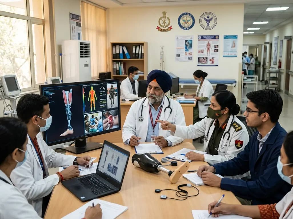
India's premier scientific bodies have joined the Armed Forces Medical Services to solve unique physiological and operational challenges faced by soldiers. The national collaboration covers combat casualty care, artificial intelligence diagnostics, bionic prosthetics, and extreme-environment physiological resilience.
Today

A retrospective cohort study evaluates the short- and long-term impacts of SARS-CoV-2 infection on patients with Graves' disease receiving antithyroid drug therapy, highlighting thyroid status destabilization, clinical symptom exacerbations, and post-viral sequelae.
Today
