.webp&w=384&q=75)

Loading, please wait...
Loading, please wait...

Advances in multimodal pediatric neuro-oncology have significantly improved survival rates for young patients with central nervous system neoplasms. However, childhood brain tumor survivors frequently experience debilitating late neurocognitive sequelae that disrupt academic success and adult independence. Furthermore, conventional structural magnetic resonance imaging cannot adequately capture the subtle microstructural disruption occurring within normal-appearing brain tissue. Therefore, clinicians urgently require sensitive neuroimaging biomarkers to monitor ongoing cerebral development and detect vulnerable patients early. Diffusion tensor imaging provides an advanced quantitative method to evaluate white matter tract integrity and subcortical microarchitecture. Specifically, this modality assesses fractional anisotropy and mean diffusivity, reflecting axonal density and cellular barriers. In a pioneering longitudinal investigation, researchers examined changes in callosal and hippocampal microstructure among childhood brain tumor survivors. Consequently, their observations elucidate why certain young patients maintain intact cognition while others experience progressive intellectual decline.
Pediatric central nervous system neoplasms represent a biologically complex group of malignancies requiring aggressive multimodal therapy. Although therapeutic interventions cure many pediatric patients, therapy frequently exposes developing neural circuits to severe cellular stress. Consequently, survivors often demonstrate delayed declines in processing speed, intellectual quotient, and working memory domains. In this context, researchers sought to determine whether longitudinal variations in diffusion metrics correlate with distinct neurocognitive trajectories. Specifically, fractional anisotropy measures the directional preference of water diffusion along myelinated axons. Higher fractional anisotropy generally indicates well-organized, densely packed white matter tracts and mature myelination. Conversely, mean diffusivity quantifies the overall magnitude of water motion across neural tissues. Elevated mean diffusivity typically reflects cellular edema, disrupted membranes, or expanded extracellular spaces. In developing children, normal brain maturation involves progressive increases in fractional anisotropy and parallel decreases in mean diffusivity. However, cranial radiation and chemotherapy may disrupt these physiological maturational dynamics. Therefore, analyzing these parameters within the corpus callosum and hippocampus provides invaluable pathophysiological data regarding treatment toxicity versus normal neurodevelopment.
To investigate these developmental differences, researchers designed a comprehensive longitudinal investigation spanning eight clinical years between 2014 and 2022. The study cohort comprised sixty-eight pediatric brain tumor survivors who underwent serial diffusion tensor imaging paired with extensive neurocognitive evaluations. In total, the investigators analyzed four hundred sixty-four paired clinical and radiological assessments alongside eighty matched healthy control participants. Furthermore, all oncology patients were younger than eighteen years at primary diagnosis and completed at least two longitudinal follow-up evaluations. The investigators employed multitrajectory modeling to group patients into distinct developmental cognitive phenotypes. Specifically, the modeling analyzed three vital performance domains: full-scale intelligence quotient, processing speed index, and working memory performance. As a result, the statistical clustering separated the pediatric cohort into two primary clinical phenotypes: a low-performance group and a normal-performance group. Patients in the low-performance group displayed deteriorating cognitive scores falling significantly below age-matched population norms over time. In contrast, patients within the normal-performance group sustained stable cognitive abilities comparable to healthy individuals. Subsequently, linear mixed models evaluated how these distinct developmental trajectories correlated with longitudinal imaging parameters.
The imaging analyses revealed profound divergences in white matter microstructure between the two patient trajectory groups. Notably, patients in the normal-performance group exhibited progressive increases in fractional anisotropy across successive scans. In addition, these normal-performance survivors demonstrated a corresponding longitudinal decrease in mean diffusivity within major cerebral pathways. Most importantly, this directional change closely mirrored the physiological neurodevelopmental trajectory observed in healthy control participants. In stark contrast, survivors belonging to the low-performance cluster exhibited a completely opposite neuroimaging pattern over time. These low-functioning patients showed declining fractional anisotropy and rising mean diffusivity throughout critical cerebral white matter tracts. For instance, the corpus callosum demonstrated marked microstructural disruption, reflecting ongoing axonal degradation and demyelination. Furthermore, these microstructural abnormalities were not isolated to callosal pathways but extended across widespread white matter networks throughout the cerebrum. Consequently, these findings indicate that childhood cancer therapy can severely perturb systemic neurodevelopmental timelines. While some young brains manage to recover and resume normal myelination, others suffer persistent microstructural injury that manifests as measurable cognitive impairment.
Beyond widespread white matter pathways, the study identified critical microstructural alterations within subcortical gray matter structures, particularly the hippocampus. Because the hippocampus plays a central role in memory consolidation and intellectual function, hippocampal microstructural integrity directly influences learning capacity. In this investigation, the low-performance group exhibited persistently elevated mean diffusivity within the bilateral hippocampi over time. Conversely, normally developing controls and normal-performance survivors displayed progressive hippocampal diffusivity reductions with age. Therefore, elevated hippocampal mean diffusivity serves as a direct radiological surrogate of impaired neurogenesis and dendritic rarefaction. Similarly, the corpus callosum exhibited distinct differences in axial and radial diffusivity between the two clinical groups. Because the corpus callosum coordinates interhemispheric communication, callosal axonal loss severely hinders information processing speed. In addition, cerebellar peduncles demonstrated strong correlations with working memory and processing speed metrics among survivors. Specifically, reduced fractional anisotropy in cerebellar pathways corresponded with impaired executive functioning and attention. Consequently, monitoring these specific callosal and subcortical biomarkers allows clinicians to detect subclinical neurotoxicity long before school performance falters.
These groundbreaking longitudinal observations carry profound clinical implications for contemporary pediatric oncology and radiation therapy practice. Historically, clinical surveillance after brain tumor treatment relied solely on standard macroscopic structural imaging to exclude tumor recurrence. However, gross structural scans remain entirely blind to microstructural injury and axonal degeneration. Therefore, integrating diffusion tensor imaging into routine neuro-oncology survivorship protocols can bridge this critical diagnostic gap. In clinical practice, identifying adverse white matter changes early enables oncologists to refer patients for prompt neurocognitive rehabilitation. Furthermore, cognitive remediation programs and academic accommodations show greater efficacy when initiated during early stages of cerebral plasticity. In addition, radiation oncologists can utilize substructure-specific diffusion sensitivity metrics to optimize advanced treatment planning. Specifically, radiation oncologists can contour the hippocampus, corpus callosum, and cerebellar peduncles as distinct avoidance structures during proton therapy or intensity-modulated photon radiotherapy. As a result, clinicians can reduce unnecessary radiation doses to sensitive neurocognitive tracts without compromising oncological control. Ultimately, incorporating diffusion tensor biomarkers into routine care transforms childhood neuro-oncology by preserving vital developmental potential.
Diffusion tensor imaging detects neurocognitive vulnerability by measuring water diffusion across cerebral tissues. Specifically, it assesses fractional anisotropy and mean diffusivity within white matter tracts. These quantitative parameters reflect axonal organization, myelin density, and cellular architecture, revealing microscopic neurotoxic injury well before gross structural lesions appear on conventional neuroimaging.
Longitudinal studies demonstrate that the corpus callosum and hippocampus exhibit the strongest associations with cognitive decline. In addition, the middle and inferior cerebellar peduncles strongly influence processing speed and working memory. Microstructural disruption within these interhemispheric and subcortical pathways impairs neural transmission, leading to delayed academic and executive deficits.
Yes, diffusion tensor imaging findings directly influence modern radiation therapy planning. Specifically, radiation oncologists can designate sensitive white matter tracts, including the corpus callosum and bilateral hippocampi, as avoidance structures. By limiting radiation doses to these vulnerable microstructures during proton or photon therapy, clinicians preserve essential long-term neurocognitive function.
Disclaimer: This content is for informational and educational purposes only and is not intended as medical advice, endorsement, or recommendation. Healthcare professionals should make clinical decisions based on their independent medical judgment and patient-specific factors. Refer to the latest local and national guidelines for clinical practice.
References

Read summarized clinical updates, watch expert medical content, and earn CME certifications right from your smartphone.
Open your smartphone camera to scan and install.





A longitudinal study utilizing diffusion tensor imaging reveals distinct neurocognitive trajectories in pediatric brain tumor survivors. Alterations in fractional anisotropy and mean diffusivity within the corpus callosum and hippocampus identify patients at risk for late intellectual decline.
Today

A crossover study demonstrates that locally deployed large language models with exact-match knowledge augmentation significantly elevate outpatient prescription review accuracy to 97.2%, curbing hallucinations and securing patient data without requiring complex cloud infrastructure.
Today

A new scoping review protocol systematically maps global evidence on how climatic shifts, ambient heat, and pollution impact human fertility. Discover key physiological mechanisms, clinical preconception strategies, and critical public health alignments under Sustainable Development Goals 3 and 13.
Today

Pharmacogenomics and artificial intelligence are revolutionizing cardiology by tailoring therapies to individual genetic profiles and clinical data, reducing adverse drug events, and improving cardiovascular patient outcomes.
Today
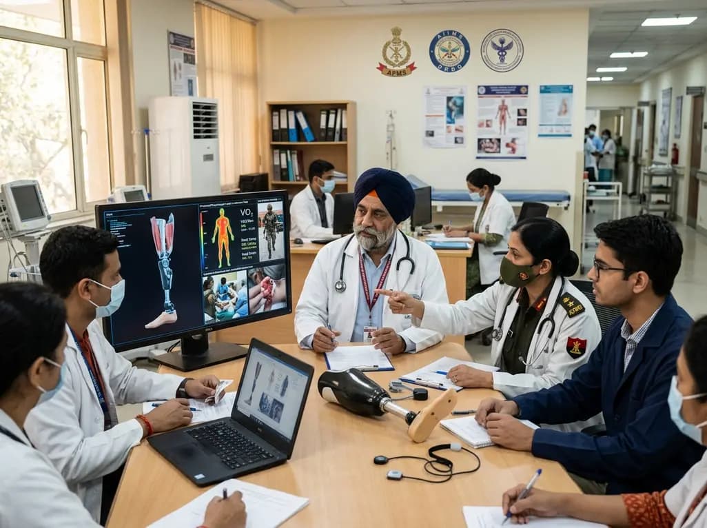
India's premier scientific bodies have joined the Armed Forces Medical Services to solve unique physiological and operational challenges faced by soldiers. The national collaboration covers combat casualty care, artificial intelligence diagnostics, bionic prosthetics, and extreme-environment physiological resilience.
Today

A retrospective cohort study evaluates the short- and long-term impacts of SARS-CoV-2 infection on patients with Graves' disease receiving antithyroid drug therapy, highlighting thyroid status destabilization, clinical symptom exacerbations, and post-viral sequelae.
Today
