

Loading, please wait...
Loading, please wait...

Glioblastoma remains the most lethal primary central nervous system malignancy in adult patients. Within IDH-wildtype glioblastoma, molecular profiling provides essential guidance for clinical prognostication and therapeutic decision-making. Specifically, the epigenetic silencing of the O6-methylguanine-DNA methyltransferase gene dictates tumor sensitivity to alkylating chemotherapy such as temozolomide. However, current clinical practice depends entirely upon invasive surgical resection or stereotactic biopsy to acquire tissue specimens for molecular assays. To address procedural risks, investigators actively pursue non-invasive radiogenomic tools. Advanced machine learning models aim to infer epigenetic alterations directly from diagnostic neuroimaging. Consequently, non-invasive MGMT promoter methylation prediction using multiparametric magnetic resonance imaging has emerged as an active frontier in modern neuro-oncology. A major multi-institutional study led by Leone and colleagues now provides critical insights into the real-world performance of these computational architectures.
In contemporary neuro-oncology, establishing MGMT promoter status directly dictates systemic treatment regimens. When promoter methylation silences the repair enzyme, alkylating agents such as temozolomide induce cytotoxic DNA cross-links and trigger apoptosis in neoplastic cells. Conversely, unmethylated tumors rapidly repair alkylator-induced DNA lesions, leading to therapeutic resistance and shorter overall survival. For elderly or frail patients, knowing this status preoperatively can alter surgical goals and spare individuals toxic regimens lacking therapeutic benefit. Unfortunately, molecular assays require stereotactic tissue acquisition, which carries surgical risks including hemorrhage, neurological deficit, and anesthesia complications. Furthermore, spatial intratumoral heterogeneity often confounds focal tissue biopsies, yielding unrepresentative genetic profiles. Preoperative neuroimaging captures the complete three-dimensional tumor volume, offering a comprehensive assessment of phenotypic heterogeneity. Therefore, non-invasive imaging biomarkers provide an attractive method to stratify patients before initiating interventions. Robust computational algorithms could expedite personalized oncologic planning, particularly in centers with limited molecular pathology resources.
Public neuroimaging datasets, notably the Brain Tumor Segmentation challenge, serve as the primary proving ground for radiogenomic architectures. Researchers frequently utilize these curated repositories to develop three-dimensional convolutional neural networks. In the multi-institutional investigation by Leone and colleagues, investigators analyzed 949 glioblastoma cases to evaluate model stability. Under controlled benchmark conditions within the BraTS dataset, whole-brain deep learning architectures demonstrated encouraging discriminative capacity. Specifically, a three-dimensional DenseNet121 architecture achieved an area under the receiver operating characteristic curve of 0.80. This level of accuracy generated substantial optimism for non-invasive molecular characterization. Because benchmark datasets feature standardized voxel spacing, uniform skull-stripping, and curated image contrasts, algorithms easily identify reproducible feature sets. Consequently, algorithms achieve high statistical performance within these idealized algorithmic sandboxes. These results initially suggested that end-to-end neural networks could bypass complex manual tumor delineation and reliably predict epigenetic markers directly from entire brain volumes.
Despite remarkable accuracy on public benchmarks, deep learning architectures frequently experience performance collapse when deployed in real-world clinical environments. In the study, investigators systematically tested the trained neural models on an independent, held-out multicenter patient cohort. Strikingly, the whole-brain models exhibited marked performance deterioration when encountering uncurated clinical scans. Real-world hospital networks utilize diverse magnetic resonance scanners from different manufacturers, operating at varying magnetic field strengths. Furthermore, clinical acquisition protocols introduce subtle discrepancies in repetition times, echo times, slice thicknesses, and contrast injection delays. Neural networks inadvertently latch onto non-biological imaging artifacts and scanner-specific signatures instead of genuine tumor physiology. As a result, models trained on homogeneous benchmark repositories fail to generalize across external hospital networks. This performance degradation highlights the fundamental danger of deploying radiogenomic tools into clinical workflows without rigorous multicenter cross-validation.
To mitigate generalizability failures, the researchers compared whole-brain classification models against segmentation-guided region-of-interest frameworks. The team deployed an nnU-Net framework paired with expert quality control to delineate precise anatomical tumor boundaries across multiparametric sequences. In contrast to whole-brain models, segmentation-guided region-of-interest pipelines maintained superior stability on the held-out multicenter test cohort. Restricting feature extraction to explicit tumor margins substantially reduced false-positive classifications. Whole-brain models often extract spurious correlations from normal brain parenchyma or non-tumor background tissues. Conversely, targeted segmentation forces the network to evaluate biologically meaningful areas, such as enhancing margins, necrotic cores, and peritumoral edema. Therefore, explicit lesion localization prevents algorithms from memorizing scanner-specific background noise. These results demonstrate that upstream methodological choices remain vital determinants of real-world model reliability. While automated segmentation requires computational resources, it provides a necessary guardrail against domain shift.
Translating artificial intelligence from academic benchmarking into everyday neurosurgical oncology requires addressing several technical and regulatory barriers. First, researchers must incorporate robust domain-shift harmonization techniques to standardize diverse magnetic resonance scans before neural processing. Advanced algorithms, such as generative adversarial networks, can align imaging distributions across heterogeneous scanners. Second, integrating multimodal data streams could enhance diagnostic precision. Combining radiomic features with clinical parameters and circulating liquid biopsy markers may yield superior diagnostic accuracy compared to imaging alone. Third, clinical software must incorporate uncertainty-aware deep learning metrics. Quantifying algorithmic confidence enables clinicians to identify borderline predictions and defer to conventional tissue sampling when uncertainty is elevated. Ultimately, prospective multicenter clinical trials across diverse global populations, including Indian healthcare centers, remain imperative. Only through rigorous prospective validation can radiogenomic tools earn regulatory approval and provide reliable clinical guidance for brain tumor patients.
For practicing clinicians, these findings deliver crucial lessons regarding medical artificial intelligence adoption. Clinicians should view current artificial intelligence predictions as experimental decision-support tools rather than definitive diagnostic surrogates. When interpreting radiogenomic literature, neurosurgeons and radiologists must scrutinize validation methodologies carefully. Studies claiming exceptional accuracy on single-center or public benchmark datasets often overstate clinical readiness. Instead, true clinical utility demands testing on heterogeneous external cohorts with varied scanner hardware. Furthermore, multidisciplinary collaboration between radiologists, neurosurgeons, and data scientists remains essential to ensure algorithmic transparency. Clinicians must actively participate in annotating training datasets and validating region-of-interest segmentations to eliminate biological inaccuracies. By recognizing the strengths and limitations of radiogenomic models, the medical community can foster responsible technology adoption that enhances diagnostic workflows while maintaining patient safety.
MGMT promoter methylation silences an essential DNA repair enzyme, leaving glioblastoma cells susceptible to alkylating chemotherapy such as temozolomide. Consequently, methylated status predicts superior response to standard chemoradiotherapy and prolonged overall survival. Unmethylated tumors frequently exhibit intrinsic chemoresistance, prompting oncologists to consider alternate clinical trials or modified radiotherapy schedules.
Real-world datasets introduce significant domain shift from diverse scanner manufacturers, varied magnetic field strengths, and inconsistent imaging protocols. Benchmark datasets feature uniform preprocessing, whereas clinical scans contain variable noise and resolution. Consequently, models trained on homogeneous benchmarks often fail to generalize when encountering unfamiliar real-world institutional imaging artifacts.
Segmentation-guided modeling restricts deep learning feature extraction to explicit anatomical tumor compartments rather than the entire brain. This targeted approach prevents the algorithm from learning spurious correlations in normal brain tissue or background imaging noise. Consequently, lesion-focused analysis substantially reduces false-positive classifications and enhances model robustness across multicenter cohorts.
Disclaimer: This content is for informational and educational purposes only and should not be used as a substitute for professional medical advice, diagnosis, or treatment. Always seek the advice of your physician or other qualified health provider with any questions you may have regarding a medical condition. Refer to the latest local and national guidelines for clinical practice.
References

Read summarized clinical updates, watch expert medical content, and earn CME certifications right from your smartphone.
Open your smartphone camera to scan and install.





A landmark multicenter study evaluates deep learning for non-invasive MGMT promoter methylation prediction in glioblastoma, comparing benchmark models against real-world hospital cohorts to highlight generalizability challenges and the value of segmentation-guided workflows.
Today

A validated natural language processing pipeline leverages clinical notes and intermediate annotations to identify risky alcohol use across surgical patients, offering automated preoperative risk stratification.
Today

The GESS-AEEM consensus establishes a clear clinical implementation framework for testosterone therapy in postmenopausal women with hypoactive sexual desire disorder, focusing on transdermal dosing, precise laboratory monitoring, and safety communication.
Yesterday
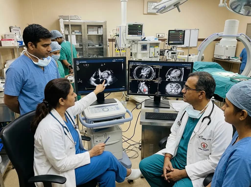
A rare case of an asymptomatic left ventricular hemangioma highlights the vital role of multimodal imaging in differentiating vascular tumors from thrombus. Complete surgical resection under transesophageal echocardiography guidance prevents thromboembolism and delivers optimal outcomes.
Today

A landmark study evaluates long-term quality of life in adolescents and adults with brachial plexus birth injury. Severe functional limitation and limited access to care significantly worsen outcomes, underscoring the urgent need for persistent, multidisciplinary management across adult healthcare transitions.
Today

An analysis of 1,000 posts by macro- and mega-influencers on Instagram and TikTok revealed that 78.5% contained nutrition misinformation, while only 24% of creators held valid credentials. This article examines the clinical risks of viral dietary fads and provides actionable guidance for healthcare professionals.
Today
