

Loading, please wait...
Loading, please wait...

For decades, neurosurgeons regarded microsurgical clipping as a definitive cure for vascular lesions. However, modern imaging reveals that clipped intracranial aneurysms require far more vigilance than previously thought. Historical data on recurrence rates primarily relied on conventional two-dimensional digital subtraction angiography. Unfortunately, conventional planar angiography fails to identify subtle morphological flaws along the clip construct. Consequently, clinical teams frequently conflate true de novo recurrences with preexisting unclipped tissue remnants. To resolve this longstanding ambiguity, researchers synthesized comprehensive volumetric imaging datasets. A seminal systematic review analyzed thirty-eight studies encompassing thousands of treated vascular patients. The investigators established three-dimensional digital subtraction angiography as the definitive reference standard. Through this rigorous meta-analysis, clinicians gained unprecedented insights into surgical obliteration quality. The findings demonstrate that post-clipping remnants occur far more frequently than historical studies reported. Moreover, these newly recognized baseline defects carry distinct biomechanical risks over time. Vascular specialists must therefore update their clinical paradigms to reflect high-resolution diagnostic realities. Embracing advanced rotational angiography allows neurosurgical teams to establish accurate post-operative baselines. Ultimately, precise anatomical visualization improves long-term patient counseling and surgical audit quality.
Evaluating post-clipping vascular anatomy demands exceptional visual resolution. Historically, two-dimensional digital subtraction angiography served as the diagnostic benchmark for surgical durability. However, planar projections suffer from severe vessel overlap and clip-induced metallic artifacts. In contrast, rotational three-dimensional angiography reconstructs intricate vascular geometries without substantial obscuration. The recent landmark meta-analysis uncovered startling discrepancies between these two imaging modalities. According to three-dimensional datasets, surgeons leave an anatomical remnant in approximately 23.5 percent of cases. Even more critically, two-dimensional angiography demonstrated an overall false-negative rate exceeding fifty-one percent. When assessing small neck remnants under two millimeters, planar imaging missed nearly seventy-nine percent of defects. Therefore, relying solely on standard two-dimensional imaging gives surgical teams a false sense of security. Small dog-eared residuals often escape detection during routine post-operative planar assessments. Consequently, clinicians incorrectly classify subsequent remnant expansion as de novo aneurysm formation. This diagnostic blind spot distorts long-term epidemiological data and skews risk stratification models. Neurovascular centers must recognize these technological limitations when reviewing post-clipping angiograms. Hence, adopting volumetric imaging proves essential for accurate postoperative evaluation in modern neurosurgical centers.
Understanding which residual vascular defects will progress remains a vital neurosurgical objective. The meta-analytic evidence shows that completely clipped lesions demonstrate extraordinary long-term durability. In fact, fully obliterated aneurysms rarely exhibit true recurrence during extended clinical follow-up. Conversely, incompletely clipped intracranial aneurysms display substantial vulnerability to progressive remnant expansion. The meta-analysis established that incomplete occlusion significantly elevates the relative risk of vascular recurrence. Furthermore, specific baseline patient demographics and morphological variables dictate long-term stability. Remnant size serves as the most potent independent predictor of subsequent enlargement. Residual necks larger than two millimeters experience markedly higher hemodynamic shear stress. Consequently, larger remnants show an accelerated tendency toward structural destabilization and growth. In addition, younger patient age substantially increases the cumulative lifetime risk of remnant enlargement. Younger individuals maintain active arterial wall remodeling and endure decades of continuous pulsatile stress. Interestingly, presentation status during initial surgery also shapes subsequent structural risk profiles. Ruptured lesions that leave postoperative remnants frequently exhibit more unpredictable biological behavior. Therefore, neurovascular specialists must stratify surveillance intensity according to these precise anatomical and demographic determinants.
These compelling radiological findings demand a fundamental realignment in post-operative patient surveillance strategies. Historically, many neurosurgical protocols discharged clipped patients after an initial unremarkable planar angiogram. However, the high false-negative rate of conventional angiography renders that traditional practice obsolete. Clinicians must realize that standard imaging misses nearly four out of five small remnants. Therefore, vascular teams should obtain high-resolution volumetric imaging before declaring complete aneurysm obliteration. When surgeons confirm complete neck occlusion with rotational angiography, they can safely reassure patients. These completely cured individuals require minimal repeated radiological surveillance over subsequent years. On the other hand, patients harboring verified residual necks require structured, lifelong monitoring protocols. Neurosurgeons must explain this distinction transparently during postoperative counseling sessions. Patients must understand that a stable remnant behaves differently from a completely obliterated lesion. Furthermore, interdisciplinary neurovascular boards should review cases showing significant remnant expansion. When residual pockets expand beyond critical thresholds, clinicians must consider timely secondary interventions. Therapeutic options include microsurgical re-clipping, coil embolization, or flow-diverting stent reconstruction. Consequently, diligent follow-up prevents catastrophic delayed subarachnoid hemorrhage and ensures patient safety.
Implementing advanced post-clipping surveillance in India presents unique opportunities and notable infrastructural challenges. Tertiary neurosurgical centers across metropolitan hubs frequently possess cutting-edge rotational angiography equipment. However, resource-constrained secondary hospitals in semi-urban areas rely almost exclusively on standard planar digital angiography. Consequently, neurosurgeons practicing in Indian environments must exercise heightened clinical judgment when reviewing local imaging. If high-resolution rotational angiography is unavailable locally, clinicians should utilize dedicated contrast-enhanced computed tomography angiography. This non-invasive modality effectively detects larger residual pouches while minimizing procedure costs. Furthermore, Indian patients face distinct socioeconomic barriers to longitudinal hospital follow-up. Long travel distances and out-of-pocket healthcare expenses frequently lead to patient attrition over time. Therefore, surgical teams must optimize surveillance intervals to maximize compliance and diagnostic yield. Specialists should reserve intense radiological monitoring for younger patients with verified remnants or large ruptured aneurysms. Conversely, completely clipped elderly individuals can avoid burdensome recurring imaging schedules. Clinicians should also educate primary physicians across district clinics regarding delayed warning symptoms. Thus, a tiered surveillance protocol optimizes institutional resources while safeguarding vulnerable neurosurgical patients.
Conventional two-dimensional digital subtraction angiography flattens three-dimensional vascular structures into planar projections. Consequently, dense metallic clips frequently cast artifacts and obscure underlying vessel anatomy. Furthermore, tortuous parent arteries often overlap the surgical neck, concealing small residual pouches. Recent systematic review data show that planar angiography misses nearly seventy-nine percent of remnants under two millimeters. In contrast, three-dimensional rotational angiography provides comprehensive geometric reconstructions that reveal occult dog-eared remnants clearly.
Remnant size and patient age represent the primary clinical determinants of post-clipping remnant growth. Residual neck outpouches exceeding two millimeters experience heightened wall shear stress, making them prone to structural enlargement. Furthermore, younger patients face elevated lifetime growth risks due to enduring decades of continuous arterial pulsation and vascular remodeling. In addition, aneurysms that initially presented with acute rupture exhibit less predictable wall stability, demanding more rigorous surveillance protocols over time.
Managing an expanding remnant requires multidisciplinary evaluation by vascular neurosurgeons and interventional neuroradiologists. Clinicians thoroughly assess remnant geometry, patient comorbidities, and previous surgical approaches. If the remnant poses substantial re-rupture danger, treatment options include surgical re-clipping, coil embolization, or flow-diverting stent reconstruction. In contrast, small, asymptomatic, and stable residual rests under two millimeters often warrant conservative observation with serial non-invasive imaging, reserving active invasive interventions for documented anatomical progression.
Disclaimer: This content is for informational and educational purposes only... Refer to the latest local and national guidelines for clinical practice.
References

Read summarized clinical updates, watch expert medical content, and earn CME certifications right from your smartphone.
Open your smartphone camera to scan and install.





A systematic review reveals that 3D-DSA detects remnants in 23.5% of clipped intracranial aneurysms, while 2D-DSA carries a 51.7% false-negative rate. Complete clipping prevents recurrence, whereas incompletely clipped lesions require vigilant long-term surveillance for remnant growth.
Today
A national cross-sectional survey reveals marked disparities in infectious disease diagnostic capacity across hospital tiers, underscoring the urgent need for integrated referral networks and enhanced point-of-care testing to improve outbreak detection and patient outcomes.
Today

The GESS-AEEM consensus establishes a clear clinical implementation framework for testosterone therapy in postmenopausal women with hypoactive sexual desire disorder, focusing on transdermal dosing, precise laboratory monitoring, and safety communication.
Yesterday
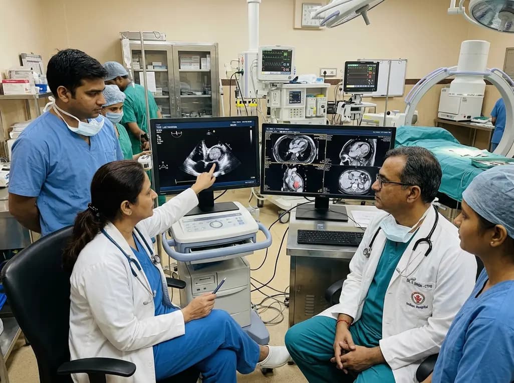
A rare case of an asymptomatic left ventricular hemangioma highlights the vital role of multimodal imaging in differentiating vascular tumors from thrombus. Complete surgical resection under transesophageal echocardiography guidance prevents thromboembolism and delivers optimal outcomes.
Today
A landmark study evaluates long-term quality of life in adolescents and adults with brachial plexus birth injury. Severe functional limitation and limited access to care significantly worsen outcomes, underscoring the urgent need for persistent, multidisciplinary management across adult healthcare transitions.
Today
An analysis of 1,000 posts by macro- and mega-influencers on Instagram and TikTok revealed that 78.5% contained nutrition misinformation, while only 24% of creators held valid credentials. This article examines the clinical risks of viral dietary fads and provides actionable guidance for healthcare professionals.
Today
