.webp&w=384&q=75)

Loading, please wait...
Loading, please wait...

The management of central nervous system secondary malignancies has evolved rapidly with the expanding adoption of immunotherapy. Evaluating brain metastases MRI phenotypes offers clinicians noninvasive physiological insights into therapeutic response and intratumoral microenvironmental changes. Conventional anatomical neuroimaging frequently struggles to differentiate between treatment-induced inflammatory changes and true tumor progression. Consequently, oncologists and neuroradiologists increasingly explore quantitative functional metrics, such as apparent diffusion coefficient and dynamic contrast subtraction. Recent investigational evidence confirms that distinct voxel-level imaging patterns correlate directly with immune checkpoint inhibitor therapy and histopathological inflammatory cell infiltration. Therefore, adopting multiparametric profiling can enhance diagnostic accuracy in neuro-oncology practice.
Immune checkpoint inhibitors stimulate antitumor immune responses by releasing T cells from immunosuppressive signaling pathways. However, these physiological mechanisms cause profound alterations within the delicate intracranial tumor microenvironment. Activated lymphocytes cross the disrupted blood-brain barrier to infiltrate metastatic tumor nests, generating local inflammatory cascades. In response, local microvessels show increased permeability, which manifests as prominent gadolinium extravasation on contrast-enhanced scans. Furthermore, therapeutic cytotoxic activity induces cell lysis, necrosis, and expanded extracellular volume. These cellular changes noticeably alter water molecule diffusivity within the lesion. Standard magnetic resonance imaging captures gross morphological alterations, but it fails to isolate specific cellular responses. In contrast, quantitative imaging captures these delicate pathophysiological processes simultaneously. Investigators specifically employ bivariate histograms of apparent diffusion coefficients and contrast-enhancement subtraction maps to categorize individual tumor voxels. These analytical models resolve the intracranial lesion into functional spatial domains. As a result, clinicians gain a more objective window into treatment efficacy before gross dimensional changes emerge. Multiparametric profiling thus provides a mechanistic link between systemic immunotherapy and local intracranial tissue architecture.
Multiparametric neuroimaging relies heavily on apparent diffusion coefficient mapping and contrast-enhancement quantification to characterize brain lesions. The apparent diffusion coefficient specifically measures the unconstrained Brownian motion of water molecules within tissue compartments. Densely packed malignant cells restrict water movement, resulting in low values across diffusion-weighted sequences. In contrast, tumor necrosis and edema increase extracellular space, which produces elevated apparent diffusion values. Concurrently, digital subtraction techniques applied to pre-contrast and post-contrast T1-weighted images isolate true gadolinium extravasation. This subtraction workflow effectively removes confounding intrinsic T1 hyperintensity caused by intratumoral hemorrhage or melanin deposits. By pairing these complementary physiological metrics, clinicians construct comprehensive bivariate voxel distributions. Specifically, researchers classify metastatic tissue into distinct functional quadrants based on these physiological parameters. Lesions exposed to immune checkpoint inhibitors demonstrate a substantial voxel fraction residing within the high-diffusion and high-enhancement quadrant. This unique imaging signature directly reflects simultaneous vasodilation, heightened vessel permeability, and inflammatory cell-mediated tumor disruption. Therefore, quantitative voxel mapping presents an objective methodology to monitor intracranial therapeutic responses.
Histopathological correlation confirms the biological validity of functional imaging observations in metastatic intracranial lesions. Researchers evaluated surgical tissue specimens to assess leukocyte infiltration following immune checkpoint inhibitor exposure. Specifically, investigators quantified leukocyte common antigen CD45-positive cell density within resected brain metastasis samples. The resulting pathological data demonstrated a robust, statistically significant relationship with quantitative imaging phenotypes. Lesions demonstrating high voxel concentrations in the high-diffusion and high-enhancement domain exhibited markedly higher CD45-positive immune cell density. Consequently, this observation confirms that prominent contrast extravasation accompanied by high water diffusivity signals active immune infiltration rather than rapid malignant proliferation. Conversely, uninhibited tumor progression typically produces dense cellularity, which markedly restricts diffusion and generates low apparent diffusion coefficients. Thus, advanced magnetic resonance mapping bridges the traditional gap between noninvasive imaging and microscopic immune biology. Clinicians can utilize these multiparametric signatures to confirm whether systemic checkpoint inhibitors effectively penetrate intracranial compartments. Furthermore, this imaging strategy provides reassurance when confronted with lesions showing increased contrast uptake during early treatment phases.
Corticosteroids remain a foundational therapy for mitigating peritumoral edema and managing neurological symptoms in patients with intracranial metastases. However, their potent anti-inflammatory properties exert pronounced physiological effects that dramatically alter neuroimaging appearances. Glucocorticoids rapidly restore blood-brain barrier integrity, which substantially decreases vessel permeability and contrast enhancement across T1-weighted sequences. Furthermore, steroids suppress local lymphocyte trafficking, potentially blunting the very inflammatory reaction that checkpoint inhibitors aim to stimulate. In clinical imaging analyses, multivariate models accounting for steroid exposure revealed an opposing radiological phenotype. Steroid-treated lesions demonstrated a marked reduction in contrast extravasation and significant shifts away from the high-enhancement quadrants. Therefore, administering dexamethasone can easily mask immunotherapy-induced inflammatory changes on routine MRI scans. Clinicians must interpret post-treatment scans with caution when patients receive concurrent corticosteroid therapy. Moreover, oncologists should record exact steroid dosages at the time of each imaging acquisition. Accounting for these pharmacological interactions ensures accurate differentiation between actual therapeutic failure and steroid-mediated imaging modulation.
Accurate response assessment remains one of the most formidable challenges in modern neuro-oncology. Immune checkpoint inhibitors frequently induce pseudoprogression, a phenomenon where lesions transiently enlarge due to intense inflammatory edema and leukocyte influx. Standard anatomical response criteria, such as RECIST or RANO, rely primarily on bidimensional contrast measurements. Consequently, conventional criteria frequently misinterpret therapeutic inflammatory infiltration as overt disease progression. This diagnostic error can lead oncologists to prematurely terminate effective systemic immunotherapies. By incorporating quantitative diffusion and contrast-enhancement phenotypes, multidisciplinary teams can resolve these clinical dilemmas more reliably. For example, a growing intracranial lesion that exhibits elevated apparent diffusion alongside contrast enhancement points toward immune infiltration. In contrast, progressive neoplastic growth demonstrates restricted diffusion driven by dense cellular packing and high nuclear-to-cytoplasmic ratios. Integrating advanced voxel-level phenotyping into routine clinical practice empowers clinicians to make confident management decisions. Therefore, patient care improves because oncologists can distinguish genuine therapeutic resistance from beneficial inflammatory flare.
The translation of quantitative magnetic resonance phenotypes into routine neuro-oncology workflows promises to transform brain metastasis management. Currently, widespread clinical adoption requires standardized post-processing algorithms and vendor-neutral acquisition protocols. Automated software pipelines can facilitate voxel-by-voxel bivariate analysis, generating intuitive color-coded spatial maps for reporting radiologists. Furthermore, combining diffusion and perfusion metrics with artificial intelligence models could refine predictive performance. Deep learning architectures can integrate multiparametric imaging with circulating tumor DNA and clinical biomarkers to forecast individual intracranial response trajectories. Longitudinal validation across larger multicenter prospective cohorts will solidify the clinical utility of these imaging biomarkers. In addition, future clinical trials should incorporate quantitative diffusion metrics into primary response evaluation frameworks. As systemic targeted therapies and immune combinations continue to expand, noninvasive physiological imaging will become indispensable. Ultimately, standardizing multiparametric imaging phenotypes will allow clinicians to tailor personalized therapies, minimize unnecessary neurosurgical interventions, and optimize clinical outcomes for patients facing complex metastatic cancer.
Brain metastases MRI phenotypes reflect immunotherapy activity by capturing shifts in tissue microstructure and vascular permeability. Lesions exposed to checkpoint inhibitors exhibit high water diffusivity and elevated contrast enhancement. This distinctive pattern correlates with cytotoxic tumor destruction, increased blood-brain barrier leakage, and dense CD45-positive leukocyte infiltration within the tumor.
Corticosteroids reduce peritumoral edema and rapidly restore blood-brain barrier integrity. Consequently, steroid therapy decreases contrast extravasation on post-contrast imaging and suppresses local leukocyte trafficking. This pharmacological suppression masks immunotherapy-induced inflammatory changes on routine MRI scans, potentially causing clinicians to misjudge the true biological response to checkpoint inhibitor therapy.
Multiparametric MRI differentiates pseudoprogression by evaluating apparent diffusion coefficients and contrast enhancement simultaneously. Pseudoprogression features elevated diffusion and high contrast leakage caused by inflammatory edema and leukocyte influx. Conversely, true tumor progression exhibits restricted water diffusion due to high cellular density, allowing clinicians to distinguish active immune infiltration from disease progression.
Disclaimer: This content is for informational and educational purposes only... Refer to the latest local and national guidelines for clinical practice.
References

Read summarized clinical updates, watch expert medical content, and earn CME certifications right from your smartphone.
Open your smartphone camera to scan and install.





Quantitative MRI phenotypes combining diffusion and contrast enhancement correlate with immune checkpoint inhibitor exposure and immune cell infiltration in brain metastases, offering clinicians a noninvasive tool to monitor intracranial immunotherapy responses.
Today

A crossover study demonstrates that locally deployed large language models with exact-match knowledge augmentation significantly elevate outpatient prescription review accuracy to 97.2%, curbing hallucinations and securing patient data without requiring complex cloud infrastructure.
Today

A new scoping review protocol systematically maps global evidence on how climatic shifts, ambient heat, and pollution impact human fertility. Discover key physiological mechanisms, clinical preconception strategies, and critical public health alignments under Sustainable Development Goals 3 and 13.
Today

Pharmacogenomics and artificial intelligence are revolutionizing cardiology by tailoring therapies to individual genetic profiles and clinical data, reducing adverse drug events, and improving cardiovascular patient outcomes.
Today
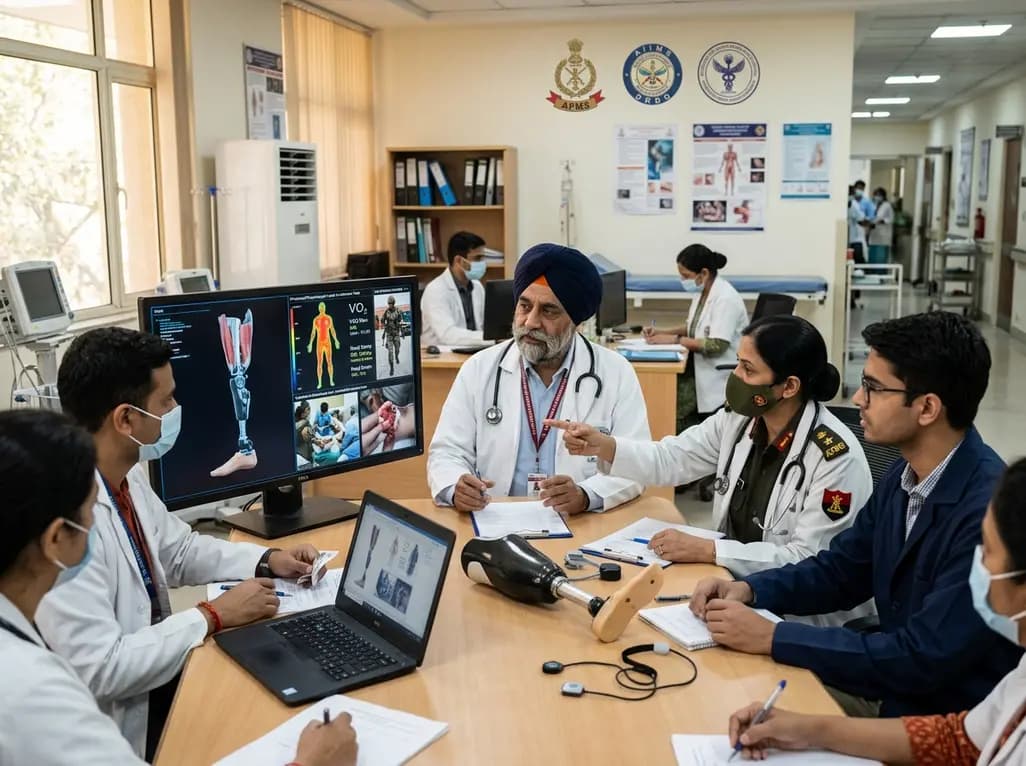
India's premier scientific bodies have joined the Armed Forces Medical Services to solve unique physiological and operational challenges faced by soldiers. The national collaboration covers combat casualty care, artificial intelligence diagnostics, bionic prosthetics, and extreme-environment physiological resilience.
Today

A retrospective cohort study evaluates the short- and long-term impacts of SARS-CoV-2 infection on patients with Graves' disease receiving antithyroid drug therapy, highlighting thyroid status destabilization, clinical symptom exacerbations, and post-viral sequelae.
Today
