.webp&w=384&q=75)

Loading, please wait...
Loading, please wait...
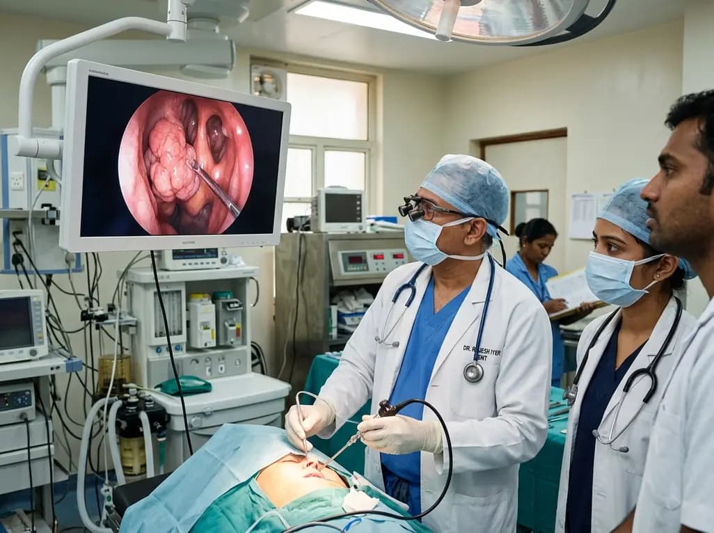
Benign sinonasal tumors represent a clinically diverse group of neoplasms arising within the nasal cavity and paranasal sinuses. Although these lesions lack malignant metastatic capabilities, their intimate proximity to the orbit and skull base poses formidable surgical challenges. Historically, surgeons relied primarily on open transfacial approaches to achieve complete clearance. Over the past two decades, however, endonasal endoscopic surgery has revolutionized operative outcomes. A comprehensive 17-year study by Kara and colleagues evaluated 368 patients surgically treated for benign sinonasal tumors. Their extensive real-world findings clarify histopathological patterns, surgical approaches, complications, and long-term recurrence risks.
In this expansive cohort, Kara and colleagues reported a median patient age of 42.5 years and a significant male predominance of 70.7 percent. This male predilection stems largely from specific underlying pathologies. Histopathologically, sinonasal papillomas formed the single largest diagnostic cohort, accounting for 34.2 percent of all treated cases. Among these, inverted papilloma represented the predominant subtype. These Schneiderian neoplasms demand careful attention because they cause local osteolysis and carry an inherent risk of malignant transformation. Additionally, osseous lesions constituted the second most frequent histological category at 22.6 percent. This group predominantly encompassed frontoethmoidal osteomas, fibrous dysplasia, and ossifying fibromas. Furthermore, juvenile nasopharyngeal angiofibromas comprised 19.0 percent of the surgical population, appearing almost exclusively in young male patients. Other less common benign entities included hemangiomas, schwannomas, and pleomorphic adenomas. Understanding this diverse histopathological distribution assists surgeons in anticipating clinical behavior, tailoring anatomical exposure, and optimizing therapeutic strategies.
Patients with sinonasal neoplasms frequently present with non-specific rhinologic symptoms that mimic common inflammatory conditions. Consequently, definitive diagnosis often experiences significant delays. Progressive unilateral nasal obstruction remains the most frequent presenting complaint. Additionally, recurrent unprovoked epistaxis strongly suggests hypervascular pathology such as juvenile nasopharyngeal angiofibroma or lobular capillary hemangioma. In contrast, expansile lesions like inverted papillomas often provoke secondary rhinosinusitis, facial pressure, and hyposmia. Cross-sectional imaging provides critical diagnostic insights prior to surgical intervention. High-resolution computed tomography meticulously delineates osseous anatomy, cortical erosion, and site-specific hyperostosis. Moreover, contrast-enhanced magnetic resonance imaging differentiates neoplastic tissue from retained sinonasal secretions while assessing intraorbital or intracranial invasion. Preoperative catheter angiography plays an essential role in vascular lesions. Specifically, preoperative embolization drastically minimizes intraoperative hemorrhage. Office biopsies require extreme caution, however, because inadvertently incising vascular masses can cause life-threatening bleeding.
Surgical techniques for sinonasal pathology have undergone a remarkable paradigm shift toward minimally invasive modalities. In their 17-year analysis, Kara and colleagues demonstrated that surgical approach differed significantly based on tumor histopathology and anatomical extent. Today, transnasal endoscopic resection serves as the gold standard for most benign neoplasms. High-definition endoscopes and angled micro-instruments afford pristine visualization of complex sinus architecture. Consequently, surgeons achieve complete clearance of sinonasal papillomas while preserving physiological sinonasal mucosa. Nevertheless, certain complex or extensive lesions still require open or combined approaches. Massive juvenile angiofibromas extending into the infratemporal fossa or middle cranial fossa often necessitate external corridors to secure arterial control. Similarly, far lateral frontal sinus osteomas may require an osteoplastic flap approach. Therefore, surgical teams must evaluate lesion geometry, vascularity, and skull base involvement when choosing the surgical corridor. The ultimate goal remains radical excision without creating debilitating aesthetic or functional defects.
Achieving negative margins around sinonasal tumors while preventing neurovascular complications requires exceptional surgical precision. In their cohort, Kara and colleagues observed that residual disease correlated with aggressive histology and difficult anatomic locations. Hypervascular tumors present substantial intraoperative bleeding risks. Therefore, surgeons utilize hypotensive anesthesia, preoperative embolization, and precise bipolar cautery to maintain adequate visibility. Major complications include cerebrospinal fluid leaks, orbital hematomas, and cranial nerve deficits. When tumors erode the skull base, endoscopic multilayer duraplasty using vascularized mucosal flaps repairs cerebrospinal fluid leaks effectively. Orbital violations require immediate recognition and prompt intervention to prevent permanent vision loss. Furthermore, minor postoperative complications include synechiae formation, transient infraorbital hypesthesia, and prolonged mucosal crusting. Meticulous postoperative endoscopic debridement and regular saline irrigations significantly reduce these minor morbidities. Ultimately, thorough preoperative planning and strict anatomic orientation safeguard patients against severe perioperative harm.
Recurrence remains a formidable concern during the postoperative management of benign sinonasal tumors, especially inverted papillomas. Incomplete resection of the hyperostotic tumor pedicle represents the primary cause of disease recurrence. Therefore, surgeons must aggressively drill the underlying bone to eradicate microscopic tumor nests. In their 17-year analysis, Kara and colleagues highlighted that distinct histological types display varying recurrence patterns. This variability reinforces the necessity of structured, histology-specific follow-up protocols. Otolaryngologists recommend regular nasal endoscopy every three months during the first postoperative year. Thereafter, clinicians transition patients to bi-annual examinations for the subsequent year, followed by annual reviews. Because inverted papillomas can recur after several years, long-term surveillance remains vital. Moreover, vigilant surveillance detects metachronous malignant transformations into squamous cell carcinoma early. Baseline cross-sectional imaging obtained several months post-surgery helps clinicians distinguish normal cicatricial changes from true tumor recurrence.
Clinicians differentiate benign sinonasal tumors from inflammatory polyps through detailed endoscopy and cross-sectional imaging. Unilateral obstruction, persistent epistaxis, facial numbness, or visual alterations strongly indicate neoplastic pathology rather than bilateral inflammatory disease. Computed tomography highlights bone remodeling, hyperostosis, or focal cortical erosion. Furthermore, contrast-enhanced magnetic resonance imaging distinguishes true neoplasm from entrapped sinus secretions. Ultimately, a definitive histological diagnosis obtained via careful biopsy remains mandatory prior to planned surgical resection.
Intraoperative bone drilling is necessary because inverted papillomas originate from a distinct mucosal root that invades underlying bony channels. Simple mucosal stripping frequently leaves microscopic neoplastic rests within Haversian systems, leading to persistent tumor recurrence. Therefore, surgeons must identify the primary attachment site and thoroughly drill the underlying cortical bone using diamond burrs. This meticulous clearance eradicates microscopic tumor cells, substantially reduces local recurrence rates, and preserves surrounding critical anatomical boundaries.
Preoperative embolization provides vital hemodynamic benefits during juvenile nasopharyngeal angiofibroma surgery. Interventional radiologists embolize distal branches of the external carotid artery twenty-four to forty-eight hours before operative resection. Consequently, this intervention dramatically decreases intraoperative blood loss, improves surgical field visibility, and shortens operative duration. Furthermore, reducing tumor vascularity allows safer endoscopic dissection around critical skull base structures, minimizing the need for emergency blood transfusions and reducing overall perioperative morbidity.
Disclaimer: This content is for informational and educational purposes only and does not substitute for professional medical advice, diagnosis, or treatment. Qualified healthcare providers should exercise their independent clinical judgment. Refer to the latest local and national guidelines for clinical practice.
References

Read summarized clinical updates, watch expert medical content, and earn CME certifications right from your smartphone.
Open your smartphone camera to scan and install.





A 17-year single-center study evaluates 368 patients surgically treated for benign sinonasal tumors, highlighting the distribution of sinonasal papillomas, osseous lesions, and angiofibromas, alongside endoscopic surgical outcomes, residual disease, and long-term surveillance protocols.
Today

Preclinical research reveals that the hydrophilic extract of the inner bran fraction of rice attenuates skeletal muscle proteolysis in protein-deficient rats, restoring glutathione and polyamine metabolites while downregulating Atrogin-1 and FoxO1 expression.
Today

A recent qualitative study evaluates institutional gaps across health, police, and justice sectors in supporting survivors of intimate partner violence. Healthcare providers must bridge procedural fragmentation with empathetic, trauma-informed care and cross-sector coordination to safeguard vulnerable patients.
Today
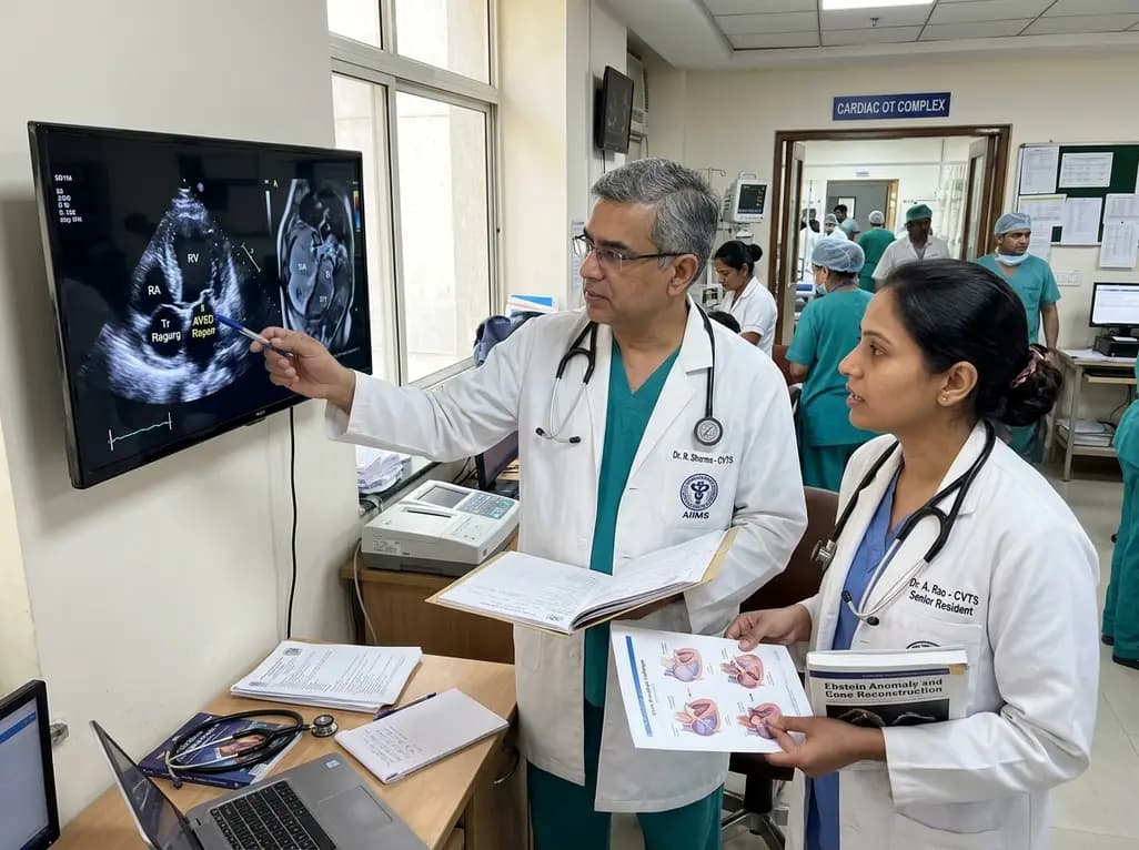
A review of the Cone procedure for severe right atrioventricular valve insufficiency in atypical Ebstein anomaly following partial AVSD repair, emphasizing operative technique and ventricular recovery.
Today

A Level I trauma center study shows a brief bedside mental health intervention boosts 30-day follow-up screening adherence by 84% in nonviolent injury patients. However, outcomes for violence survivors reveal critical care gaps, underscoring the urgent need for specialized community violence intervention programs.
Today
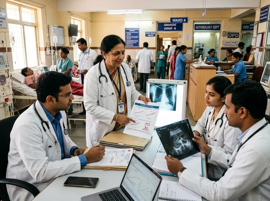
Recent data from Karnataka reveals a threefold surge in genitourinary and renal deaths over ten years. Driven by uncontrolled diabetes, hypertension, and emerging lifestyle hazards, this epidemiological shift highlights an urgent need for proactive screening and multidisciplinary nephrology management in primary care.
Today
